Glucose 6 phosphatase glucose G6 P F6 P


![Regulation of glycogen synthase Allosteric: G 6 P Phosphorylation : inhibits [Gsa] R active](https://slidetodoc.com/presentation_image/50894fa69f89f06b01215e81253f3755/image-9.jpg "Regulation of glycogen synthase Allosteric: G 6 P Phosphorylation : inhibits [Gsa] R active")


 G 6 P, ATP phosphorylation:")
 Phos kinase (liver, muscle)")


- Slides: 21
Glucose 6 phosphatase glucose G-6 -P F-6 -P fructose 1, 6 bis phosphatase F-1, 6 -bis. P DHAP G-3 -P 1, 3, bis-PG 3 -P-G 2 -P-G 1. pyruvate carboxylase 2. phosphoenol pyruvate carboxy kinase PEP Pyruvate
from lactate from pyruvate OAA pyruvate PEP pyruvate Asp malate mitochondrial matrix cytosol pyruvate Asp malate pyruvate lactate OAA alanine PEP G 3 P dehydro glucose
from lactate pyruvate from pyruvate Pyruvate carboxylase malate NADH dehydrog. PEPCK transaminase NAD PEP Asp malate OAA pyruvate mitochondrial matrix cytosol pyruvate LDH NADH lactate malate pyruvate malate transaminase NADHtransaminase dehydro. Asp OAA PEPCK PEP G 3 P dehydro glucose alanine
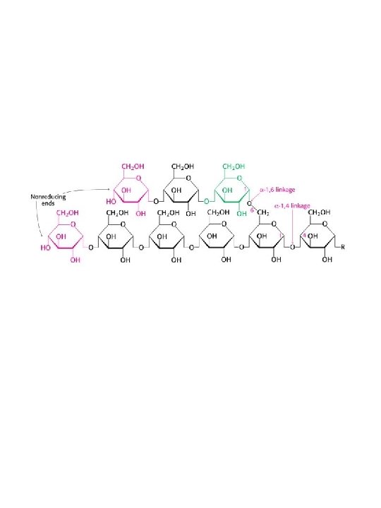
Cellulose b 1 -4 glucose Plants Animals Starch • amylose • amylopectin Glycogen a 1 -4 + a 1 -6 glucose
Structure of Glycogen A A A A B B B A • • A B B A A each B-chain has two branch points all chains have the same length of 14 residues the material is distributed at 50% between A- and B-chains. a molecule of glycogenin, a protein that acts as a primer, is located at the centre of the structure. See also amylopectin: fig 8 -24 in Horton amylopectin
Structure of Glycogen A A A B B 1 B B A B 2 3 5 4 A A B B B A A • • each B-chain has two branch points all chains have the same length of 14 residues the material is distributed at 50% between A- and B-chains. a molecule of glycogenin, a protein that acts as a primer, is located at the centre of the structure. • because synthesis and degradation takes place at non-reducing ends, branching provides more sites for these processes occur. See also amylopectin: fig 8 -24 in Horton amylopectin
After a meal as much as 10% by weight of the liver may be glycogen
Regulation of glycogen synthase Allosteric: G 6 P Phosphorylation : inhibits [Gsa] R active PKA PP 1 [GSb-PO 4]T inactive G 6 P [GSb-PO 4]R active
Phosphorylase cleaves a 1 -4 bonds from the non-reducing termini of glycogen to yield Glucose -1 -P
Glycogen debranching enzyme
Glycogen degradation: combined action of phosphorylase and debranching enzyme
a 1 6 branching enzyme a 1 6
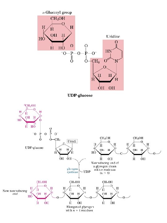
Glycogen synthesis: glycogen synthase and branching enzyme a 1 -4 linkage UDP-glucosyltransferase autocatalysis several cycles glycogen synthase several cycles Amylo(1, 4 1, 6) transglycosylase (branching enzyme) a 1 -6 linkage glycogen synthase/ branching enzyme multiple cycles
Regulation of phosphorylase Allosteric: AMP (muscle) G 6 P, ATP phosphorylation:
Regulation of phosphorylase: part 2 2 ATP T AMP (muscle) Phos kinase (liver, muscle) Phos b inactive G 6 P ATP R Phos b active 2 ADP 2 Pi PP 1 PO 4 T PO 4 Phos a inactive glucose (liver) PO 4 R PO 4 Phos a active
Co-ordinated regulation of phosphorylase and glycogen synthase by PKA GSb-PO 4 GSa active inactive PP 1 active PKA PP 1 -PO 4 inactive Phos kinaseb inactive See fig 13. 7 Horton Phos kinasea-PO 4 Ca 2+ active Phosb Phosa-PO 4 inactive
Gb, g G-protein linked receptor Ga Adenylate cyclase GDP GDP GTP GTP Pi ATP GDP c. AMP + PPi
3’ 5’ cyclic AMP Adenyl cyclase ATP 3’ 5’ cyclic AMP + PPi Cyclic nucleotide phosphodiesterase AMP 3’ 5’ cyclic AMP
Regulation of PKA R C inactive 4 c. AMP R R C C active