
GLOMERULAR DISEASES MED 341 Nov 27 2016 Objectives
GLOMERULAR DISEASES MED 341 Nov 27, 2016
Objectives 1 - To understand the pathophysiology of Glomerular Diseases. 2 - To correlate the clinical findings with the underlying renal pathology. 3 - To recognize the important features of Nephrotic syndrome. 4 - Learn the most common causes of NS in adults. 5 - To recognize the most important Glomerular diseases that cause Nephritic pattern ( Glomerulonephritis).

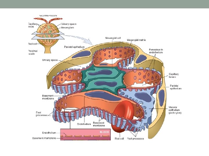
3 Renal cortex is the most important functional part of the kidney, because it has the Glomeruli Nephron (zoom)
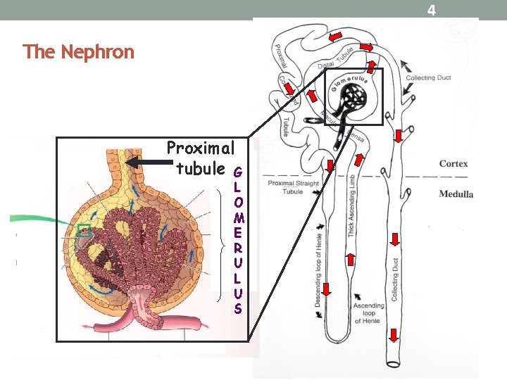
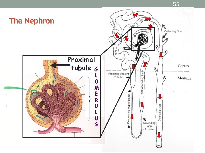
4 The Nephron Proximal tubule G L O M E R U L U S
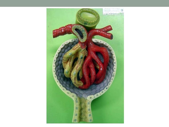
Microscopy Light Microscope 2000 x EM 10, 000
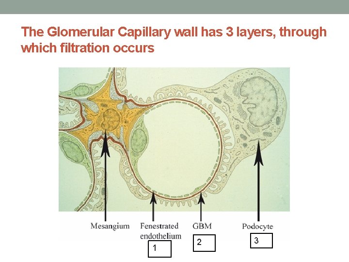
The Glomerular Capillary wall has 3 layers, through which filtration occurs 1 2 3
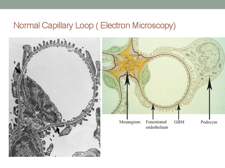
Normal Capillary Loop ( Electron Microscopy)
Normal Glomerular structure is needed to: • Keep the glomerular filtration normal, thus maintains normal kidney function. • keeps the urine volume maintained; so preventing fluid retention in the body which causes edema and high blood pressure. • Prevents the blood components (cells, proteins) from leaving the blood stream and appearing in the urine.
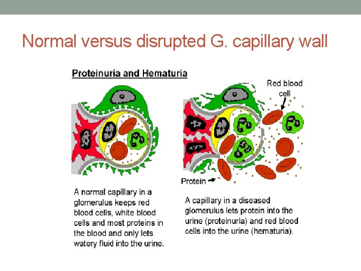
Normal versus disrupted G. capillary wall
So normal urine will have: • NO PROTEIN. • NO RED BLOOD CELLS ( Accept: <2 RBCs/High power field) • NO HEME. • NO CELLULAR CASTS. • No fat • No sugar
How glomerular diseases start? • Here we are talking about primary glomerular diseases that are mostly caused by immune system dysfunction. • Auto-antibodies targeting glomerular structure or immune- complexes (antigen-antibody) depositing and traumatizing the glomerular components.
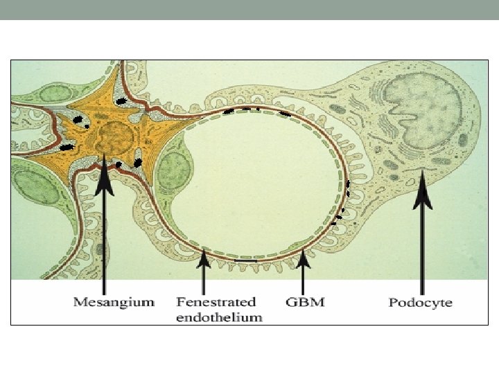
How glomerular diseases start? Most important to recognize: • The manifestations of a glomerular disease are usually indicative of which components of glomerular capillary wall was affected at the most. if Podocytes are the main target of the disease process >>>> mainly proteinuria ( at large amount) will manifest; thus Nephrotic Syndrome will be the main finding. if endothelial cells, Mesangial cells or GBM are affected>>>> mainly hematuria and abnormal renal function will manifest because of disruption in glomerular filtration wall; thus Nephritic pattern of renal disease will manifest. Proteinuria is always present in this kind of Glom injury as well.
Another important things to remember; >> Glomerular diseases are named based on their histopathological characteristics seen under the microscope >> So, almost always a kidney biopsy is needed to diagnose any suspected primary glomerular disease
• But to make things easier, we can put Glomerular diseases in two main clinical categories (clinical i. e. the symptoms, signs and laboratory abnormalities) >>> Nephrotic ( due to Podocytes dysfunction, so heavy proteinuria will be present) >>> Nephritic ( due to glomerular mesangial cells proliferation glomerular capillaries inflammation; so hematuria, impaired renal function and variable amount of proteinuria will be present)
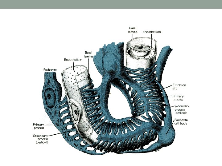
Nephrotic Syndrome (NS) • Podocytes abnormality is the primary finding in NS. • Podocytes will sustain a structural dysfunction; making them lose their Foot-processes, but the cells bodies are intact. • This will lead to significant amount of protein appearing in the urine (Proteinuria).
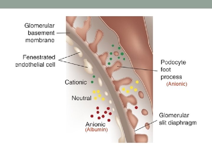
(Anionic) (Albumin)
Nephrotic Syndrome It refers to a constellation of clinical and laboratory features of renal disease: Hypoalbuminemia (serum Albumin <30 g/L) Normal serum Alb: 35 -55 g/L Ø Heavy proteinuria ( > 3. 5 g/24 hours of urine collection) Ø Peripheral or generalized edema Ø Hyperlipidemia Ø
Complications of Nephrotic Syndrome Infections & sepsis. Ø Thrombosis. Ø Acute kidney injury. Ø ESRD if heavy proteinuria does not resolve. Ø
Proteinuria How many milligrams of proteins are normally secreted in the urine per-day? • < 150 mg/day of all kinds of proteins. • Including on average 4 -7 mg/day of Albumin that are secreted in the urine normally.
Urine Analysis in Nephrotic Syndrome will show: • Lots of protein (Proteinuria) or called Nephrotic range proteinuria (>3. 5 g/24 h urine) • No RBCs ( some times few are occasionally seen) • No RBCs casts • Fat (Lipiduria)( Fatty casts, oval fat bodies & fat droplets) • No WBCs ( few may be seen)
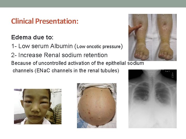
Clinical Presentation: Edema due to: 1 - Low serum Albumin (Low oncotic pressure) 2 - Increase Renal sodium retention Because of uncontrolled activation of the epithelial sodium channels (ENa. C channels in the renal tubules)
Clinical Presentation Patients also get: Ø Fatigue Ø Frothy urine (froth persists for long time after voiding) Ø Anorexia Ø Nausea & vomiting Ø Abdominal pain Ø Weight gain due to fluid retention Ø Shortness of breath if having pleural effusion Ø Signs & symptoms of DVT, PE
Glomerular Diseases that present as Nephrotic Syndrome 1 -Focal Segmental Glomerulo. Sclerosis (FSGS) 2 - Minimal Change Disease (MCD) 3 - Membranous Nephropathy (MN)

Focal Segmental Glomerulo. Sclerosis (FSGS) The primary type seen on light microscopy as: • Focal: some glomeruli are affected by sclerosis (the rest of them look normal) • Segmental: sclerosis only involves a segment of each glomerulus that is affected. • But most important; all glomeruli (the ones affected by sclerosis and the ones that are not affected) will have a diffuse foot processes effacement (thus Nephrotic syndrome appears)
Focal Segmental Glomerulo. Sclerosis (FSGS) • A common cause of Nephrotic syndrome in adults. • Causes 12 – 35 % of the cases in adults.
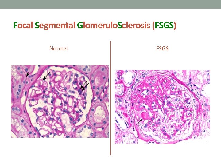
Focal Segmental Glomerulo. Sclerosis (FSGS) Normal FSGS
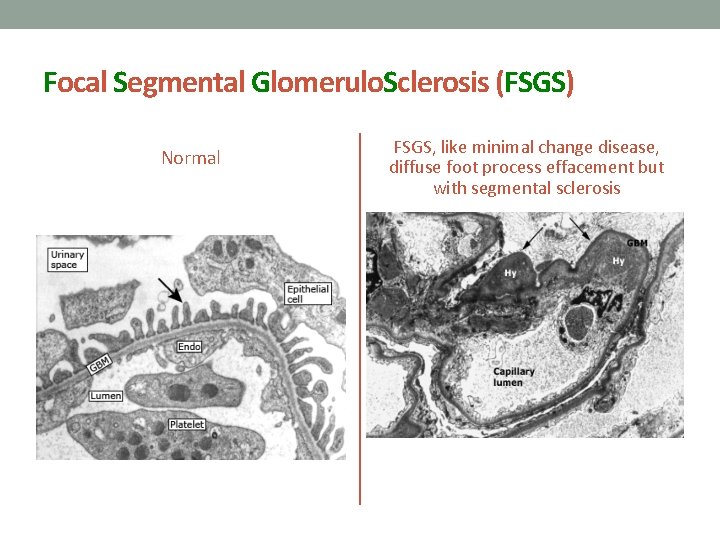
Focal Segmental Glomerulo. Sclerosis (FSGS) Normal FSGS, like minimal change disease, diffuse foot process effacement but with segmental sclerosis
Focal Segmental Glomerulo. Sclerosis (FSGS) Can be: Primary FSGS: Ø Has sudden onset of heavy proteinuria and other manifestations of nephrotic syndrome. Ø Usually treated with corticosteroids and other immunosuppressing medications.
Focal Segmental Glomerulo. Sclerosis (FSGS) Or can be Secondary FSGS: -Proteinuria is less heavy than other causes of nephrotic syndrome. -Serum Albumin is not very low like the primary type. -Renal impairment is commonly seen with the secondary FSGS and this is not a good prognostic sign
Focal Segmental Glomerulo. Sclerosis (FSGS) Possible causes of Secondary FSGS: - Obesity - Nephron loss ( > 75% of renal mass) e. g renal agenesis - Reflux nephropathy - Healing of prior GN (example Ig. A ) - Anabolic steroid abuse - Severe preeclampsia - Drugs: Interferon, Pamidronate, Heroin - Infections: HIV
Focal Segmental Glomerulo. Sclerosis (FSGS) Immunosuppressive therapy is indicated in most patients with primary FSGS - First line: corticosteroids - Second line: cyclosporine or tacrolimus Secondary FSGS: not typically treated with Immunosuppression, treat the primary cause and add supportive measures to protect the kidneys, e. g. keeping blood pressure well controlled with ACEi.

Minimal Change Disease (MCD) Called minimal because: - light microscopy: is typically showing normal glomeruli So called: nil disease. BUT: - electron microscopy: shows diffuse effacement of the epithelial cells’ foot processes only. - So the most important difference between MCD and the FSGS is the presence of glomerular sclerosis in FSGS - Here there is no sclerosis i. e. in MCD.
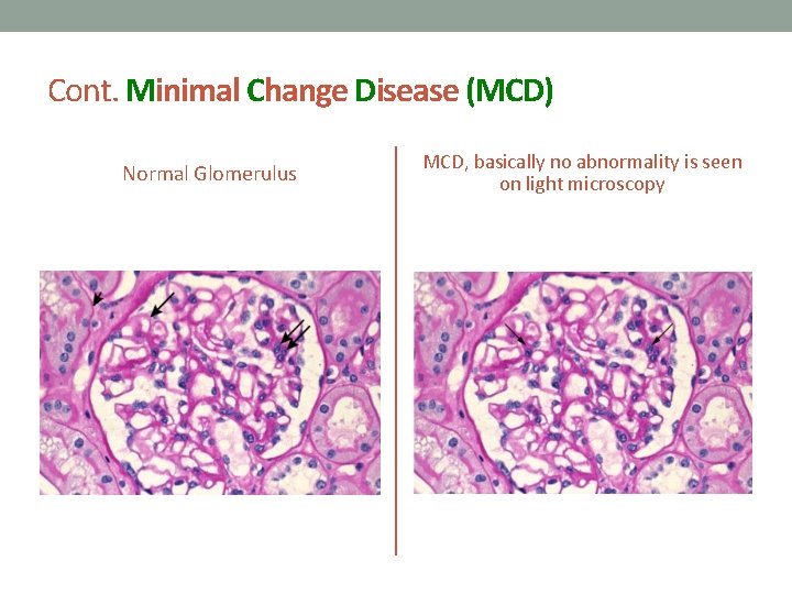
Cont. Minimal Change Disease (MCD) Normal Glomerulus MCD, basically no abnormality is seen on light microscopy
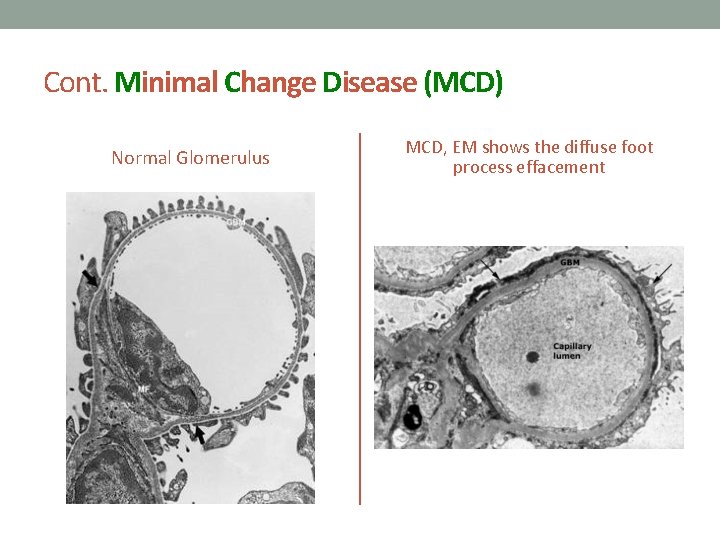
Cont. Minimal Change Disease (MCD) Normal Glomerulus MCD, EM shows the diffuse foot process effacement
Cont. Minimal Change Disease (MCD) It is the main cause of Nephrotic syndrome in children: - The cause in 90 % of cases in children < 10 years old. - > 50 % of cases in older children In children; typically is corticosteroid responsive in > 90%, thus kidney biopsy is commonly not done and treatment is given empirically for such cases. So, usually nephrotic syndrome in a child < 10 years old is MCD until proven otherwise. - It causes 10 -25 % of Nephrotic syndrome cases in adults
Cont. Minimal Change Disease (MCD) Can be : Primary (Idiopathic) or Secondary (much less common): Ø Drugs ( NSAIDs, Lithium, Sulfasalazine, Pamidronate, Dpenicillamine, some antibiotics) Ø Neoplasm ( Hodgkin Lymphoma, non-Hodgkin lymphoma, and leukemia) Ø Infections ( TB, syphilis ) Ø Allergy
Cont. Minimal Change Disease (MCD) Clinical presentation: Ø Typically has a sudden onset Edema Ø BP may be normal or slightly elevated Ø Heavy proteinuria (Nephrotic range) Ø Lipiduria Ø Hypoalbuminmia (usually very low serum Albumin) Ø Hyperlipidemia Ø Creatinine is always within the normal range or slightly elevated and normalizes with remission
Cont. Minimal Change Disease (MCD) Diagnosis: Must do kidney biopsy in adult patients with this presentation, It shows Diffuse effacement of foot process. Treatment: First line: Corticosteroids, given x 3 -4 months then taper over 6 months Second line: oral Cyclophosphamide, Cyclosporin
Membranous Nephropathy (MN) • Most common cause of Primary nephrotic syndrome in adults (15% and 33%) • Mostly secondary in children (hepatitis B antigenemia) • Presentation: slowly developing nephrotic syndrome
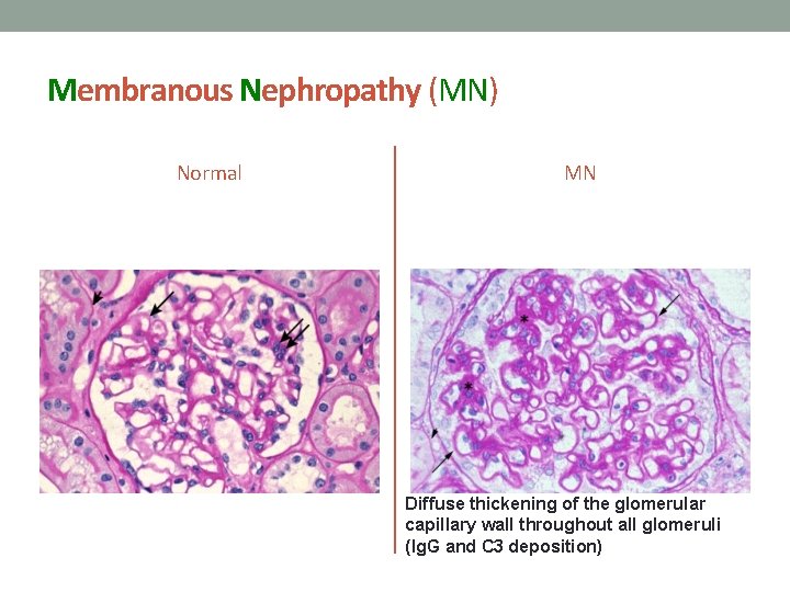
Membranous Nephropathy (MN) Normal MN Diffuse thickening of the glomerular capillary wall throughout all glomeruli (Ig. G and C 3 deposition)
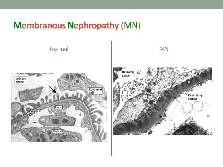
Membranous Nephropathy (MN) Normal MN
Membranous Nephropathy (MN) Etiology: Ø Primary (Idiopathic) In approximately 75% of cases in adults.
Membranous Nephropathy (MN) Secondary: causes of MN: Systemic lupus erythematosus (SLE) Class V Lupus Nephritis (10 -20%) Ø Drugs: penicillamine, IV gold salts, high dose Captopril, and NSAIDs, Anti-TNF. Ø Infections: Hepatitis B, Hepatitis C, syphilis Ø Malignancy: solid tumors prostate, lung, or GI track Ø
Membranous Nephropathy (MN) Treatment of Primary MN - Corticosteroids plus - Cyclophosphamide or cyclosporine - May be Rituximab Secondary MN - Mainly target the primary disease that caused MN, and treat the Nephrotic syndrome manifestations.
Other important secondary causes of Nephrotic syndrome in adults: • Diabetes Mellitus • Amyloidosis • Ig. A Nephropathy • MPGN
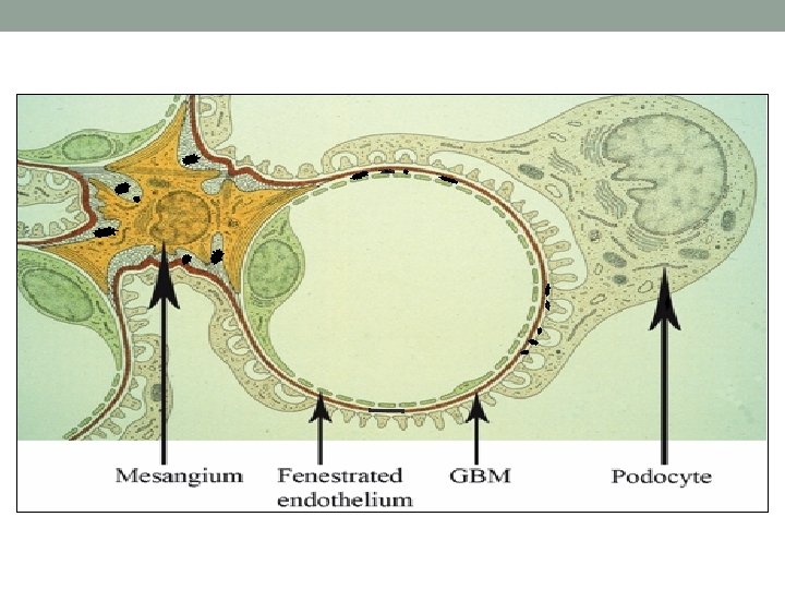
Nephritic Glomerular diseases When we say Nephritic; it means a clinical pattern of presentation for a group of GNs, and not a syndrome like what we saw in Nephrotic causes. The Nephritic pattern is always indicative of underlying inflammatory process in the glomeruli; causing inflammatory modulators attraction, cellular proliferation and eventually glomerular permanent dysfunction if left untreated. The Glomerular mesangium, endothelium and GBM components of the Glomerulus are likely going to be targeted because of their proximity to blood circulation.
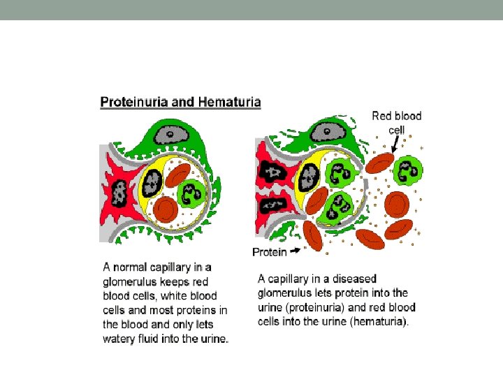
Nephritic urine analysis shows: • Red Blood Cells (RBCs) • RBCs casts, or cellular casts • Dysmorphic RBCs (RBCs lose their smooth surface passing through the cracks in inflamed glomerular basement membrane) • Protein (at variable amount from mgs to grams per day) Those are called Active Urinary Sediments ( Active = is indicative of underlying glomerular inflammatory process; requiring urgent medical attention)

RBCs cast formed by naturally occurring Tamm-Horsfall mucoprotein in the distal tubules & collecting ducts when they become loaded with RBCs coming from the inflamed Glomerulus (due to GN)
55 The Nephron Proximal tubule G L O M E R U L U S
Nephritic clinical manifestations: • AKI (Acute Kidney Injury) =Acute Renal impairment or Failure= elevated Creatinine) • Decreased Urine output • Edema • High Blood Pressure • May have other manifestations of systemic vasculitis since some GN types are actually vasculitis (e. g. skin rash, pulmonary hemorrhage, etc) • Positive immune markers: ANA, Anti-DNA, low complements, +ve ANCA (depends on the cause)
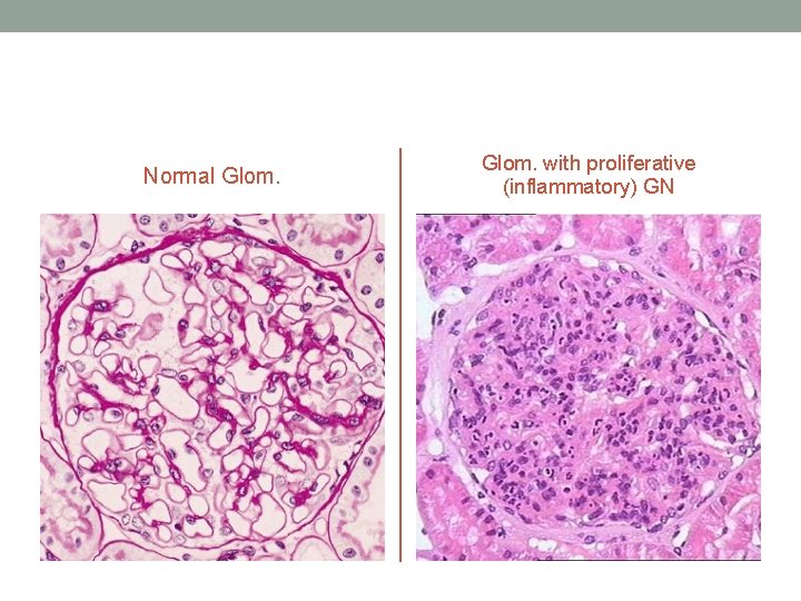
Normal Glom. with proliferative (inflammatory) GN
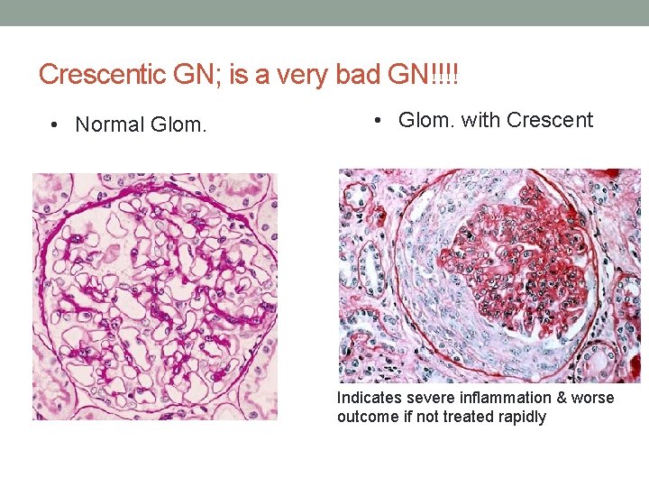
Crescentic GN; is a very bad GN!!!! • Normal Glom. • Glom. with Crescent Indicates severe inflammation & worse outcome if not treated rapidly
Nephritic Glomerular diseases Here; they are called GN=Glomerulonephritis Renal Diseases that can present with Nephritic picture: Ø Ig. A Nephropathy / HSP (Henoch-Schönlein purpura) Ø Post streptococcal glomerulonephritis (PSGN) Ø Lupus Nephritis Ø Anti-GBM (Goodasture’s disease) Ø ANCA vasculitis ( e. g. Wegner’s Granulomatosis) Ø Membranoproliferative GN (MPGN)
Ig. A Nephropathy • Most common type of Primary GN in developed countries • Can present as dark urine (hematuria) 1 -3 days after upper respiratory tract infection. (< one week of URT infection) • A lot of times it gets picked up incidentally by finding abnormal urine analysis (Hematuria+/- Proteinuria) done for other reasons with no symptoms. • It has a chronic course that can progress to ESRD. • Needs kidney biopsy to reach the diagnosis. • The diagnosis is made by finding abnormal deposition of Ig A immunoglobulin in the Glomeruli, it elicit a local inflammatory response in the Glom mesangium (mesangial expansion)
• It is thought to be secondary to altered mucosal immunity that leads to excessive Ig. A synthesis followed by deposition in the gloms. • There is really no effective immunosuppressing therapy except in severe cases where it can be tried. • Most important treatment is to control the blood pressure which also decreases the proteinuria. • HSP (Henoch-Schönlein purpura) is a systemic vasculitis caused by immune deposition of Ig. A in different organs; typically skin, bowel and kidneys.
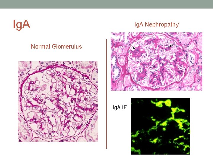
Ig. A Nephropathy Normal Glomerulus Ig. A IF

Post streptococcal glomerulonephritis (PSGN) • Typically caused by throat infection with Gram positive cocci • • (Group A beta-hemolytic Streptococcus (GAS). But also can be caused by Staphylococcus soft tissue or bone infection in adults. Bacterial Antigen cross react with Glom antigens, or may be an immune-complex (Antigen-antibody) response that is responsible. Patients present with frank hematuria usually after one week and up to 3 weeks from the start of infection. Serum will show positive Antistreptolysin (ASO) titer. Low C 3, Normal or slightly low C 4 in the serum. May have positive throat culture. Children have better and faster recovery than adults. Treatment is usually supportive= wait and see.
Lupus Nephritis • Lupus (SLE): The Disease with a Thousand Faces • Kidneys can be affected by SLE like other organs. • The degree of involvement can be from mild (or even not visible to the physician) to a very severe one causing ESRD in few months. • Most important in dealing with these cases is having high suspicion of its presence and to start immediate workup & referral for diagnosis and treatment.
Lupus Nephritis • Kidney biopsy is mandatory to make the diagnosis. • Low complements (C 3, C 4) level along with the positive Lupus markers, abnormal urine analysis & abnormal renal function should make you think of its presence. • Lupus Nephritis treatment depends on the findings in renal biopsy. • It usually involves high degree of immunosuppressing medications.
ANCA vasculitis • Autoimmune disease that involves the presence of Neutrophils adhesion enhancing molecule called ANCA=Anti-neutrophil cytoplasmic antibody Two types of ANCA: 1 - C-ANCA= Cytoplasmic type, more commonly causing Granulomatous Polyangiitis = old name Wegner’s Granulomatosis ( so a granuloma forming disease) Angiitis: means small vessels vasculitis 2 - P-ANCA= Perinuclear type, more commonly associated with Microscopic Polyangiitis & Churg-Strauss syndrome
ANCA vasculitis • Upper airways and lung involvement is common and patients can present with renal and pulmonary manifestations (GN + Pulmonary hemorrhage: hemoptysis) • Diagnosis is made by kidney biopsy and positive ANCA titer in the serum. • It is usually an aggressive disease that should be treated with potent immunosuppressing medications. (high dose corticosteroids & cyclophosphamide).
Anti-GBM antibody disease (Anti Glom Basement Membrane) • Due to autoantibody against (alpha-3 chain) of type IV Collagen that is found in Glomerular & alveolar (lungs) basement membrane. • So the manifestations will be: 1 - GN (can be the only presenting finding) & 2 - Pulmonary hemorrhage (if with GN; is called Goodpasture’s disease = Lungs + renal involvement. 3 - positive test for Anti-GBM antibodies in the serum 4 - Kidney biopsy shows the diagnostic Immunofluorescence pattern : Linear stain of Ig. G and C 3
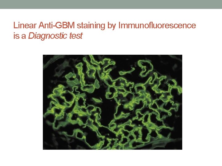
Linear Anti-GBM staining by Immunofluorescence is a Diagnostic test
Anti-GBM • Treatment is always started immediately to remove the antibodies by Plasmapheresis and preventing further antibodies production by giving heavy immunosuppression that includes corticosteroids and cyclophosphamide.
Membranoproliferative GN (MPGN) It is a pathological description & has multiple causes. It may present with Nephritic picture or Nephrotic syndrome The primary (idiopathic) MPGN is mainly seen in children. The secondary type is seen in adults due to: - Hepatitis B and C - Endocarditis - Lupus and Sjogren’s syndrome - Cancer - Complement deficiency
- Slides: 71