Glomerular diseaseincludes glomerulonephritis i e inflammation ofthe glomeruli

. �Rapidly progressive glomerulonephritis. �Asymptomatic urinary abnormality (haematuria,")


 • • • Epithelial cell")


 � proliferation of cells within the glomeruli, accompanied by leukocyte")


, �nephritic (edemas, hypertension, gross haematuria, proteinuria), �nephrotic (edemas, proteinuria, hypoproteinemia, hypercholesterolemia),")
. �non-nephrotic range proteinuria (<2 g")
: cast) a) Urine microscopy")
 are used to control")


 type II (intramembranous deposits)")


 �Hypertensive (increased blood pressure) �Hematuric �Nephrotic (edemas, proteinuria, hypoproteinemia, hypercholesterolemia),")


 Corticosteroids induce remission in >90%of children and 80%of adults (slowerresponse). Indications for immunosuppression:")
. Cyclophosphamide or ciclosporin (=cylosporin) may be used in")


 is a disease of the kidney that results in a rapid decrease")


 anti-GBM antibody disease (approximately 3%of cases),")
.")


- Slides: 56
�Glomerular diseaseincludes glomerulonephritis, i. e. inflammation ofthe glomeruli andglomerulopathies whenthere is no evidenceofinflammation. �Glomerulonephritis is asubsetof glomerulopathies
�Nephrotic syndrome. �Acute glomerulonephritis (Acute nephritic syndrome). �Rapidly progressive glomerulonephritis. �Asymptomatic urinary abnormality (haematuria, proteinuria or both).
� Primary – confinedto thekidney � Secondary –dueto asystemicdisease
� Proteinuria – asymptomatic � Haematuria – asymptomatic � Hypertension � Nephrotic syndrome � Nephritic syndrome � Acute renal failure � Rapidly progressive renalfailure � End stage renalfailure
� Presence of glomerular disease as opposed to tubulointersititial or vascular disease is suspectedfrom history � Haematuria (especially dysmorphic redcells) � Red cell casts � Lipiduria (glomerular permeability must be increased toallow the filtration oflarge lipoproteins) � Proteinuria (may be in nephrotic range of >3. 5 g/24 hours)
Immune complex disease Complement- dependent • C₅- C₉ (MAC) • • • Epithelial cell detachment. (+)epithelial &mesangial cells to secrete damaging chemical mediators. Neutrophils: Protease • O₂ free readicals AA metabolites • Upregulates TGF receptors onepithelial cells, excessive synthesis of extracellular matrix which leads to GBM thickening • GBM degradation cell damage • ↓ GFR Complement-leukocyte- mediated mechanism Activation of the complement pathway Recruitment of neutrophils and monocytes
�Namedaccording to �etiology �microscopic findings �clinical syndrome �Most common clinicalpresentations �acute nephritic syndrome �nephrotic syndrome �Most common cause isautoimmune
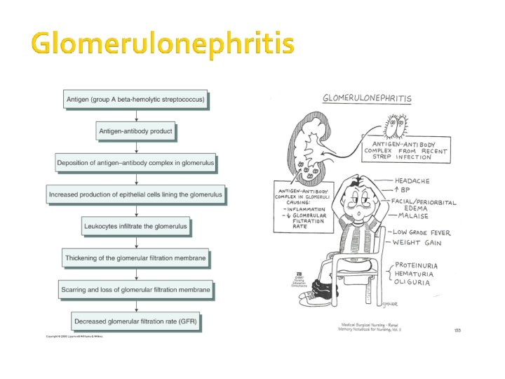
� Autoimmune injury initiated bybeta-hemolytic streptococcus � akaacuteproliferativeglomerulonephritis � Presents as acute nephriticsyndrome � hematuria � HT � increased urea & creatinine low � urineoutput � edema � Antibodies produced by strep throat deposit in glomerulus � Most fully recover but about 10%evolve into rapidly progressive glomerulonephritis
� � � Unknown causesor secondaryto poststreptococcal glomerulonephritis Autoimmune akacrescentric glomerulonephritis Some present as acutenephritic syndrome & others as renal failure Caused by deposition of. An-Ab complexes All but a few progress to renal failure
� Autoimmune � Most common cause of nephrotic syndrome in adults � About 10%proceed torenal failure within 10 yrs, 25%recover completely, most progress slowly with proteinuria, HTN, loss of renalfunction
�Incidental discovery of occult proteinuria or. HTN �Usually presents as chronic renal failure oroccult proteinuria �Glomerulus has scartissue �Dialysis &transplant
�Diabetes most commoncause �most common cause of renalfailure �glycoproteins deposit in basementmembrane � Vascular disease � atherosclerosis �HTN �vascultitis
Heavy proteinuria Proteinuria & haematuria Predominant haematuria Minimal Change Lupus nephritis Acute post strep Focal sclerosis Membranoproliferative Crescentic (RPGN) Endocarditis Haemolytic uraemic syndrome Membranous Diabetes Mellitus Amyloidosis Henoch- Schonlein purpura
�Acute glomerulonephritis is the inflammationof the glomeruliwhichcauses the kidneys to malfunction �Itis also called Acute Nephritis, Glomerulonephritis and. Post-Streptococcal Glomerulonephritis �Predominantly affects children from agesto 2 12 �Incubation period is 2 to 3 weeks
�Infectious �Streptococcal �Nonstreptococcal postinfectious glomerulonephritis ▪ Bacterial ▪ Viral ▪ Parasitic �Noninfectious �Multisystemicdiseases �Primary glomerular diseases
Previously M-protein ofthe organism was felt to be responsible for PSGN. Recently, nephritis-associated streptococcal cationic protease andits zymogen precursor (NAPR) has been identified as aglyceraldehyde-3 -phosphate dehydrogenase that functions as a plasmin(ogen) receptor.
Diffuse proliferative GN (PGN) � proliferation of cells within the glomeruli, accompanied by leukocyte filtrate � typical features of immune complex disease : - hypocomplimentemia - granular deposits of Ig. G & complement on GBM � Implicated antigens seem to be endostreptosin and nephritisplasmin– binding ptn
� Fever � Headache � Malaise � Anorexia � Nausea and vomiting � High blood pressure � Pallor due to edema and/oranemia � Confusion � Lethargy � Loss of muscletissue � Enlargement of theliver
�Hematuria: dark brown or smokyurine �Oliguria: urine output is <400 ml/day �Edema: starts in the eye lids and face thenthe lower and upper limbs thenbecomes generalized; maybemigratory �Hypertension: usually mild to moderate
�urinary (haematuria, proteinuria), �nephritic (edemas, hypertension, gross haematuria, proteinuria), �nephrotic (edemas, proteinuria, hypoproteinemia, hypercholesterolemia), �mixed.
�Abruptonset of: � glomerular haematuria (RBCcasts or dysmorphic. RBC). �non-nephrotic range proteinuria (<2 g in 24 hrs). � oedema (periorbital, sacral). � hypertension. � transient renal impairment (oliguria, uraemia).
Base line measurements: - ↑ Urea - ↑Creatinine Urinalysis (MSU): cast) a) Urine microscopy (red cell b) proteinuria COMPLICATION Hypertensive encephalopathy, heart failure and acute pulmonary edema may occur in severe cases Acute renal necrosis due to injury of capillary or capillary thrombosis
� Treattheunderlyinginfections whenacute. GNisassociatedwith chronicinfections. Antimicrobialtherapy � Antibiotics (eg, penicillin) are used to control local symptoms and to prevent spread of � infection toclose contacts. � Antimicrobial therapy does not appear to prevent the development of GN, except ifgiven within the first 36 hours. Loopdiuretictherapy � Loop diuretics maybe required in patients whoare edematous and hypertensive in order to remove excess fluid and to correct hypertension. � Relieves edema and controls volume, thereby helping to control volume-related elevation in BP. � Vasodilatordrugs(eg, nitroprusside, nifedipine, hydralazine, diazoxide) maybe used ifsevere hypertension or encephalopathy is present � Diet: � Sodium and fluid restriction � Protein restriction forazotemic patients � Activity: Recommend bed rest until signs of glomerular inflammation and circulatory congestion subside.
Post streptococcal GN - Has a GOOD prognosis. - Supportive measures until spontaneous recovery. - Control HT. - Fluid balance. - Oliguric with fluid overload. -GN complicating SLE or systemic vasculitides: immunosuppression with prednisolone, cyclophosphamide or azathioprine/MMF.
The condition is characterized by irreversible and progressive glomerularand tubulointerstitial fibrosis. ->ultimately leading to a reduction in the glomerular filtration rate (GFR) andretention of uremictoxins. ->If disease progression is nothalted with therapy, the net result is chronic kidney disease (CKD), end-stage renal disease (ESRD), and cardiovasculardisease
Nearly all forms of acute glomerulonephritis have a tendency to progress tochronic glomerulonephritis. The progression from acute glomerulonephritisto chronic glomerulonephritis isvariable. Whereas complete recovery of renal function is the rule for patients with poststreptococcal glomerulonephritis, severalother glomerulonephritides, such as immunoglobulin. A (Ig. A) nephropathy, often have a relativelybenign course and manydo not progress to. ESRD.
Reduction in nephron massfrom the initialinjury reduces the GFR. This reduction leads to hypertrophy and hyperfiltration of the remaining nephrons and to the initiation of intraglomerular hypertension. These changes occur in order to increase the GFR of the remaining nephrons, thus minimizing the functional consequences of nephronloss. The changes, however, are ultimatelydetrimental because they lead to glomerulosclerosis andfurther nephron loss.
fusion of podocytes on electronmicroscopy
Segmentalareasof glomerular sclerosis, hyalinization of glomerular capillaries
proliferation and ‘double’ BM. 2 histological types: type I(subendothelial deposits) type II (intramembranous deposits)
thickened BM, IF +vefor Ig. G &C 3 and subepithelial deposits on. EM
Hypercellularity, mesangial proliferation, inflammatory cell infiltrate, positive IF for Ig. G and C 3 and subepithelial deposits on EM.
Most common cause of GN in Asia but uncommon in Sth America or. Africa � 15 -40%of allbiopsy proven GN � Male >Females � 2 nd-3 drdecade � Most commonly asymptomatic with serendipitous findingof haematuria and mild proteinuria � Another classic presentation is macroscopic haematuria in conjunction with aviralinfection � Renal function is usually normal but occasionally a patient will present with acute renal failure due to acute tubular necrosis secondary to the grosshaematuria � Biopsy – mild to moderate mesangial cell proliferation, Ig. Adeposits in the mesangium on immunofluorescence, often with C 3 deposition also �
�Slowly progressive �By 20 years, 50%have end stage kidney disease �Worse prognosis if >1 g/dayproteinuria, hypertension, increased creatinine of glomerular fibrosis at biopsy, onpresentation
� Uremia-specific findings � Edemas � Hypertension � Jugular venous distension (if severe volume overload s ipresent) � Pulmonary rales (if pulmonary edema ispresent) � Pericardial friction rub inpericarditis � Tenderness in the epigastric region or blood in the stool (possible indicators foruremic gastritis or enteropathy) � Decreased sensation and asterixis (indicators for advanced uremia)
�Latent (changes in urine) �Hypertensive (increased blood pressure) �Hematuric �Nephrotic (edemas, proteinuria, hypoproteinemia, hypercholesterolemia), �Mixed
� Urinalysis � Urinary proteinexcretion � Serumchemistry � Serumcreatinineandureanitrogenlevelsareelevated. � Impairedexcretionof potassium, freewater, andacidresults inhyperkalemia, hyponatremia, andlowserumbicarbonate levels, respectively. Impairedvitamin. D-3 productionresultsinhypocalcemia, � hyperphosphatemia, andhighlevelsof parathyroid hormone. Lowserumalbuminlevelsmaybepresentif uremia interfereswith nutritionorif thepatientisnephrotic. �
�Renal ultrasonogram �Obtain a renal ultrasonogram to determine renal size, to assess for the presence of both kidneys, and to exclude structural lesions that maybe responsible for azotemia. �Small kidneys often indicate an irreversible process. �Kidney biopsy
� The target pressurefor patients with proteinuria greater than 1 g/d is less than 125/75 mm. Hg; for patients with proteinuria lessthan 1 g/d, the target pressure is lessthan 130/80 mm. Hg. � Angiotensin-convertingenzymeinhibitors(ACEIs) angiotensin � II receptorblockers(ARBs) combination therapy with ACEIs � and. ARBs. � Diuretics � Beta-blockers, � calciumchannelblockers, � centralalpha-2 agonists(eg, clonidine), alpha-1 � antagonists � directvasodilators(eg, minoxidil, nitrates) maybe used toachieve the targetpressure.
� Renal osteodystrophy can be managed early by replacing vitamin D and by administeringphosphate binders. � Seek and treat nonuremic causes of anemia, suchas iron deficiency, before institutingtherapy with erythropoietin. � Discuss options for renal replacement therapy (eg, hemodialysis, peritoneal dialysis, renal transplantation). � Treat hyperlipidemia (if present) � Expose patients to educational programs for early rehabilitation from dialysis ortransplantation.
Minimalchangeglomerulonephritis(MCGN) Corticosteroids induce remission in >90%of children and 80%of adults (slowerresponse). Indications for immunosuppression: (cyclophosphamide, ciclosporin (=cylosporin)): early/ frequent relapses; steroid SEs/dependence. Prognosis: 1%progress to. ESRF.
Focalsegmentalglomerulosclerosis Poor response to corticosteroids(10– 30%). Cyclophosphamide or ciclosporin (=cylosporin) may be used in steroid-resistant cases. Prognosis: 30– 50%progress to ESRF.
Mesangiocapillary. GN Treatment: None is ofproven benefit. Prognosis: 50%develop ESRF.
Membranousnephropathy Ifrenal function deteriorates, consider corticosteroids andchlorambucil. Prognosis: Untreated, 15%complete remission, 9%ESRF at 2– 5 yrs and 41%at 15 yrs.
Mesangialproliferative. GN Antibiotics, diuretics, and antihypertensives as necessary. Dialysisis rarely required. Prognosis: Good.
�Management �Aggressive control of blood pressure and proteinuria with ACEI’s or AR 2 B’s �Corticosteroids +/-azathiprine – varied schools ofthought �However if rapidly progressive GN with crescent deposition treatment should be aggressive with high dose steroids andcyclophosphamide �Consult the Nephrologist
glomerulonephritis (RPGN) is a disease of the kidney that results in a rapid decrease inthe glomerular filtration rate of at least 50%over a short period, from a few days to 3 months.
� Classic – haemoptysisafter upperrespiratory infection and have nephritic urinary sediment � History of smoking or hydrocarbon exposure is common � CXR – pulmonaryhaemorrhage � Lab- iron deficiency anaemia and renal dysfunction, circulating anti-GBM antibodies � Kidney biopsy crescentic GN with linear staining Ig. G and C 3 along the glomerular basementmembrane 50
More than 80%of patients with pauciimmune RPGN were subsequently found to have circulating antineutrophil cytoplasmic antibodies (ANCA), and thus, this form of RPGN is now termed ANCA-associated vasculitis.
RPGN is classified pathologically into 3 categories: �(1) anti-GBM antibody disease (approximately 3%of cases), �(2) immune complex disease (45%of cases), �(3) pauci-immune disease (50%of cases).
�Symptoms and signs of renalfailure, �pain, �haematuria, �systemic symptoms (fever, malaise, myalgia, weight loss).
• � Laboratory studies theof following: The most important requirementinclude in the diagnosis antineutrophil cytoplasmic antibodies (ANCA) ANCA-associated diseaseis ahigh index of suspicion. Rapiddiagnosisisessentialfor organmost preservation. �Routine chemistry: The commonabnormality is anincreasedserum creatininelevel. �Urinalysis withmicroscopy: �Antinuclear antibody (ANA)titer: �ANCA.
High-dose corticosteroids; cyclophosphamide ±plasma exchange/ renal transplantation. Prognosis: Poor if initial serum creatinine >600µmol/L.
THANK YOU