Globular Proteins Respiratory Block 1 Lecture Objectives n

Globular Proteins Respiratory Block | 1 Lecture

Objectives n To describe the globular proteins using common examples like hemoglobin and myoglobin. n To study the structure and functions of globular proteins liken n n Hemoglobin (a major globular protein) Myoglobin, and g-globulins (immunoglobulins) n To know the different types of hemoglobin and difference between normal and abnormal hemoglobin n To understand the diseases associated with globular proteins

Globular proteins n Amino acid chains fold into shapes that resemble spheres are called globular proteins n This type of folding increases solubility of proteins in water Polar groups on the protein’s surface n Hydrophobic groups in the interior n n Fibrous proteins structural proteins are mainly insoluble

Globular proteins n Hemoglobin: oxygen transport function n Myoglobin: oxygen storage/supply function in heart and muscle n a 1, a 2, b-globulins: various functions n g-globulins (immunoglobulins): immune function n Enzymes: catalysis of biochemical reactions

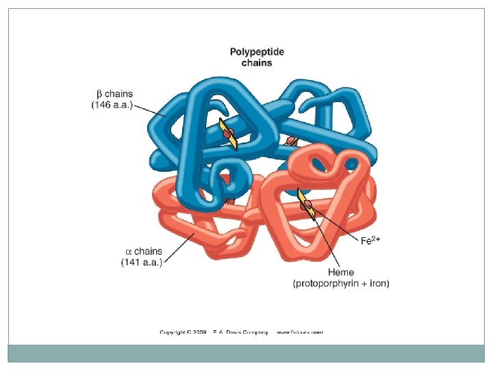
Hemoglobin �A major globular protein in humans �Composed of four polypeptide chains: Two a and two b chains �Contains two dimers of ab subunits Held together by non-covalent interactions �Each chain is a subunit with a heme group in the center that carries oxygen �A Hb molecule contains 4 heme groups and carries 4 molecules of O 2
 Hb. A 2 (2%) Hb. F")
Types of Hb Normal: Abnormal: Hb. A (97%) Hb. A 2 (2%) Hb. F (1%) Hb. A 1 c Carboxy Hb Met Hb Sulf Hb

Hb. A structure Oxygen binding to hemoglobin

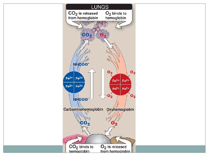
Hemoglobin function n Carries oxygen from the lungs to tissues n Carries carbon dioxide from tissues back to the lungs n Normal level (g/d. L): • • Males: 14 -16 Females: 13 -15
: n Major hemoglobin found in the fetus")
Types of hemoglobin Fetal hemoglobin (Hb. F): n Major hemoglobin found in the fetus and newborn n Tetramer with two a and two g chains n Higher affinity for O 2 than Hb. A n Transfers O 2 from maternal to fetal circulation across placenta

Types of hemoglobin Hb. A 2: n Appears ~12 weeks after birth n Constitutes ~2% of total Hb n Composed of two a and two d globin chains

Types of hemoglobin Hb. A 1 c: n Hb. A undergoes nonenzymatic glycosylation n Glycosylation depends on plasma glucose levels n Hb. A 1 c levels are high in patients with diabetes mellitus NH 2 groups of the N-terminal valines of β chains

Abnormal Hbs Unable to transport O 2 due to abnormal structure: n Carboxy-Hb: CO replaces O 2 and binds 200 X tighter than O 2 (in smokers) n Met-Hb: Contains oxidized Fe 3+ (~2%) that cannot carry O 2 n Sulf-HB: Forms due to high sulfur levels in blood (irreversible reaction)

Hemoglobinopathies n Disorders of hemoglobin caused by: n Synthesis of structurally abnormal Hb n Synthesis of insufficient quantities of normal Hb n Combination of both
 disease �Caused by a single mutation in b-globin gene")
Hemoglobinopathies Sickle cell (Hb. S) disease �Caused by a single mutation in b-globin gene �Glutamic acid at position 6 in Hb. A is replaced by valine �The mutant Hb. S contains bs chain �The shape of RBCs become sickled � Causes sickle cell anemia

Hemoglobinopathies Hemoglobin C disease: �Caused by a single mutation in b-globin gene �Glutamic acid at position 6 in Hb. A is replaced by lysine �Causes a mild form of hemolytic anemia
 state n")
Hemoglobinopathies Methemoglobinemia: n Caused by oxidation of Hb to ferric (Fe 3+) state n Methemoglobin cannot bind oxygen n Caused by certain drugs, reactive oxygen species and NADH-cytochrome b 5 reductase deficiency n Chocolate cyanosis: brownish-blue color of the skin and blood

Hemoglobinopathies Thalassemia: n Defective synthesis of either a or b-globin chain due to gene mutation n a-thalassemia: n n Synthesis of a-globin chain is decreased or absent Causes mild to moderate hemolytic anemia n b-thalassemia: n n n Synthesis of b-globin chain is decreased or absent Causes severe anemia Patients need regular blood transfusions

Myoglobin �A globular hemeprotein in heart and muscle �Stores and supplies oxygen to the heart and muscle only �Contains a single polypeptide chain forming a single subunit with eight a-helix structures �The interior of the subunit is composed of nonpolar amino acids

Myoglobin n The charged amino acids are located on the surface n The heme group is present at the center of the molecule n Myoglobin gives red color to skeletal muscles n Supplies oxygen during aerobic exercise

Structure of myoglobin

Myoglobin in disease n Myoglobinuria: Myoglobin is excreted in urine due to muscle damage (rhabdomyolysis) n May cause acute renal failure n Specific marker for muscle injury n Less specific marker for heart attack

Immunoglobulins n Defensive proteins produced by the B-cells of the immune system n Y-shaped structure with 2 heavy and 2 light polypeptide chains n Neutralize bacteria and viruses n Types: Ig. A, Ig. D, Ig. E, Ig. G, Ig. M

Take Home Messages �Amino acid chains fold into shapes that resemble spheres are called globular proteins. �Fibrous proteins are mainly insoluble, while globular proteins are soluble structural proteins. �Hb, Myoglobin, globulines and enzymes are examples of globular proteins. �Functionally, Hb is for O 2 and CO 2 transport. �Hb. A, Hb. A 2 and Hb. F are examples of normal Hb, in which the tetrameric structure is composed of 2α constant subunits with 2 changeable β subunits according to Hb type.

Take Home Messages n Hb. A 1 C is a Hb. A which undergoes non-enzymatic glycosylation, depending on plasma glucose levels. n Carboxy-Hb, Met-Hb and Sulf-Hb are examples of abnormal Hb, in which O 2 molecules are not transported due to abnormal Hb structure. n Disorders of Hb caused by synthesis of structurally abnormal Hb and/or insufficient quantities of normal Hb. n Sickle cell (Hb. S) and Hb. C diseases are caused by a single mutation in β-globin gene.

Take Home Messages �Glu 6 in Hb. S is replaced by Val, while it is replaced by Lys in Hb. C. �Methemoglobinemia is caused by oxidation of Hb, inhibiting O 2 binding leading to chocolate cyanosis. �Thalassemia is caused by a defect in synthesis of either α- or β-globulin chain, as a result of gene mutation. �α-Thalassemia causes less sever anemia than βThalassemia. �Myoglobin is a globular hemeprotein, which stores and supplies O 2 to the heart and muscle only.
, while Myoglobin is composed")
Take Home Messages �Hb is composed of 4 chains (subunits), while Myoglobin is composed of a single chain. �Myoglobinuria is a specific marker for muscle injury and may cause acute renal failure. �Immunoglobulins are defensive proteins produced by the B-cells. �Immunoglobulins consist of 5 types: Ig. A, Ig. D, Ig. E, Ig. G and Ig. M.

References n Illustrations in Biochemistry by Lippincott 6 th edition.
- Slides: 29