Glands Most glands are formed during development by

Glands Most glands are formed during development by proliferation of epithelial cells so that they project into the underlying connective tissue. q Some glands retain their continuity with the surface via a duct and are known as EXOCRINE GLANDS. q Other glands lose this direct continuity with the surface when their ducts degenerate during development. These glands are known as ENDOCRINE glands. q MIXOCRINE : Both endocrine + Exocrine e. g. Pancreas , Secretes digestive enzymes in the intestine & insulin hormone to the blood

Patterns of Hormone Action q Endocrine: circulated by blood to distant target cells. q Paracrine: Hormones that affect neighboring cells q Autocrine: Hormones that act on the cells that secrete them q Neuroendocrine q Neurotransmitter
 are secreted")
Endocrine Glands • Endocrine glands do not have ducts. Their secretions (hormones) are secreted into the blood stream. • The secretory cells of endocrine glands are therefore always have a rich network of blood vessels. • The released - hormones, are usually released by exocytosis, by the secretory cells, into the interstitial spaces and pass through fenestrated cpaillaries to enter the blood stream and move to target organs. • The target organs will have specific receptors for the hormone, and can respond when the hormone binds to it.
 can be chemically classified into: q Proteins : A. A. peptides")
Hormones (chemical messanger) can be chemically classified into: q Proteins : A. A. peptides , polypeptide e. g. pituitary gland q Lipids : Hormones that are lipids synthesized from cholesterol (steroids). • E. g. adrenal cortex 1 2 1. Protein secreting cells v Basophilic cytoplasm v Numerous ribosomes & r. ER v Secretory granules stored hormones 2. Steroid secreting cells Ø Acidophilic cytoplasm Ø Numerous s. ER Ø numerous mitochondria Ø Numerous lipid droplets Not stored hormones

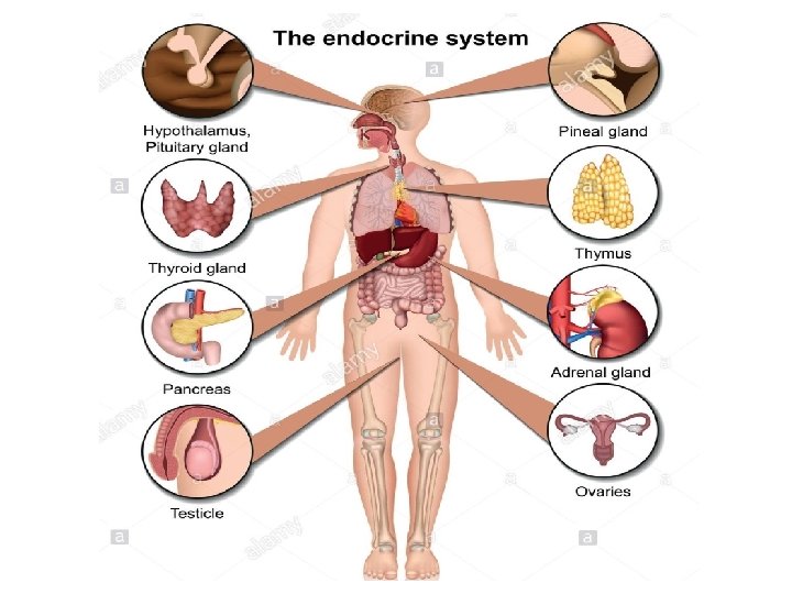
Introduction to the Endocrine System The endocrine glands v Principle glands : q q q q Hypothalamus Pituitary gland Pineal body Thyroid gland & Parathyroid Suprarenal gland (adrenal ) Pancreas Gonads (testis, ovary) v Others Ø Kidney (erythrop + renin) Ø heart(ANF) Ø Thymus (hormone stimulate T cell maturation) Ø adipose (leptin) v Local Hormones q GIT (Diffuse neuroendocrine system) q Neurotransmitter (nerve ending)
 • The pituitary gland is found in the inferior part")
Pituitary gland (Hypophysis Cerebri) • The pituitary gland is found in the inferior part of the brain and is connected by the pituitary stalk so it is called (Hypophysis Cerebri) • The pituitary gland consists of two major regions: v Anterior pituitary gland (anterior lobe or adenohypophysis v Posterior pituitary gland (posterior lobe or neurohypophysis . • The Anterior pituitary is involved in sending hormones that control all other hormones of the body so referred to as the : master gland.
 Neurohypophysis (nervous part) Derived from Oral ectoderm pinched off Rathke’s pouch")
Adenohypophysis (glandular part) Neurohypophysis (nervous part) Derived from Oral ectoderm pinched off Rathke’s pouch Neural ectoderm 3 rd ventricle Is connected with the brain by neural stalk (infundibulum) Stain Dark Pale Consist of Glandular epithelium Nerve fibers in the form of irregular branching cords
 q The pituitary gland (hypophysis) is attached to the bottom")
Pituitary gland (Hypophysis Cerebri) q The pituitary gland (hypophysis) is attached to the bottom of the hypothalamus by a slender stalk called the infundibulum. q size of a pea /0. 5 gm / protrusion off the hypothalamus, Lies in the sella turcica q (bony cavity of sphenoid) covered by diaphragma sellae (fold of dura mater)

Histological structure of Pituitary gland q Stroma : surrounded by a thin connective tissue capsule/ loose connective tissue between the capsule and the periosteum. q Parenchyma : it has plexus of thin-walled veins & capillaries + v epithelial component: adenohypophysis (anterior pituitary) v neural component: Neurohypophysis (posterior pituitary).

Adenohypophysis 1. Pars Tuberalis 2 - Pars Intermedia 3. Pars distalis 1. Pars Tuberalis q Highly vascular region containing the veins of the hypophyseal portal system and wraps the pituitary stalk (infundibulum). q Principal cells of the pars tuberalis are low columnar.

2 - Pars intermedia contains basophilic and chromophobic cells surrounding colloid-filled cysts In humans In animals Development rudimentary Well developed Arrangement cords Layers and cysts Cells Cuboidal Faint basophilic Function Non specific and unknown MSH (melanocytesstimulating hormone)

3 - Pars distalis q Stroma : CT capsule, reticular fibers. q Parenchyma Cells cords of epithelial cells staining characteristics the cells are of two types : 1. chromophobes 52% not stain intensely 2. chromophils 48% densly stained cytoplasmic granules q basophils 11% (darkly pink stained) q acidophils 37% (eosinophilic or reddish stained) + Blood vessels fenestrated capillaries (sinusoids)

Cells of pars distalis Chromophils Function Endocrine cells Chromophobes Stem cells or exhausted chromophils Affinity for Great stain Large , Size & shape polyhedral Weak Small, rounded Percentage 48% 11% basophils, 37% acidophils 52% Granules protein secretion Granulated Degranulated cells or reserve cells Identification of the cells Ø Ø Routine stains Special stain Immuno-histochemistry Transmission electron microscope

Adenohypophysis hormones Cells % Chromophobes Function 52% FSH LH gonadotropic cells Basophils thyrotropic cells 11% GH or somatotropic hormone (STH) somatotropic cells mammotropic cells = Erdheim cells crinophagy TSH ACTH corticotropic cells Acidophils degranulated cell, , They are now thought to represent acidophil and basophilic cells in degranulated stage 37% Prolactin (PR) or lactogenic hormone (LTH) Large in lactation & small in males & non pregnant

Immunohistochemical localization of growth hormone
 Two parts Infundibulum, a slender stalk of nerve tissue that suspends")
Neurohypophysis (Pars nervosa) Two parts Infundibulum, a slender stalk of nerve tissue that suspends the pituitary gland from the base of the brain pituitary stalk The Pars Nervosa, is connected directly with the Hypothalamus of the brain by axons . components 1 - unmyelinated axons: – of neurosecretory cells present in supraoptic and paraventricular nuclei of hypothalamus – Transmit secretion through hypothalamohypophyseal tract. 2 - Herring bodies: homogeneous red bodies stored in dilated terminal ends of these axons - Neurosecretory cells forms Herring bodies v Oxytocin stimulate contraction of 1. Smooth muscle of uterus 2. Myoepithelial cells of mammary glands v Antidiuretic hormone (ADH) (vasopressin) increasing reabsorption of water in renal tubule

3 -Pituicytes: modified branched glial cells having supportive , nutritive and insulating function. Pituicyte is a glial cell of the posterior pituitary. They are similar to the astrocytes /glial cells of the CNS. irregular with processes / cytoplasm contain pigment granules / function: provides metabolic support of nerve fibres 4 -Rich blood capillary plexus

Blood supply of pituitary gland Releasing and inhibiting hormones Pass to hypophyseal portal system to pars distalis

Pineal gland Structure : q Stroma: pia mater → septa ( BVs & unmyelinated nerve fibers q Parenchyma: Two types of branching cells are present: Pinealocytes Astrocytes (neuroglia) Nuclei large irregular or lobulated with prominent nucleoli elongated and denser Cytoplasm pale basophilic Function Melatonin secretion Supportive & nutritive
 q parenchyma Contains: 1. pinealocytes (95%), are large and lightly")
Pineal gland (Epiphysis cerebri) q parenchyma Contains: 1. pinealocytes (95%), are large and lightly stained nuclei secrete melatonin - is involved in daily cycles or circadian rhythms. - Levels are high at night as we grow sleepy & low at day light as we awake. The pineal body is directly light sensitive; 2. glial cells (astrocytes 5%) with small dark nuclei

Pineal gland with aging • ↑ fibrosis ↑ formation of calcified bodies = (brain sand) Brain Sand (areas of calcification) that are easily seen with the microscope. These are not a degenerative change
- Slides: 22