
Gingivectomy l The present technique of gingivectomy was

- Slides: 53
Gingivectomy l The present technique of gingivectomy was described by Goldman in 1951 l According to the world workshop of Periodontics (1989), gingivectomy is defined as “an excision of the soft tissue wall of the periodontal pocket. ”
l Gingivoplasty is defined as a procedure “ which involved reshaping of the gingiva to obtain more physiologic contour; a contour that allows a gradual rise of tissue interproximally and a fall on the labial and lingual surfaces. (Goldman 1951) l To restore a gingival contour Goldman recommended the use of 450 bevel incision to produce the shape of healthy margin
l Both gingivectomy and gingivoplasty are usually performed at the same time. l Isador Hirschfeld noted the same as Kirkland in comparison the two technique, stating that surgical procedure has the advantage of access, saving of time, and more efficient erradication of foci of infection.
Definition l Gingivectomy means excision of the gingiva. (carranza) l By removing the pocket wall, gingivectomy provides visibility and accessibility for complete calculus removal and thorough smoothening of the roots creating a favorable environment for gingival healing and restoration of a physiologic gingival contour.
l The gingivectomy technique was widely performed in the past. Improved understanding of healing mechanisms and the development of more sophisticated flap methods have relegated the gingivectomy to a lesser role in the current repertoire of available techniques. However, it remains an effective form of treatment when indicated
l Rationale : l The basic rationale for gingivectomy includes l Pocket elimination to accessibility for root instrumentation and creation of a local environment that patient could maintain with daily home care
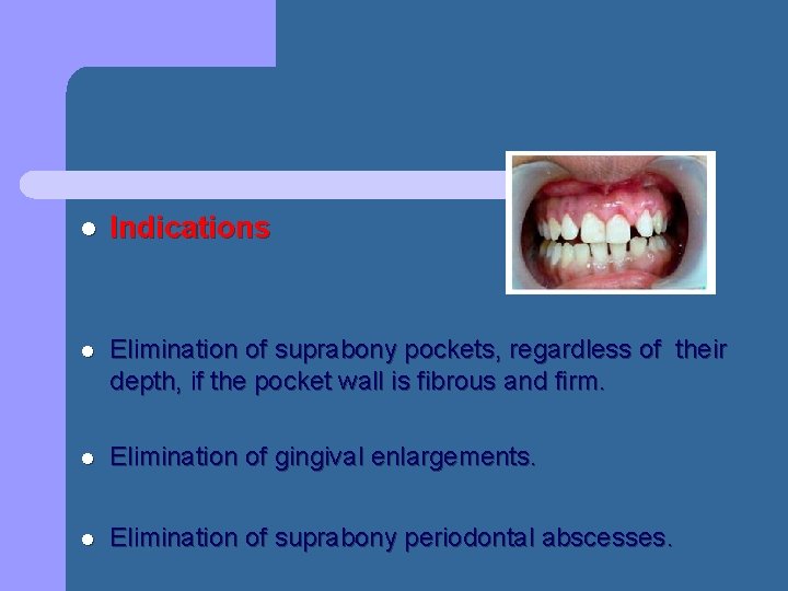
l Indications l Elimination of suprabony pockets, regardless of their depth, if the pocket wall is fibrous and firm. l Elimination of gingival enlargements. l Elimination of suprabony periodontal abscesses.
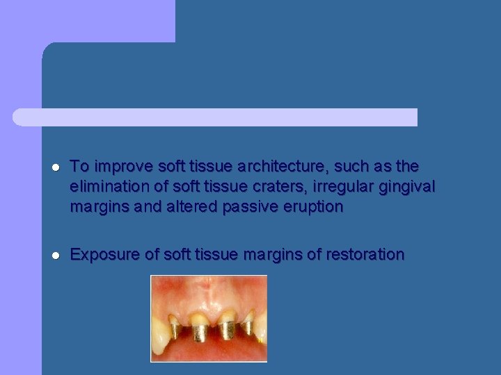
l To improve soft tissue architecture, such as the elimination of soft tissue craters, irregular gingival margins and altered passive eruption l Exposure of soft tissue margins of restoration
l Contraindications l The need for bone surgery or examination of the bone shape and morphology. l Situations in which the bottom of the pocket is apical to the mucogingival junction. l Esthetic considerations, particularly in the anterior maxilla.
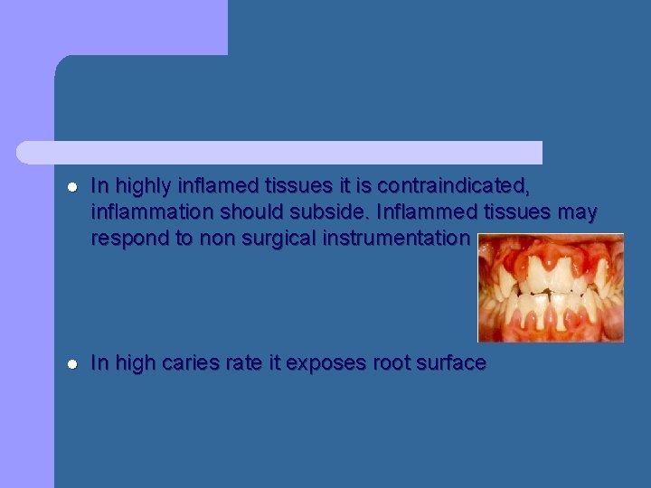
l In highly inflamed tissues it is contraindicated, inflammation should subside. Inflammed tissues may respond to non surgical instrumentation l In high caries rate it exposes root surface
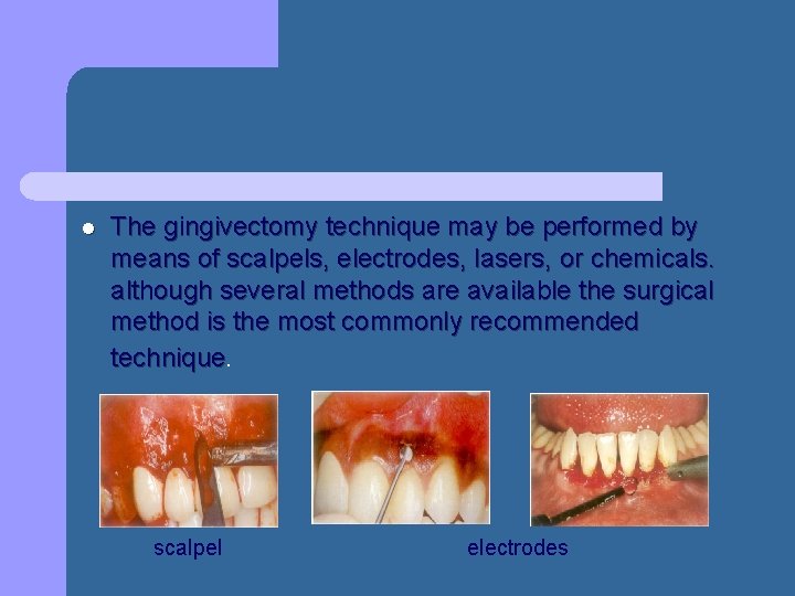
l The gingivectomy technique may be performed by means of scalpels, electrodes, lasers, or chemicals. although several methods are available the surgical method is the most commonly recommended technique. scalpel electrodes
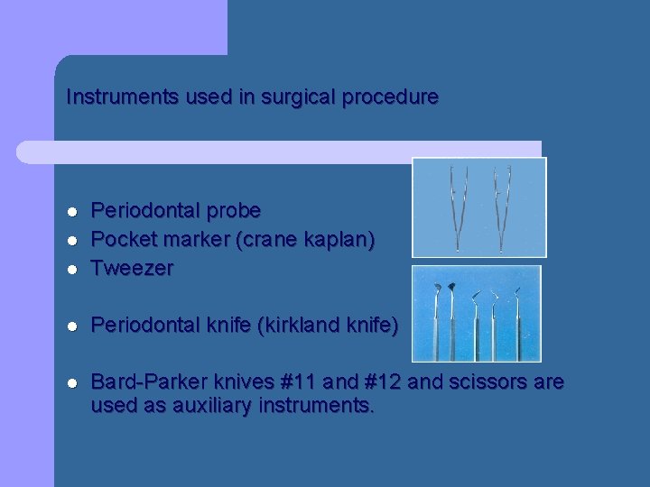
Instruments used in surgical procedure l Periodontal probe Pocket marker (crane kaplan) Tweezer l Periodontal knife (kirkland knife) l Bard-Parker knives #11 and #12 and scissors are used as auxiliary instruments. l l
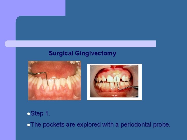
Surgical Gingivectomy l. Step l. The 1. pockets are explored with a periodontal probe.
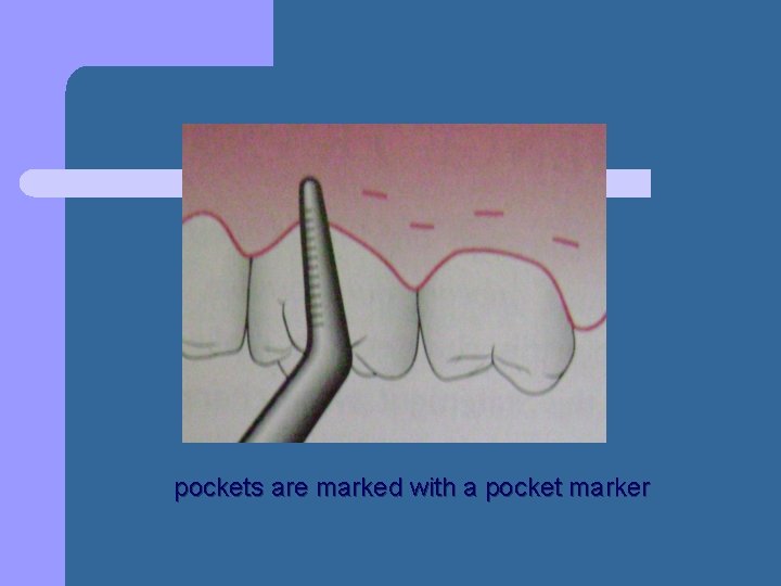
pockets are marked with a pocket marker
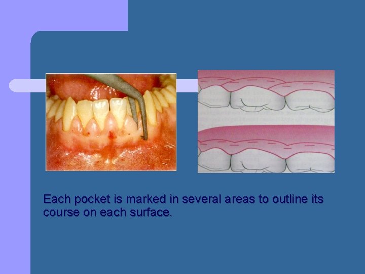
Each pocket is marked in several areas to outline its course on each surface.
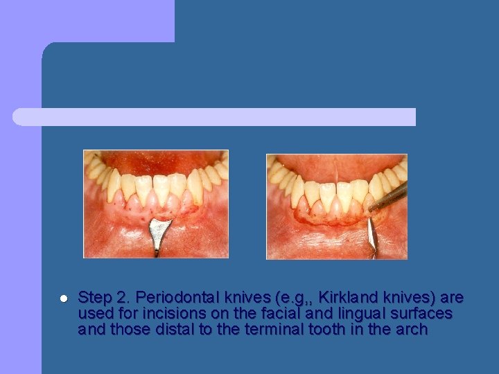
l Step 2. Periodontal knives (e. g, , Kirkland knives) are used for incisions on the facial and lingual surfaces and those distal to the terminal tooth in the arch
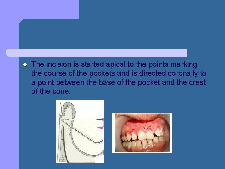
l The incision is started apical to the points marking the course of the pockets and is directed coronally to a point between the base of the pocket and the crest of the bone.
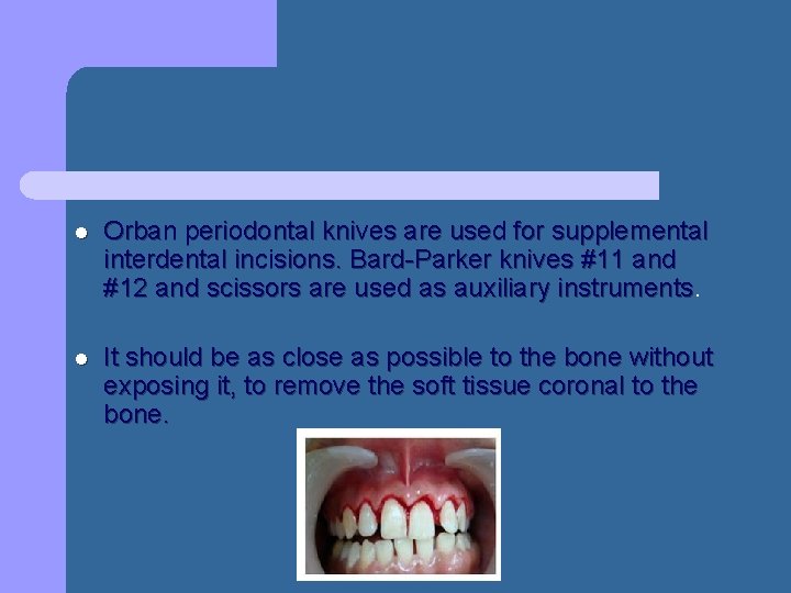
l Orban periodontal knives are used for supplemental interdental incisions. Bard-Parker knives #11 and #12 and scissors are used as auxiliary instruments l It should be as close as possible to the bone without exposing it, to remove the soft tissue coronal to the bone.
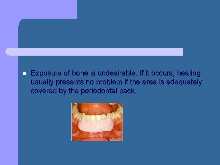
l Exposure of bone is undesirable. If it occurs, healing usually presents no problem if the area is adequately covered by the periodontal pack.
l Discontinuous or continuous incisions may be used. The incision should be beveled at 45 degrees to the tooth surface and should create, the normal festooned pattern of the gingiva. l Failure to bevel leaves a broad, fibrous plateau that takes more time than usually required to develop a physiologic contour. In the interim, plaque and food accumulation may lead to recurrence of pockets.
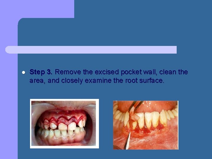
l Step 3. Remove the excised pocket wall, clean the area, and closely examine the root surface.
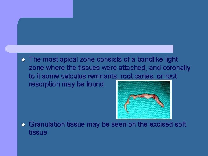
l The most apical zone consists of a bandlike light zone where the tissues were attached, and coronally to it some calculus remnants, root caries, or root resorption may be found. l Granulation tissue may be seen on the excised soft tissue
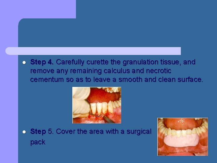
l Step 4. Carefully curette the granulation tissue, and remove any remaining calculus and necrotic cementum so as to leave a smooth and clean surface. l Step 5. Cover the area with a surgical pack
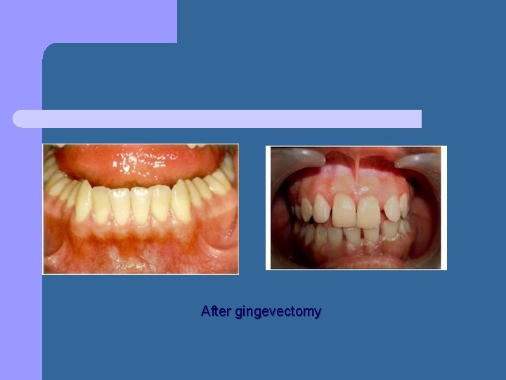
After gingevectomy
l Gingivoplasty is similar to gingivectomy, but its purpose is different. Gingivectomy is performed to eliminate periodontal pockets and includes reshaping as part of the technique. l Gingivoplasty is a reshaping of the gingiva to create physiologic gingival contours, with the sole purpose of recontouring the gingiva in the absence of pockets.
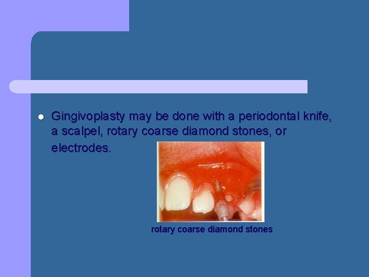
l Gingivoplasty may be done with a periodontal knife, a scalpel, rotary coarse diamond stones, or electrodes. rotary coarse diamond stones
l It consists of procedures that resemble those performed in festooning artificial dentures: tapering the gingival margin, creating a scalloped marginal outline, thinning the attached gingiva, and creating vertical interdental grooves and shaping the interdental papillae to provide sluiceways for the passage of food.
l Healing after Surgical Gingivectomy l Epithilium : the exposed connective tissue is covered by a blood clot after gingivectomy. l Within 12 -13 hrs the wound is covered by a monolayer of PMNLs. This constitutes a defence barrier which is called the “ polyband” – Ramford et al 1966. l The underlying tissue becomes acutely inflamed. An acute inflammatory infiltrate develops in connective tissue.
l After 12 to 24 hours, epithelial cells at the margins of the wound proliferate separating it from the contaminated surface layer of the clot. l The new epithelial cells arise from the basal and deeper spinous layers of the wound edge and increased cell division l Epithelial activity at the margins reaches a peak in 24 to 36 hours.
l Since the epithelium migrates at a rate of 0. 5 mm/day (Ramford et al 1966), it needs an average 1 week to reach the root surface. l As the epithelial cells migrate over the connective tissue, a new attachment is established
l After 5 to 14 days, surface epithelialization is generally complete and normal epithelium thickness is restored. l During the first 4 weeks after gingivectomv, keratinization is less than it was before surgery l Complete epithelial repair takes about 1 month.
l Connective tissue: by 24 hrs there is an increase in connective tissue cells, mainly angioblasts, beneath the surface layer of inflammation and necrosis l By the 3 rd day, numerous fibroblast are located in the area
l Capillaries derived from the blood vessels of the periodontal ligament migrate and connect with the gingival vessels l When the bone is exposed or approximated too closely an increased osteoclastic action is seen
l Vasodilation and vascularity begin to decrease after the fourth day and appear to be almost normal bv the sixteenth day. The Connective tissue repair takes about 7 weeks ( Stanton et al 1969) l The flow of gingival fluid in humans is initially increased after gingivectomy and diminishes as healing progresses. Maximal flow is reached after 1 week, coinciding with the time of maximal inflammation (Arnold et al 1966)
l l Clinical findings : Loss of attachment has been reported after gingevectomy – Waite 1976 Slight reduction in the width of attached gingiva – Glickmann 1966 An increase in tooth mobility immidiately after gingivectomy has been reported returning to pre surgical levels after healing – Burch et al 1960
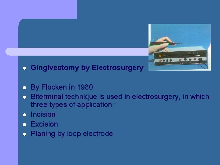
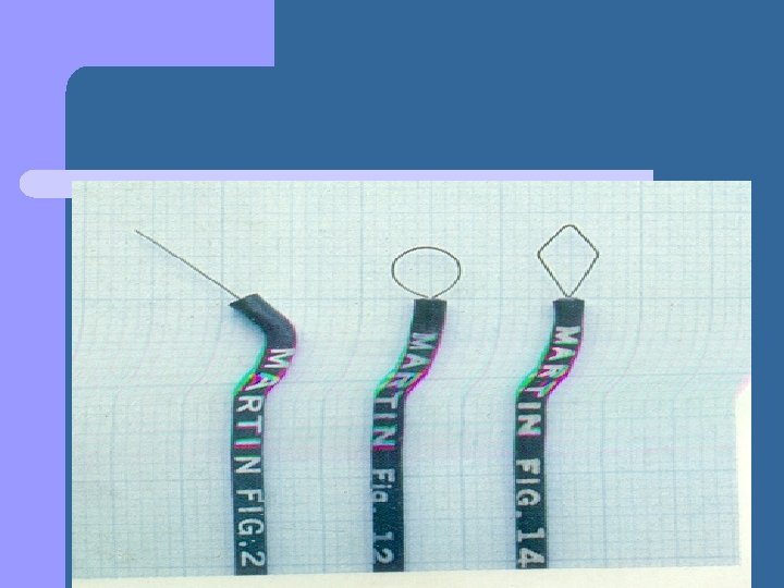
l Gingivectomy by Electrosurgery l By Flocken in 1980 Biterminal technique is used in electrosurgery, in which three types of application : Incision Excision Planing by loop electrode l l
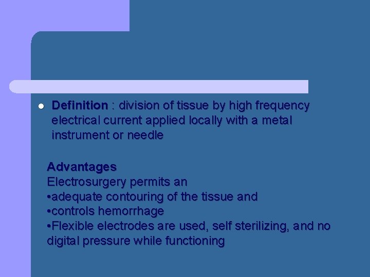
l Definition : division of tissue by high frequency electrical current applied locally with a metal instrument or needle Advantages Electrosurgery permits an • adequate contouring of the tissue and • controls hemorrhage • Flexible electrodes are used, self sterilizing, and no digital pressure while functioning
l l Provides better view to the operating field Eliminates scar formation Decreases chair side time Minimizes post operative discomfort
l In the treatment of acute periodontal abscesses, the incision to establish drainage can be made with the needle electrode without exerting painful pressure. The incision remains open because the edges are sealed by the current l For hemostasis, the ball electrode is used. Hemorrhage must be controlled by direct pressure (using air, compress, or hemostat) first; then the surface is lightly touched with a coagulating current.
l Laser Gingivectomy l The Soft tissue lasers most often used in dentistry are the carbon dioxide (CO 2) and the neodymium: yitrium-aluminum-garnet (Nd: YAG), Argon and which have wavelengths of 10, 600 nm and 1064 nm, respectively, both in the infrared range l They must be combined with other types of visible lasers for the beam to be seen and aimed.
l The CO 2 laser has been used for the excision of gingival growths, although healing is delayed compared with healing after conventional scalpel gingivectomy. l The use of a laser for oral surgery requires precautionary measures to avoid reflecting the beam on instrument surfaces, which could result in injury to neighbouring tissues and the eyes of the operator.
l Laser wound healing : l Reports suggest that laser created wounds heal more quickly and produce less scar tissue. l Pick et al (1990) suggest that wounds have less tensile strength during the early phase of healing
l Crespi et al (2002) evaluated that of CO 2 laser treatment and conclude that it causes increased fibroblast attachment on root surface. l Illria et al (2003) analyzed Er: YAG laser and concluded it promoted fibroblast adhesion
l Gingivectomy by Chemosurgery l Techniques to remove the gingiva using chemicals, such as 5% paraformaldehyde or potassium hydroxide have been described in the past but are not currently used.
l The chemical gingivectomy has the following disadvantages: l The depth of action cannot be controlled and the healthy attached tissue underlying the pocket may be injured. l Gingival remodeling cannot be accomplished effectively.
l Epithelialization and reformation of the junctional epithelium and reestablishment of the alveolar crest fiber system occur more slowly in chemically treated gingival wounds than in those produced by a scalpel.
l In general it is indicated in small areas up to six teeth of drug induced gingival enlargement with no evidence of attachment loss can be treated by gingivectomy technique
l l Errors in surgical technique: They may be caused by careless or inexact performance, failure can occur because of any of the following : Poor & inacurate probing and pocket marking that leads to incomplete pocket elimination Failure to create a proper bevel leaving blunt gingival margins
l l If dull instruments are used, chances of tissue laceration and tissue tags result If scaling is done before the process, no remnants of calculus should remain, if any should be removed at the time of procedure
l Conclusion Gingivectomy procedure has a long history of use in periodontal surgery l Current periodontal surgeries must consider : l Conservation of keratinised gingiva l Minimal gingival tissue loss so as to maintain esthetics l
l Adequate access to osseous defects for definitive defect correction l Minimal post surgical discomfort and bleeding by attempting surgical procedures that will allow healing by primary closure l The gingivectomy surgical technique has limited use in current surgical therapy because it doesnot satisfy these considerations in periodontal therapy