GILLS Histology Normal Teleost gill form anatomy Branchia

GILLS Histology
 • • ‘Branchia’ in greek = ‘gills’ In bony")
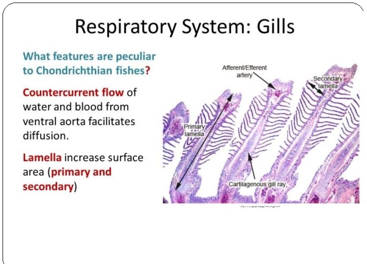
Normal Teleost gill form (anatomy) • • ‘Branchia’ in greek = ‘gills’ In bony fish (Teleosts): • Gills lie in a branchial cavity covered by the operculum: • • Usually two sets of four holobranchs Each holobranch consists of two hemibranchs (‘half gill’): • • • Anterior and posterior Hemibranchs consist of a row of long filaments (primary lamellae) with semilunar folds (secondary lamellae). Lamellae or filaments: • • Connective tissue scaffold (epithelial cells) framing a vascular network providing blood flow primarily used for gas and ion exchange. Primary and secondary.

1. gill raker; 2. mucosal epithelium; 3. basement membrane; 4. submucosa; 5. bone; 6. adipose tissue; 7. efferent branchial arterioles; 8. afferent branchial artery; 9. primary lamellae; 10. secondary lamellae.
")
Teleost gill structure Holobranch Gill filaments Hemibranch (anterior)

Gill lamellae Secondary lamellae Primary lamellae

GILLS structure 1. primary lamella; 2. secondary lamella; 3. epithelial cell; 4. mucous cell; 5. pillar cell; 6. lacuna (capillary lumen); 7. erythrocyte within capillary lumen; 8. undifferentiated

; 2. extracellular cartilaginous matrix; 3. chondrocytes; 4. secondary lamella; 5. epithelial cell; 6. mucous cell; 7. chloride cell; 8. pillar cell; 9. lacuna (capillary lumen); 10. red blood cells within lacuna.


Normal Teleost gill function �Gill functions: • • • Gaseous exchange = O 2 via 2 o lamellae. Acid-base balance = equilibrium between acidity/alkalinity. Osmoregulation = adjustment of internal osmotic pressure in relation to • • Excretion of nitrogenous waste = ammonia. Gills sensitive to a range of environmental pollutants. surrounding medium. �Gaseous exchange = secondary lamellae: • • • Consist of an envelope of epithelial cells Usually one layer thick Supported/separated by specialised cells: • Pillar cells = regulate blood flow. • Chloride cells = maintain internal ionic homeostasis.
. Histopathological")
References • Deeds, J. R. , Reimschuessel, R. and Place, A. R. (2006). Histopathological Effects in Fish Exposed to the Toxins from Karlodinium micrum. Journal of Aquatic Animal Health. 18(2), 136 -148. • Evans, D. H. , Piermarini, P. M. and Choe, K. P. (2005). The Multifunctional Fish Gill: Dominant Site of Gas Exchange, Osmoregulation, Acid-Base Regulation, and Excretion of Nitrogenous Waste. Physiological Review: 85, 97– 177. • Hallegraeff, G. , Mooney, B. and Evans, K. (2010). What triggers Fish-Killing Karlodinium veneficum Dinoflagellate Blooms in the Swan Canning River System? . Swan Canning Research and Innovation Program. • H. W. Ferguson. (2006). Systemic pathology of fish. A Text and Atlas of Normal Tissues in Teleosts and their Responses in Disease. Scotian Press, London. • Mc. Gavin, M. D. and Zachary, J. F. (2007). Pathological basis of Veterinary Disease. 4 th Ed. Mosby Elsevier, Miissouri. • Roberts. R. J. (1989). Fish Pathology. 2 nd Ed. W. B. Saunders, London. • University of Maryland College Park Campus, Aquatic Pathobiology Centre, Virginia-Maryland Regional College of Veterinary Medicine, Atlas of Flathead Minnow Normal Histology. Retreived from the World Wide Web August 2012: http: //aquaticpath. umd. edu/fhm/resp. html �
- Slides: 11