
GI 7 CASE REPORT OF PERITONEAL TUBERCULOSIS SALHI
GI 7 CASE REPORT OF PERITONEAL TUBERCULOSIS SALHI. K 1; AYAT. A 1 ; HELLARA. A 1; JERBI. S 2 ; MAHJOUB. B 1; BOUSSOFFARA. R 1; HAMZA. AH 2; SOUA. H; MT. SFAR 1. 1 Service de Pediatrie CHU Taher Sfar Mahdia 2 Service de Radiologie CHU Taher Sfar Mahdia
INTRODUCTION: Ø The peritoneum is one of the locations outside the most common pulmonary tuberculosis. Ø Peritoneal tuberculosis poses a public health problem in endemic regions of the world. Ø The diagnosis is difficult and still remains a challenge : insidious nature , variability of presentation and limitations of available diagnostic tests. Ø We report a case of an adolescent girl who was diagnostic with this disease.
Patients and Methods: Ø A 14 years old girl admitted with a chronic diarrhea since 4 months , weakness , decreased appetie and weight loss. Ø Physical examination showed : ü A pale girl. ü Fever (38. 7°). ü Painful abdomen. ü No organomegaly or lymphomegaly. We completed with a biologic and radiologic investigation.
Results: ØLaboratory investigation revealed: ü elevated erythrocyte sedimentation rate(110/130) ü anemia (Hb=8. 7 g/dl) , ü high CRP (97 mg/l) . üAll other routine biochemical tests, celiac serology , anti Dnatif , antinuclear anticorps were within the normal range in the serum. ü The BK search was negative.
Results: Ø The chest X ray was normal. Ø Abdominal ultrasonographie showed a little ascite. Ø TDM showed: ü ascite. ü small bowel thichening. ü Multiples necrotic lymphadenopathy. ü liver nodule (22 mm in the segment IV)
Results: Ølaparotomy multiple smalls nodules and fibrotic adhesive bands covering peritoneal surfaces compatible with peritoneal tuberculosis later confirmed histologically (caseating granulomas) ØThe girl was treated with quadritherapie : Rifampicine, Izoniasid, Pyrazinamide and Ethambutol during 4 months. ØThere were no clinical amelioration, and a cutaneous fistulas appeared.
Results: Ø Myelogramme was normal. Ø A second abdominal TDM: showed the persistence of the same pathologie and appearing of cutaneous fistulas. Ø we suspected a multidrug resistant tuberculosis so we added ofloxacine , Amikacine and corticotherapie. Ø After 1 month the patient became more better.
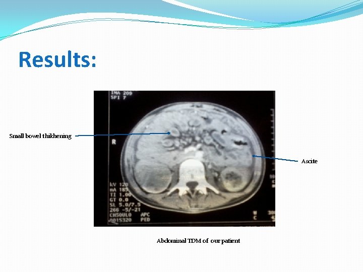
Results: Small bowel thikhening Ascite Abdominal TDM of our patient
DISCUSSION : ØPeritoneal tuberculosis is predominantly a disease of young adults between 21 -40 years old with an equal sex incidence. Ø Tuberculosis bacteria reachs the gastrointestinal tract via: ü Haematogenous spread ü Ingestion of infected sputum ü Direct spread from infected contiguous lymph nodes or fallopian tubes
DISCUSSION : Ø Peritoneal tuberculosis occurs in three forms : Wet type with ascitis+++ Dry type with adhesions. Fibrotic type with omental thickening and loculated ascites. Ø It is commonly manifested by : abdominal pain , diarrhea, fever , weight loss , and anemia. Ø Laboratory Findings are: Anemia , elevated sedimentation rate , high CRP. Elevated CA-125
DISCUSSION : Ø Chest X ray search a pulmonary tuberculosis. Ø Ultrasonographie ascites , lymphadenopathy, omental thickening and caking. Ø TDM Three main types : Wet Type Peritonitis : � Is the most common type of peritonitis (90% ). � Free or loculated ascites, � Usually slightly hyperattenuating (20– 45 HU) relative to water due to its (high protein and cellular content) .
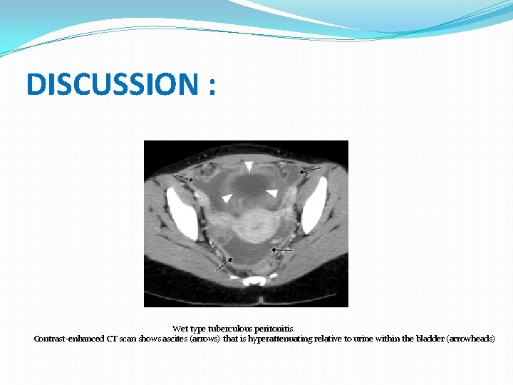
DISCUSSION : Wet type tuberculous peritonitis. Contrast-enhanced CT scan shows ascites (arrows) that is hyperattenuating relative to urine within the bladder (arrowheads)
DISCUSSION : Fibrotic Type Peritonitis: � It accounts for 60% of cases of peritonitis. � It manifests as mottled low-attenuation masses with nodular soft-tissue thickening. � Dry Type Peritonitis : � Is seen in 10% of cases. � Characterized by mesenteric thickening, fibrous adhesions, and caseous nodules. � Its imaging manifestations are highly suggestive of, but not specific for, tuberculosis.
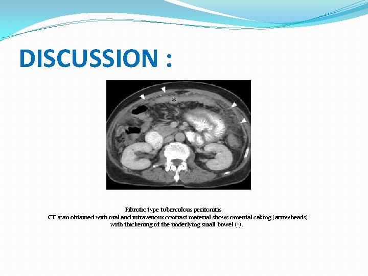
DISCUSSION : Fibrotic type tuberculous peritonitis. CT scan obtained with oral and intravenous contrast material shows omental caking (arrowheads) with thickening of the underlying small bowel (*).
DISCUSSION : Ø Peritoneal Biopsy : ü 85 -95% Sensitive ü Performed by: - laparoscopic guidance or minilaparotomy - exploratory laparotomy. • Caseating Granulomas • Langerhans Type Giant Cells. Ø Microbiology Ziehl-Neelsen Stain
Treatment Ø Same treatment as pulmonary TB � Four drug regimen: – Isoniazid – Rifampicin – Ethambutol – Pyrazinamide Ø Quadritherapie during 2 mounths than bitheraphie during 4 moutns(Isoniazide+Rifampicin)
Conclusion : ØThe diagnostic of peritoneal tuberculosis is difficult. ØIt presents with nonspecific symptoms. laboratory investigations may not be helpful. Ø Radiologic investigation and laparotomy help to get the diagnostic and to treat early affected patients.
- Slides: 17