
Gestational trophoblastic disease GTD Dr Khaldoun Khamaiseh FRCOG
Gestational trophoblastic disease (GTD) Dr Khaldoun Khamaiseh FRCOG MRCP Consultant Obstetrician & Gynecologist

GTD consists of a range of pregnancy-related disorders originating from disordered proliferation of the placental trophoblast. Distinct tumor marker……(B-h. CG). The pathogenesis of GTD is unique because the maternal tumor arises from gestational rather than maternal tissue.

Histological types of GTD • Premalignant disorders Ø Hydatidiform mole (complete or partial) • Gestational trophoblastic neoplasia (GTN) refers to malignant GTD Ø Persistent/invasive gestational trophoblastic neoplasia (GTN) Ø Choriocarcinoma Ø Placental site trophoblastic tumors GTN can progress, invade, metastasize, and lead to death if left untreated.
Epidemiology The incidence of GTD varies widely in different regions of the world. North American and European countries tend to report low or intermediate rates of disease (1 per 1000 to 1500 pregnancies), whereas Asian and Latin American nations often have high rates (1 per 12 to 500 pregnancies).
Risk Factors • Extremes of age • • The most well-established risk factor for GTD is maternal age. The risk is significantly increased in those older than age 35 and slightly increased in those under age 20. Nevertheless, most cases of GTD occur in women under age 35 because of the greater number of pregnancies among younger women. Paternal age does not appear to influence the risk of GTD. • History of previous GTD • • 1 percent chance of recurrence in subsequent pregnancies (compared to a 0. 1 percent incidence in the general population of the United States). The recurrence rate is much higher after two molar pregnancies (16 to 28 percent)
Clinical Manifestations • Common presentation is vaginal bleeding in early • • pregnancy. Other symptoms in decreasing order of frequency include: excessive uterine size (50%) prominent theca lutein cyst formation (50%) hyperemesis gravidarum (25%) early-onset pre-eclampsia (25%) hyperthyroidism (< 10%) trophoblastic pulmonary emboli causing respiratory distress (< 2%) Vaginal passage of hydropic vesicles
Theca lutein cyst • BILATERAL FUNCTIONAL OVARIAN CYST FILLED WITH CLEAR, STRAW-COLORED FLUID • RESULT FROM EXAGGERATED PHYSIOLOGICAL STIMULATION • USUALLY ASSOCIATED WITH MARKEDLY ELEVATED LEVELS OF B-HCG) • ASSOCIATED WITH GESTATIONAL TROPHOBLASTIC DISEASE DIABETES MELLITUS, RH-D ALLOIMMUNISATION , AND MULTIPLE PREGNANCY. • RESOLVES AFTER PREGNANCY
Clinical hyperthyroidism h. CG has thyroid-stimulating activity The development of hyperthyroidism requires the elevation of h. CG >200, 000 m. IU/m. L , for several weeks. GTD-associated hyperthyroidism will resolve with treatment of the GTD. Some patients will require antithyroid therapy until GTD treatment is complete.
Postpartum/post-delivery of non-molar pregnancy • GTD can occur after non-molar pregnancies. • Women with AUB after a non-molar pregnancy, including miscarriage or term delivery, should undergo a pregnancy test to exclude the possibility of persistent gestational trophoblastic neoplasia (GTN). • Persistent GTD or GTN should be considered in any woman developing acute respiratory, abdominal or neurological symptoms after any pregnancy.
Physical examination Enlarged uterus Bilateral ovarian cysts. Vaginal metastases in about 30 percent of cases; these lesions are very vascular and prone to bleeding; they may also become infected
Investigations • Baseline h. CG level should be obtained for comparison with postoperative results • Complete blood count, and renal, liver, and thyroid function tests can be used to assess for evidence of significant anemia, hyperthyroidism, electrolyte imbalance, or severe preeclampsia. • A blood type and antibody screen should also be obtained, both to prepare for heavy bleeding necessitating transfusion and to confirm the patient's Rh(D) status. • A routine chest radiograph • Pelvic Ultrasound
Diagnosis The diagnosis must be confirmed by histological examination of tissue. The accuracy of histological diagnosis is further enhanced by flow cytometry to determine the karyotype Ø It is recommended that all POC obtained after medical or surgical management of miscarriage be sent for histopathological analysis to exclude gestational trophoblastic disease (GTD)
Management • Evacuation of the uterine contents by suction curettage is indicated for: 1. pathologic confirmation of the diagnosis 2. relief of symptoms 3. to prevent complications related to molar pregnancy. Suction curettage is the preferred approach because it is less likely to result in uterine perforation or intrauterine adhesions than sharp curettage, and evacuates the uterus more completely than medical methods • • In rare circumstances, hysterectomy may be considered appropriate as an elective (e. g. women with stage 1 GTN confined to the uterus who do not seek future fertility) or emergency (e. g. life-threatening intraoperative haemorrhage) procedure. Although hysterectomy eliminates the risk of a uterine source for trophoblastic metastases, it does not eliminate an extrauterine trophoblastic source or the potential need for chemotherapy; h. CG surveillance should still be continued post hysterectomy. Theca lutein cyst managed conservatively
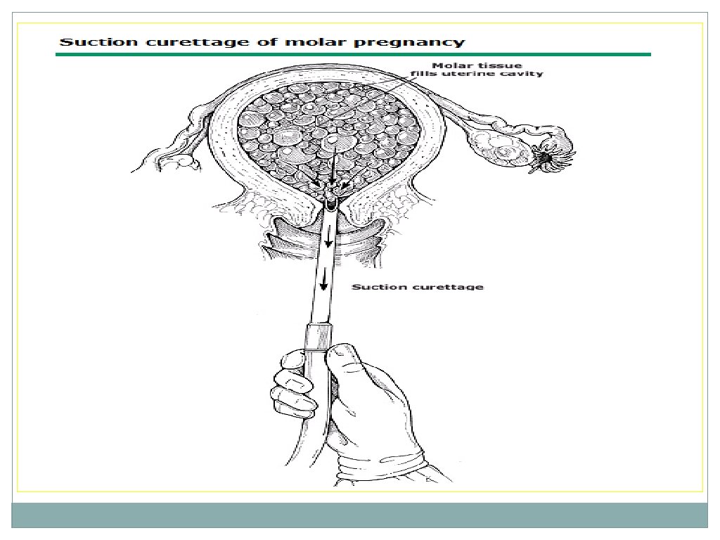
Procedure • Women with hydatidiform mole are at risk for hemorrhage during the evacuation procedure so blood should be available and adequate venous access assured. Under adequate anesthesia, the cervix is dilated to allow passage of the suction cannula. During dilation of the cervix, brisk uterine bleeding is often encountered, which generally slows significantly once suction evacuation has commenced. Heavy bleeding is nearly always self-limited, and inappropriate transfusion should be avoided. • Oxytocic agents, if required, should ideally only be commenced after completing uterine evacuation. Although there is no fetus in a complete mole, anti-Rh(D) immune globulin is administered to Rh(D) negative women with both complete and partial moles, as trophoblastic tissue expresses the Rh(D) antigen.
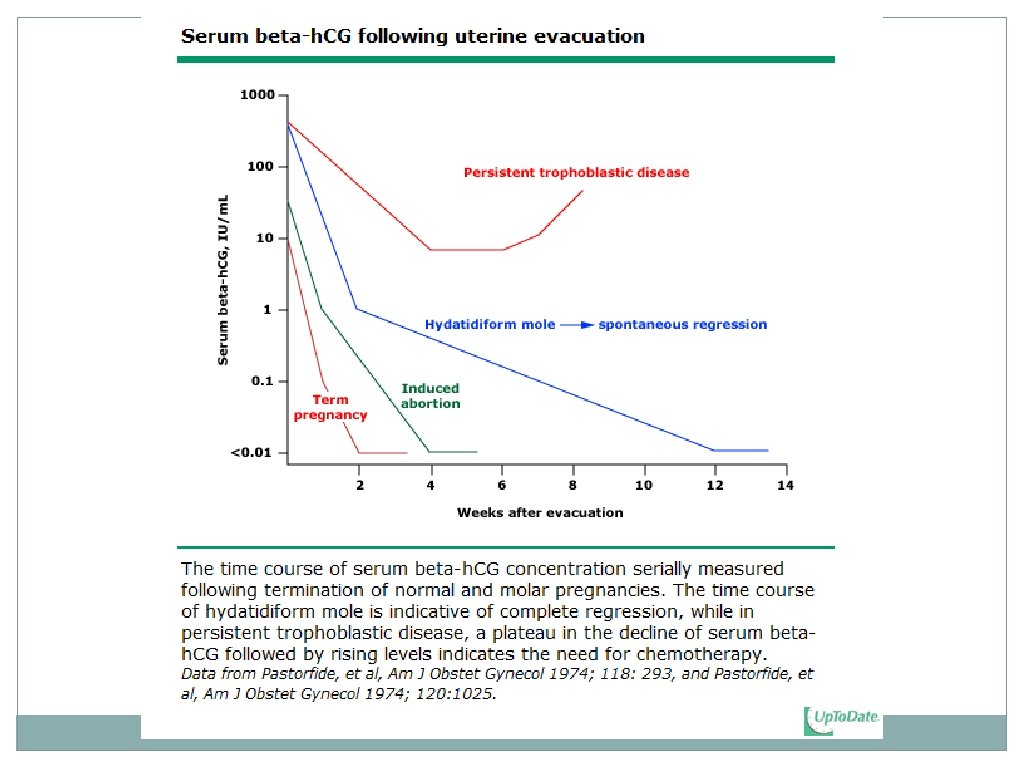
POST-MOLAR FOLLOW-UP • Patients are monitored with weekly h. CG levels until three consecutive normal values are obtained. • 50 % achieve normal h. CG levels 6 to 14 weeks after molar evacuation • plateau or rise in h. CG is indicative of the development of persistent trophoblastic disease, and necessitates chemotherapeutic treatment.
Hydatidiform mole refers to an abnormal pregnancy characterised by varying degrees of trophoblastic proliferation (both cytotrophoblast and syncytiotrophoblast) and vesicular swelling of placental villi (villous hydrops) associated with an absent or an abnormal fetus/embryo.
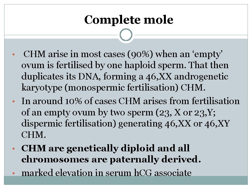
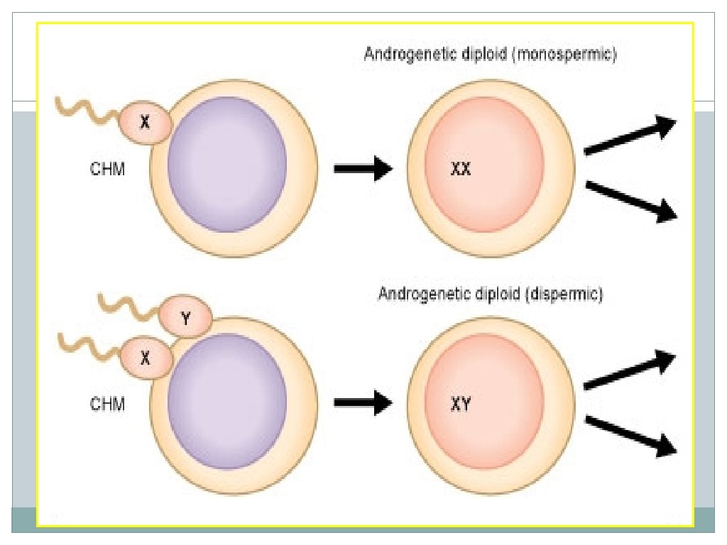
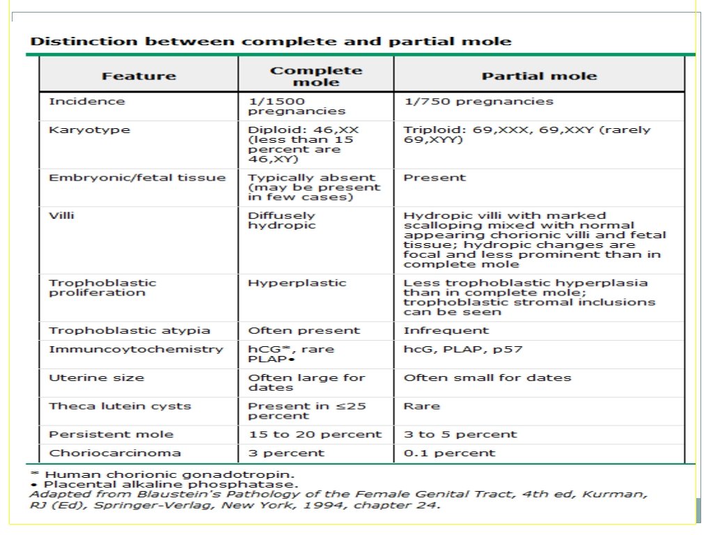
Complete mole • CHM arise in most cases (90%) when an ‘empty’ ovum is fertilised by one haploid sperm. That then duplicates its DNA, forming a 46, XX androgenetic karyotype (monospermic fertilisation) CHM. • In around 10% of cases CHM arises from fertilisation of an empty ovum by two sperm (23, X or 23, Y; dispermic fertilisation) generating 46, XX or 46, XY CHM. • CHM are genetically diploid and all chromosomes are paternally derived. • marked elevation in serum h. CG associate
The marked elevation in serum h. CG associated with: 1. Ovarian enlargement due to theca lutein cysts (multiloculated, often bilateral, and resolve a few weeks or months after treatment of GTD) 2. Hyperemesis gravidarum 3. Early development of preeclampsia (before 20 weeks of gestation) 4. Hyperthyroidism, which is most often subclinical (homology between the beta-subunits of h. CG and TSH).
Sonographic features CHM • The absence of an embryo or fetus • No amniotic fluid • Central heterogeneous mass with numerous discrete anechoic spaces, which correspond to diffuse hydatidiform swelling of the hydropic chorionic villi. This has classically been described as a "snowstorm pattern" on older ultrasounds • Theca lutein cysts
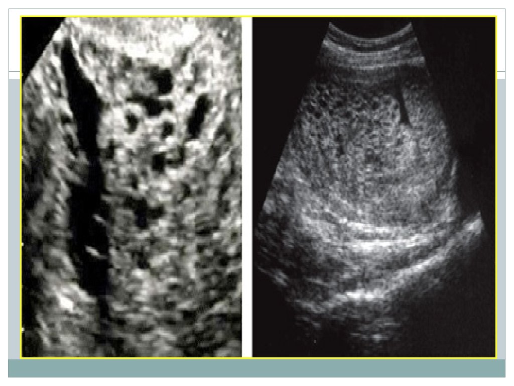
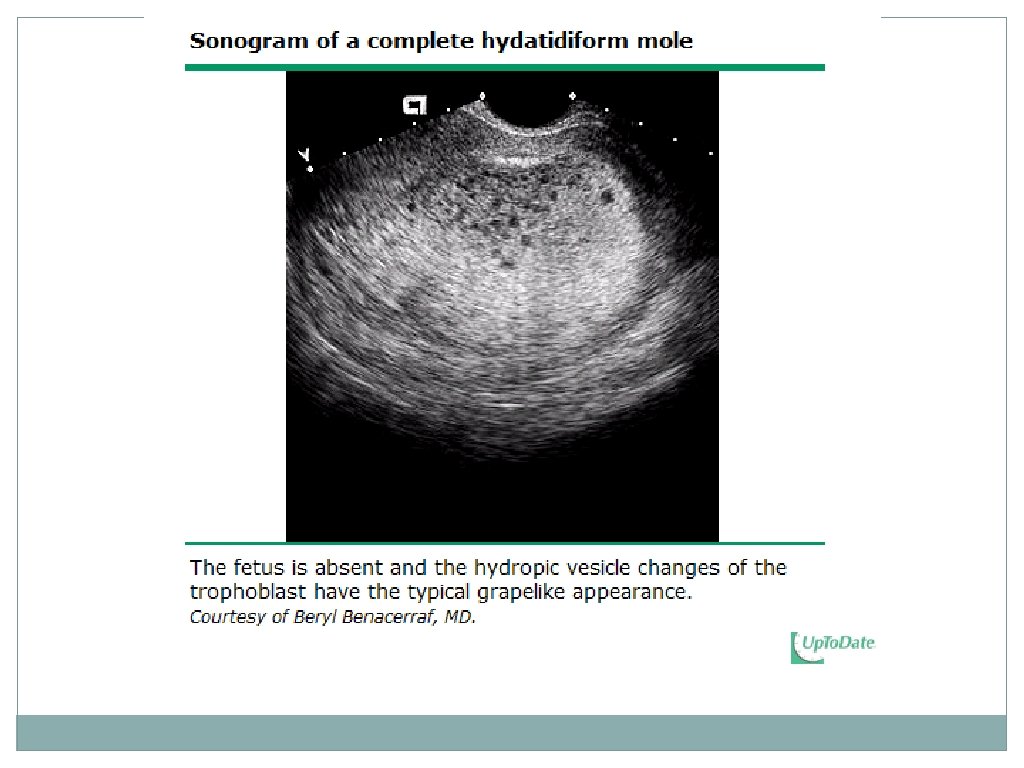
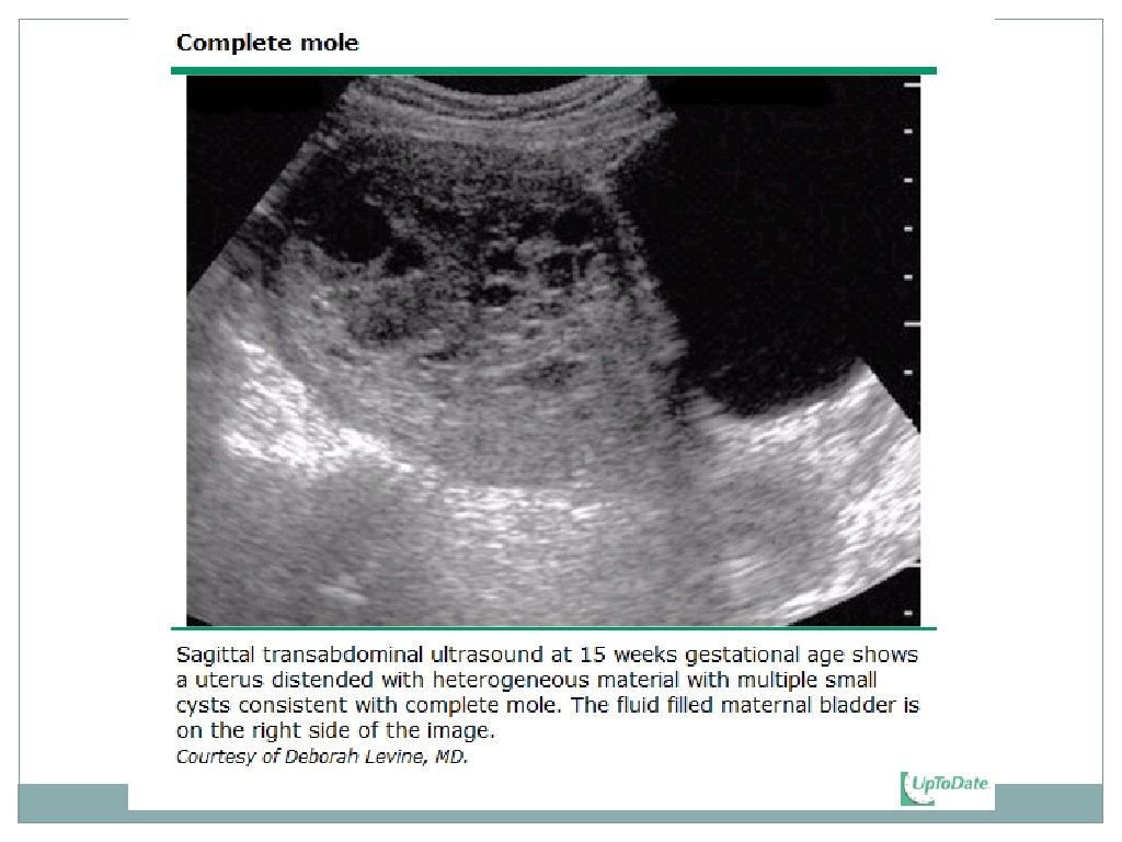
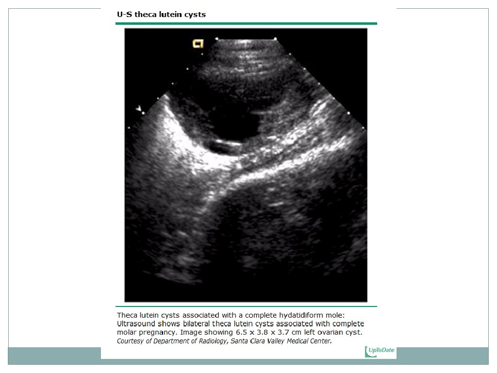
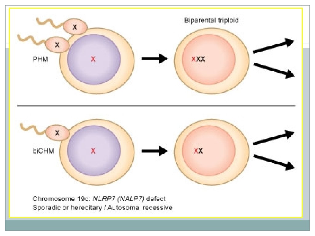
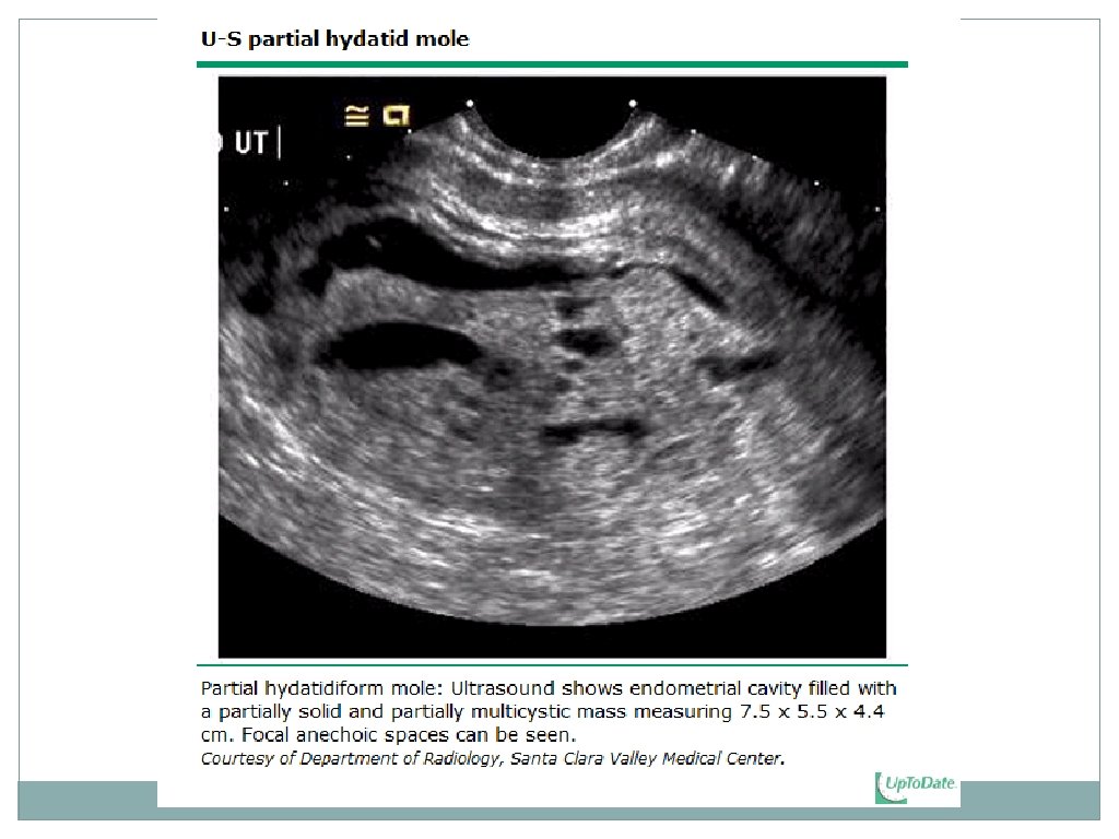
Partial Hydatidiform mole • A result of fertilization of a haploid ovum by two sperm or duplication of one sperm, resulting in a triploid karyotype (69 XXY, 69 XXX, 69 XYY). Partial moles are the only type of GTD that are o Associated with the presence of a fetus. +/- FHA o High rate of intrauterine death related to triploidy. o Often misdiagnosed as an incomplete or missed miscarriage and the correct diagnosis of GTD is made only after histologic review of the surgical specimen o Infrequently associated with excessive uterine size, ovarian enlargement, preeclampsia, hyperemesis, or hyperthyroidism because h. CG levels are generally lower than those observed with a complete mole. o Partial moles are less likely to become malignant.
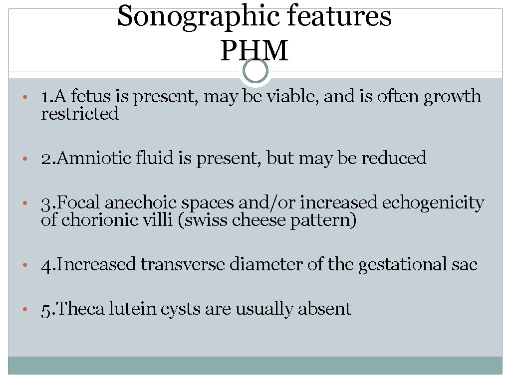
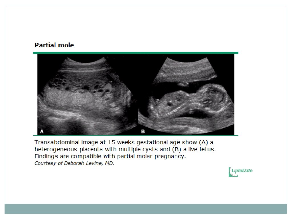
Sonographic features PHM • 1. A fetus is present, may be viable, and is often growth restricted • 2. Amniotic fluid is present, but may be reduced • 3. Focal anechoic spaces and/or increased echogenicity of chorionic villi (swiss cheese pattern) • 4. Increased transverse diameter of the gestational sac • 5. Theca lutein cysts are usually absent
Malignant GTD can develop after molar or non-molar pregnancies. After a molar pregnancy ………
Persistent GTN • 1. asymptomatic women undergoing routine h. CG monitoring after evacuation of a molar pregnancy • 2. greater than 90 % of malignant GTN. 3. Rates of persistent disease are 15 -20 % after a complete, and 3 -5 % after a partial mole. • • 4. The most common symptom is vaginal bleeding. 5. Uterine rupture resulting in hemoperitoneum is rare • 6. 15 % of patients have localized disease(invasive GTN) and 4 % have metastatic disease(choriocarcinoma) • 7. Placental site trophoblastic tumors are rare.
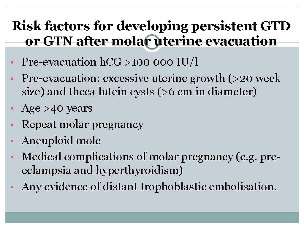
Risk factors for developing persistent GTD or GTN after molar uterine evacuation • Pre-evacuation h. CG >100 000 IU/l • Pre-evacuation: excessive uterine growth (>20 week • • • size) and theca lutein cysts (>6 cm in diameter) Age >40 years Repeat molar pregnancy Aneuploid mole Medical complications of molar pregnancy (e. g. preeclampsia and hyperthyroidism) Any evidence of distant trophoblastic embolisation.
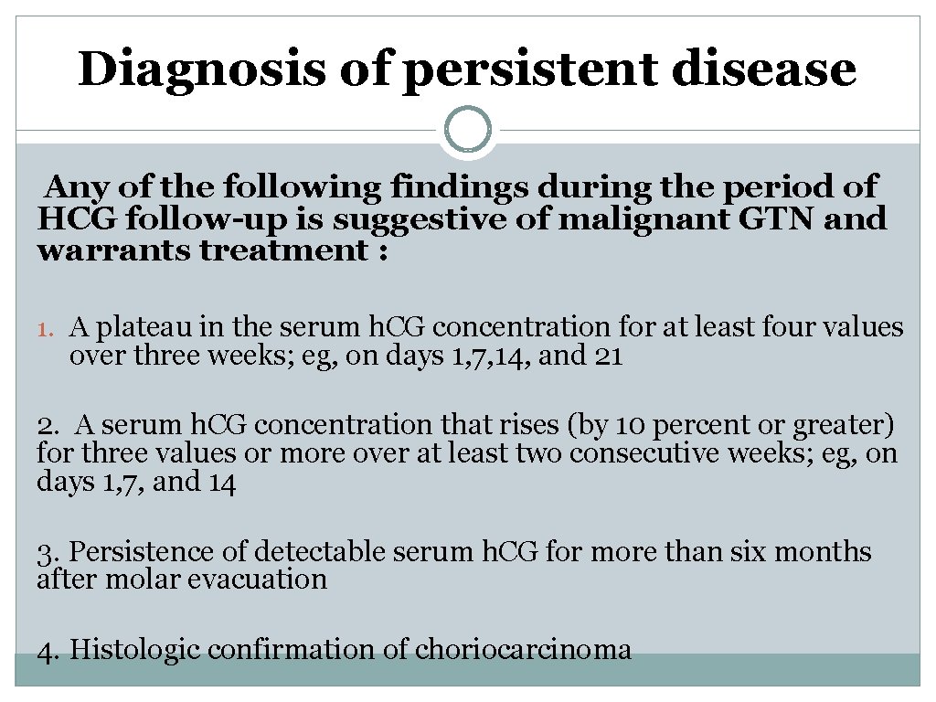
Diagnosis of persistent disease Any of the following findings during the period of HCG follow-up is suggestive of malignant GTN and warrants treatment : 1. A plateau in the serum h. CG concentration for at least four values over three weeks; eg, on days 1, 7, 14, and 21 2. A serum h. CG concentration that rises (by 10 percent or greater) for three values or more over at least two consecutive weeks; eg, on days 1, 7, and 14 3. Persistence of detectable serum h. CG for more than six months after molar evacuation 4. Histologic confirmation of choriocarcinoma
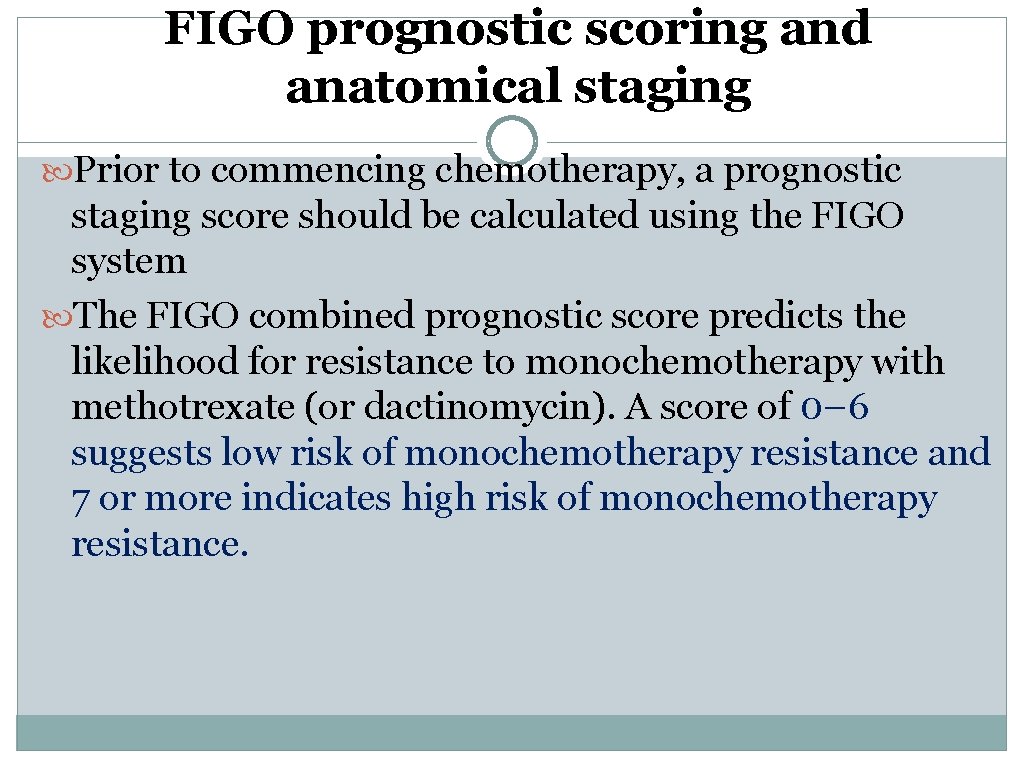
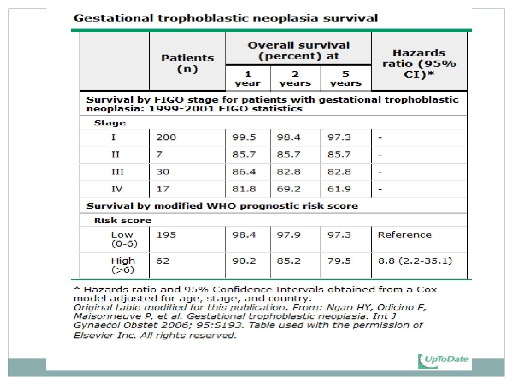
FIGO prognostic scoring and anatomical staging Prior to commencing chemotherapy, a prognostic staging score should be calculated using the FIGO system The FIGO combined prognostic score predicts the likelihood for resistance to monochemotherapy with methotrexate (or dactinomycin). A score of 0– 6 suggests low risk of monochemotherapy resistance and 7 or more indicates high risk of monochemotherapy resistance.
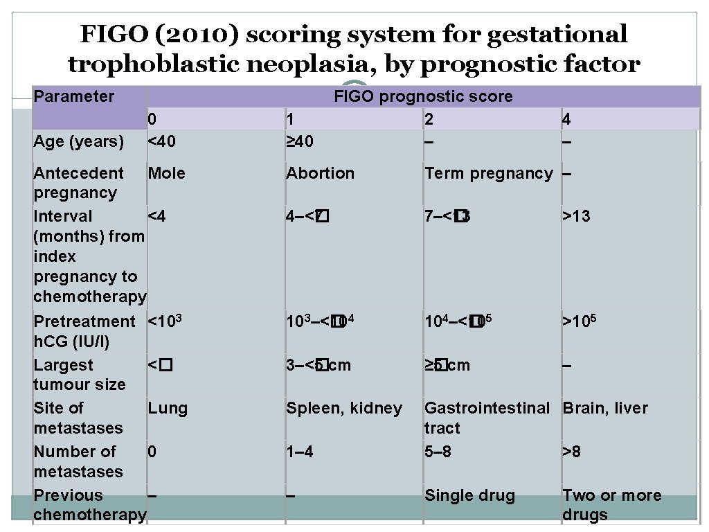
FIGO (2010) scoring system for gestational trophoblastic neoplasia, by prognostic factor Parameter Age (years) 0 <40 1 ≥ 40 FIGO prognostic score 2 – 4 – Antecedent Mole pregnancy Interval <4 (months) from index pregnancy to chemotherapy Abortion Term pregnancy – 4–<� 7 7–<� 13 >13 Pretreatment <103 h. CG (IU/l) Largest <� tumour size Site of Lung metastases Number of 0 metastases Previous – chemotherapy 103–<� 10 4 104–<� 105 >105 3–<� 5 cm ≥� 5 cm – Spleen, kidney 1– 4 Gastrointestinal Brain, liver tract 5– 8 >8 – Single drug Two or more drugs
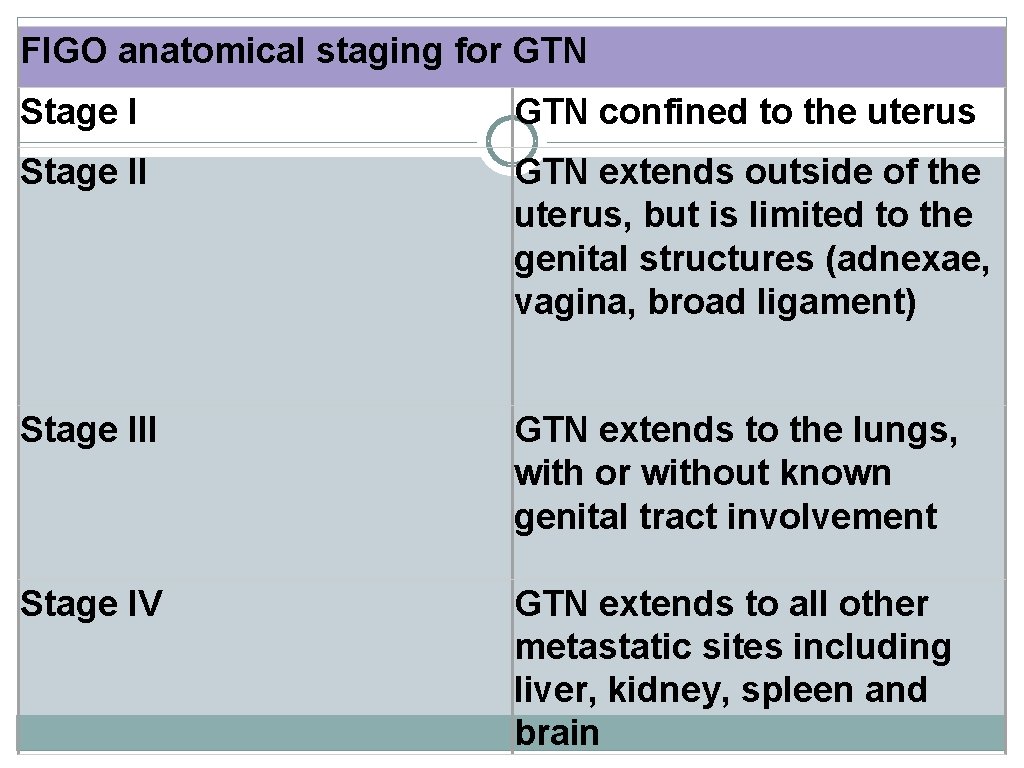
FIGO anatomical staging for GTN Stage I GTN confined to the uterus Stage II GTN extends outside of the uterus, but is limited to the genital structures (adnexae, vagina, broad ligament) Stage III GTN extends to the lungs, with or without known genital tract involvement Stage IV GTN extends to all other metastatic sites including liver, kidney, spleen and brain
Chemotherapy in low- and high-risk groups Principles of chemotherapy treatment • Inpatient • Measurements of serum h. CG is used to monitor treatment response. • An adequate response to treatment is considered to be a 50% or more reduction in consecutive weekly serum h. CG measurements. • Chemotherapy is continued until h. CG is within the normal range and then for a further 6 weeks; this helps eliminate any residual tumour cells and reduces the chance of relapse. • Most low-risk GTN presents soon after CM/PHM diagnosis. However, most high-risk GTN presents with metastases months or years after the causative pregnancy. Symptoms depend on the location of metastases; brain metastases cause seizures, headaches, or hemiparesis and lung metastases cause haemoptysis, shortness of breath or pleuritic chest pain.
Low-risk of monochemotherapy resistance (prognostic score ≤ 6) • 95% of persistent GTD/GTN cases • non-metastatic (FIGO stage I) or low-risk metastatic (FIGO stages II and III) stages. • Methotrexate (50 mg intramuscularly every 48 hours for four doses) with calcium folinate (folinic acid) rescue (15 mg orally 30 hours after methotrexate); courses are repeated every 2 weeks and survival rates approach 100%. This regimen does NOT induce hair loss, however, side effects include mucosal ulceration, conjunctivitis and, occasionally, serositis.
High-risk high risk of monochemotherapy resistance (prognostic score ≥ 7) The etoposide, methotrexate, and dactinomycin (EMA)- cyclophosphamide and vincristine (CO) regimen is recommended for high-risk cases and necessitates one overnight stay every 2 weeks. The treatment causes reversible alopecia and is myelosuppressive. Cumulative 5 -year survival of patients given EMA-CO is between 75 and 90%. EMA alternates with CO every week.
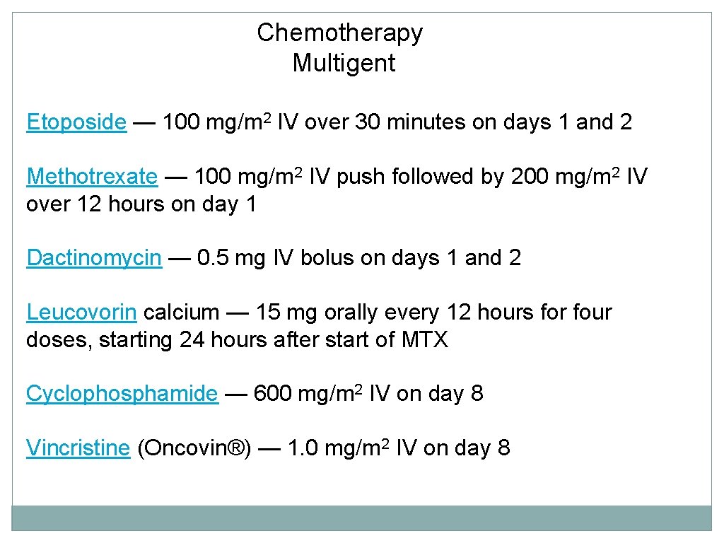
Chemotherapy Multigent Etoposide — 100 mg/m 2 IV over 30 minutes on days 1 and 2 Methotrexate — 100 mg/m 2 IV push followed by 200 mg/m 2 IV over 12 hours on day 1 Dactinomycin — 0. 5 mg IV bolus on days 1 and 2 Leucovorin calcium — 15 mg orally every 12 hours for four doses, starting 24 hours after start of MTX Cyclophosphamide — 600 mg/m 2 IV on day 8 Vincristine (Oncovin®) — 1. 0 mg/m 2 IV on day 8
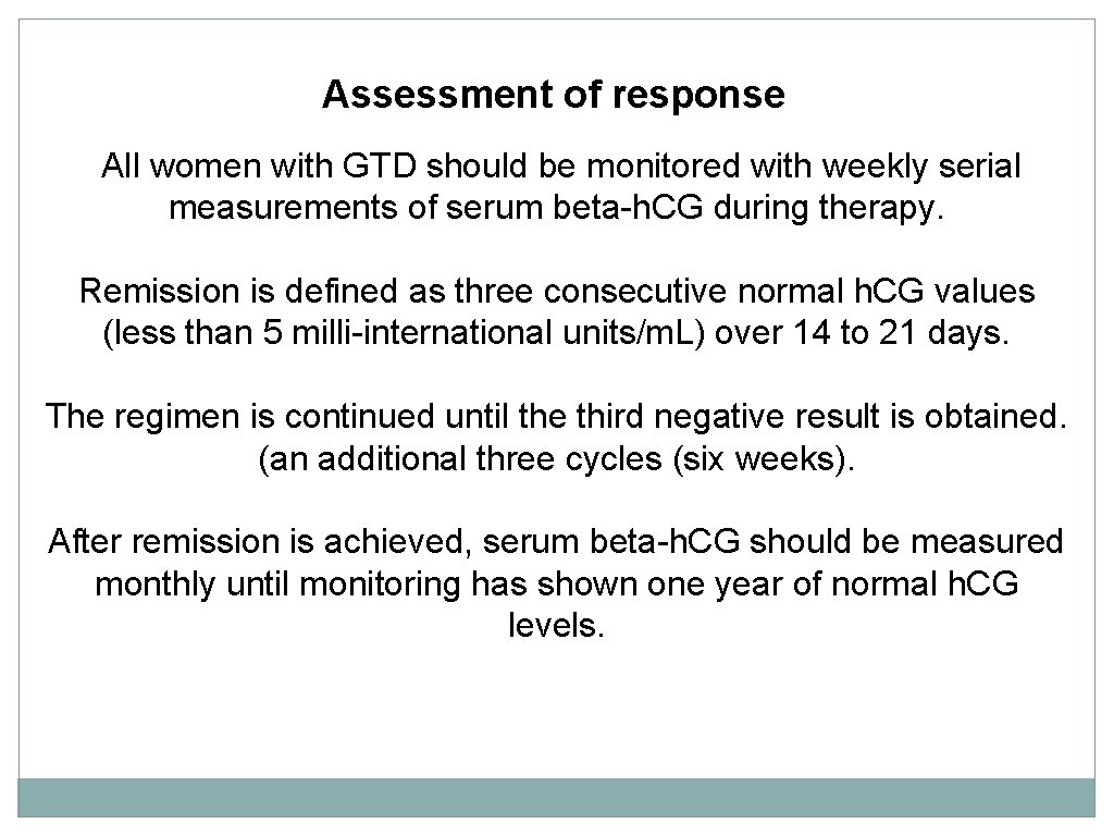
Assessment of response All women with GTD should be monitored with weekly serial measurements of serum beta-h. CG during therapy. Remission is defined as three consecutive normal h. CG values (less than 5 milli-international units/m. L) over 14 to 21 days. The regimen is continued until the third negative result is obtained. (an additional three cycles (six weeks). After remission is achieved, serum beta-h. CG should be measured monthly until monitoring has shown one year of normal h. CG levels.
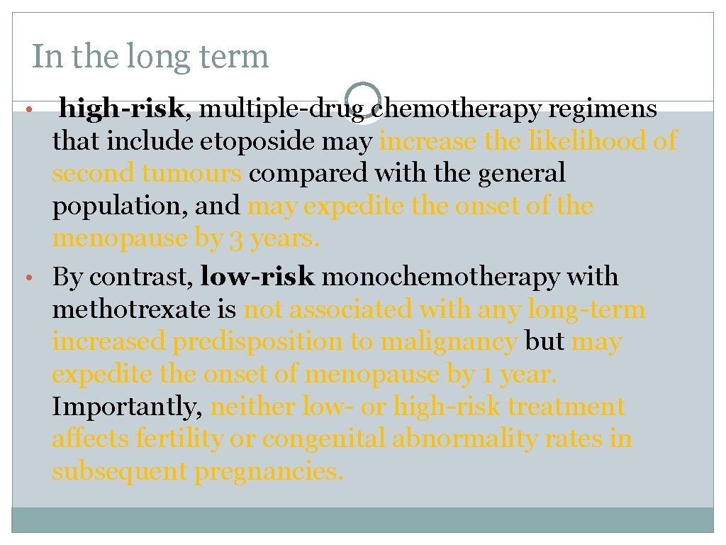
In the long term • high-risk, multiple-drug chemotherapy regimens that include etoposide may increase the likelihood of second tumours compared with the general population, and may expedite the onset of the menopause by 3 years. • By contrast, low-risk monochemotherapy with methotrexate is not associated with any long-term increased predisposition to malignancy but may expedite the onset of menopause by 1 year. Importantly, neither low- or high-risk treatment affects fertility or congenital abnormality rates in subsequent pregnancies.
Contraception and future pregnancy advice Requiring chemotherapy • GTD and GTN requiring chemotherapy should not conceive for at least 12 months from completion of their chemotherapy treatment. Not requiring chemotherapy • For patients with PHM, CHM and GTD not requiring chemotherapy, they are advised to avoid further pregnancy until their h. CG follow-up is complete (i. e. 6 months from date of uterine evacuation or normalisation of h. CG level).
Women with GTD • should be advised to use barrier methods of contraception until h. CG levels revert to normal. Oral contraceptive pills • Avoid COCPs while h. CG levels are elevated. • 2 RCTS showed no increased risk of developing GTN due to the use of COC pill, another large UK series showed a slight increase in the relative risk of 1. 19 for developing GTN. • The combined oral contraceptive pill may be used again once h. CG levels have returned to normal. • If oral contraception has been started before the diagnosis of GTD was made, the woman can be advised to remain on oral contraception, but she should be advised that there is a potential but low increased risk of developing GTN. • Intrauterine contraceptive devices should not be used until h. CG levels are normal to reduce the risk of uterine perforation.
Risk of future molar pregnancy • Women who have had a previous molar pregnancy or GTN are at an increased risk of having a second molar pregnancy or GTN. Thus, serum or urine h. CG concentrations should be checked 6 weeks and 10 weeks after every future pregnancy delivery (irrespective if live birth or non-live birth outcome) to ensure no reactivation of previous molar disease. Hormone replacement therapy • Hormone replacement therapy may be safely used once h. CG levels have returned to normal.
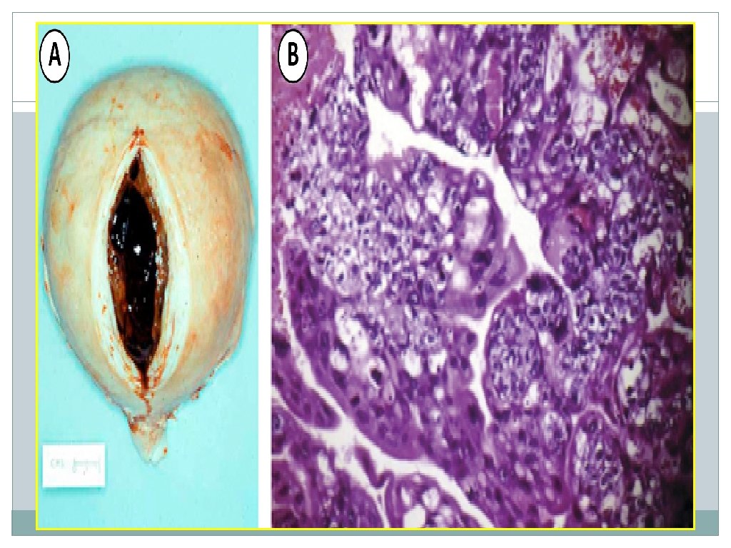
Choriocarcinoma Epidemiology • Choriocarcinoma is a highly malignant, h. CG-secreting tumour that can arise following any type of pregnancy event. Choriocarcinoma may complicate 1/ 50 000 pregnancies and 1/ 40 (3%) of hydatidiform moles. • The most aggressive GTN(early vascular invasion and widespread metastases). Pathology • The macroscopic appearance is a soft, purple, largely haemorrhagic mass. • Microscopically it appears as an abnormal trophoblastic hyperplasia and anaplasia, with the absence of chorionic villi, haemorrhage and necrosis, and with direct invasion into the myometrium and venous sinuses. • Tumour metastases to distant sites occurs by vascular spread; common sites include the lungs, brain, liver, pelvis and vagina, kidney, intestines and spleen.
Clinical presentation • The presenting features may be similar to hydatidiform moles, with vaginal bleeding, abdominal pain, a pelvic mass and symptoms due to a high serum h. CG. However, a third of women with choriocarcinoma present without gynaecological features but with features of metastases. • Primary or secondary postpartum hemorrhage. • abnormal vaginal bleeding may develop a year or more after an antecedent pregnancy. • Hemorrhage can be severe if the tumor erodes through the myometrium or uterine vessels. • Respiratory symptoms (eg, cough, chest pain, hemoptysis) or signs of gastrointestinal, urologic, and intracerebral bleeding are indicative of metastatic disease. Hepatic involvement from advanced disease may cause epigastric or right upper quadrant pain.
Management • Excision biopsy of a metastasis should be considered if this could be safely achieved. This allows both histological diagnosis and genetic analysis to establish the genetic nature of the tumour. • Often a biopsy is impossible and the diagnosis is made on clinical history and other investigational findings. Patients are then scored according to the FIGO prognostic system and treated as described for persistent gestational trophoblastic disease/gestational trophoblastic neoplasia
Placental site trophoblastic disease • • PSTT is uncommon consists predominantly of mononuclear intermediate trophoblast and syncytial elements, without chorionic villi, infiltrating in sheets between myometrial fibres. Most PSTTs arise following nonmolar pregnancies. Generally present months to years after a term gestation Produce only small amounts of h. CG and human placental lactogen (h. PL� ) tend to remain confined to the uterus late metastasis , more than 30 percent of patients already have metastases at presentation. The disorder should be suspected if h. CG concentrations are low for the volume of disease present. Histological analysis is needed to confirm the diagnosis.
PSTT Irregular vaginal bleeding and an enlarged uterus are common, amenorrhea or virilization may occur, and nephrotic syndrome has been reported. Lymph node metastases occur in 6 percent of women with PSTT
PSTT vs choriocarcinoma Compared to choriocarcinoma, PSTT is associated with less vascular invasion and necrosis and greater tendency for lymphatic spread. PSTT are relatively insensitive to chemotherapy and hysterectomy and pelvic lymphadenectomy remains the mainstay of treatment if there is residual disease confined to the uterus. Since h. CG is not a reliable marker in these patients, long-term (>5 years) clinical follow-up is recommended.
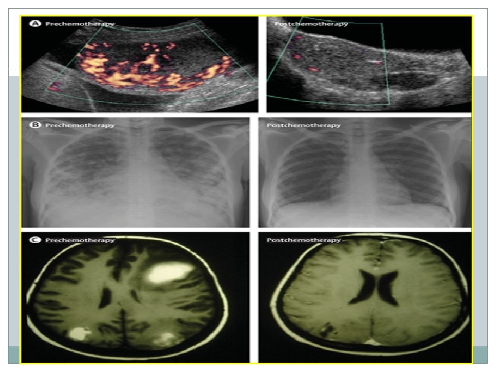
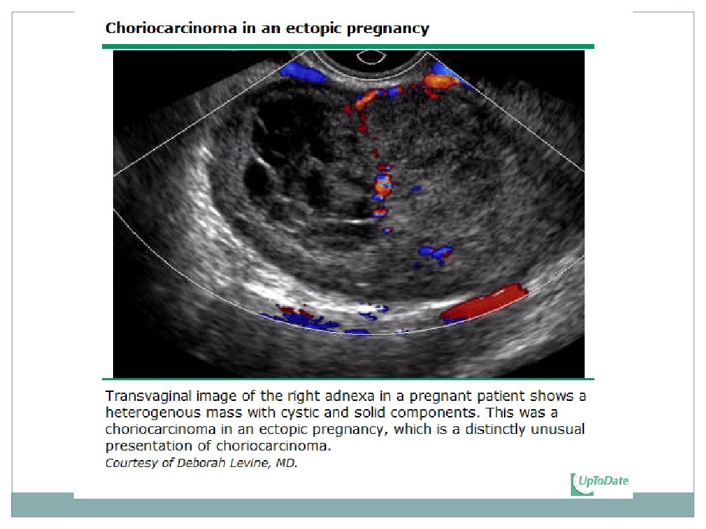
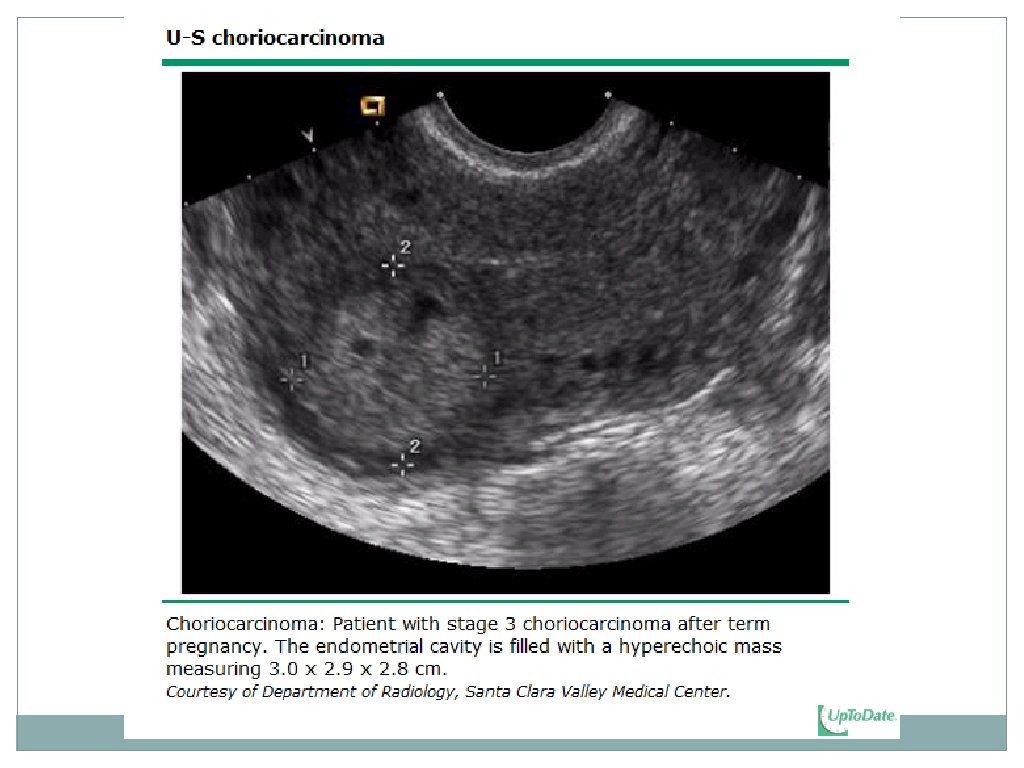
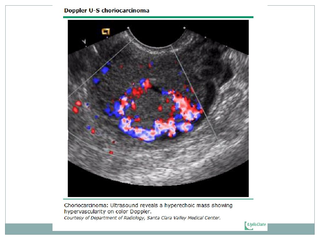
Choriocarcinoma and PSTT Sonographic features 1. A mass enlarging the uterus, with a heterogeneous appearance that correlates with areas of necrosis and hemorrhage 2. The tumor is usually markedly hypervascular on color Doppler 3. The tumor may extend into the parametrium.
Invasive mole • Arises from myometrial invasion of complete hydatidiform mole or partial hydatidiform mole via direct extension through tissue or venous channels. About 10% of hydatidiform moles may progress to become invasive moles. • By clinical presentation, 15% of invasive moles demonstrate metastases to the lung and vagina. Most often diagnosed clinically (i. e. persistent h. CG elevation after molar uterine evacuation, and/or evidence of metastatic disease) rather than pathologically (i. e. histological diagnosis), and responds to chemotherapy.
Role of uterine surgery • 1. For women with choriocarcinoma who do not desire future fertility, …. hysterectomy in addition to chemotherapy (Hysterectomy prevents the persistence of drug-resistant local disease and can shorten the duration and amount of chemotherapy required to produce remission). • 2. option of hysterectomy for women with persistent/invasive GTN that is resistant to single agent chemotherapy. • 3. For women with choriocarcinoma who wish to preserve future fertility, administer chemotherapy alone. • 4. For women with stage I and II PSTT recommend hysterectomy as the treatment of choice (PSTT is usually limited to the uterus, and the response to chemotherapy is more variable than with choriocarcinoma. ) • • 5. Hysterectomy may also be indicated in the subset of women who have chemotherapy-resistant disease, particularly if the histology is PSTT.
one of the most chemotherapy-responsive and highly curable cancers, even in the setting of widespread metastatic disease
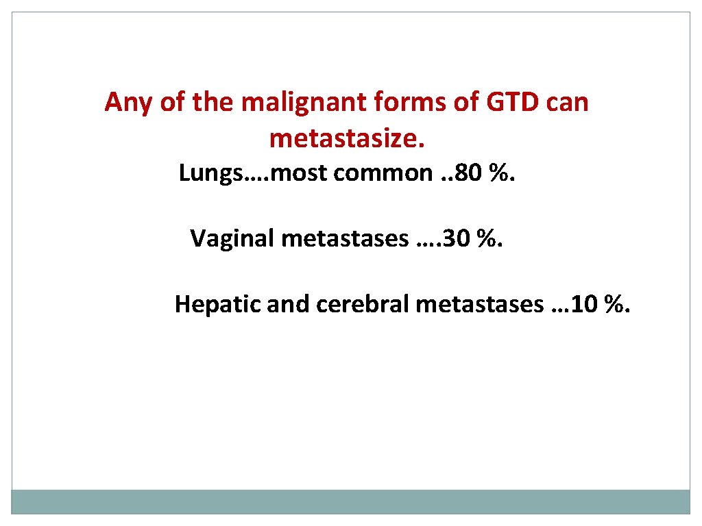
Any of the malignant forms of GTD can metastasize. Lungs…. most common. . 80 %. Vaginal metastases …. 30 %. Hepatic and cerebral metastases … 10 %.
Summary GTDs are curable with the preservation of fertility Hydatidiform moles usually present with vaginal bleeding in pregnancy Suction evacuation by an experienced gynaecologist; oxytocics should ideally be avoided Registration with a specialised centre is mandatory h. CG is an excellent marker for monitoring / the rare PSTT CHM or PHM not requiring chemotherapy, h. CG should be monitored for at least 6/12 following uterine evacuation & pregnancy avoided for this time interval Persisting GTD or GTN requiring CT, h. CG should be monitored for at least 5 years to lifetime &pregnancy avoided for 12 months after completion of chemotherapy In women with a history of molar pregnancy, serum or urine h. CG should be checked 6 weeks and 10 weeks after every future pregnancy to ensure no GTN relapse or new GTN development COCPs should not be used until h. CG levels become normal (undetectable) 15% of CHM and 0. 5% of PHM will ultimately require CT for persistent GTD/GTN CT regimen is based on a prognostic scoring system The majority of women will conceive again following GTD/GTN with no increased rate of congenital fetal malformations.
- Slides: 66