
Gestational Trophblastic Neoplasia GTN A Alobaid MBBS FRCSC
Gestational Trophblastic Neoplasia (GTN) A. Alobaid, MBBS, FRCS(C), FACOG Consultant, Gynecologic Oncology Assistant professor, KSU Medical Director, Women’s Specialized Hospital King Fahad Medical City
l The first record of gestational trophoblastic disease (GTD) probably dates to 400 BC, when Hippocrates described “dropsy of the uterus” l In 1276, the attendants of Margaret Countess of Henneberg noticed that her abnormal delivery consisted of multiple hydropic vesicles
l GTN defines a heterogeneous group of lesions that represent an aberrant fertilization event l The pathogenesis is unique because the maternal tumor arises from fetal tissue l It is the most curable gynecologic malignancy

l There are several histologically distinct types of GTD, the WHO has classified them into two premalignant diseases: Hydatidiform mole (complete or partial), and three malignant disorders: Persistent/invasive GTD Choriocarcinoma Placental site trophoblastic tumors
Epidemiology l Less than 1 in 1000 pregnancies in most of the world, 2 in 1000 in Japan (differences in diet) l Vitamin A deficiency in the rhesus monkey produces degeneration of the seminiferous epithelium with production of primitive spermatogonia and spermatocytes
Risk factors l Women <15 years or >40 years of age l Patients with previous history of molar pregnancy l Possible other factors: deficiency of animal fat and carotene, professional occupation, history of prior spontaneous abortion
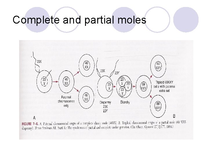
Complete and partial moles
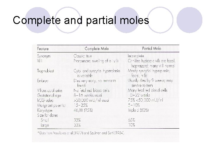
Complete and partial moles
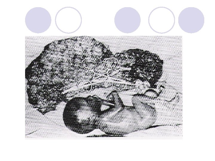
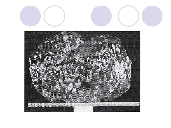
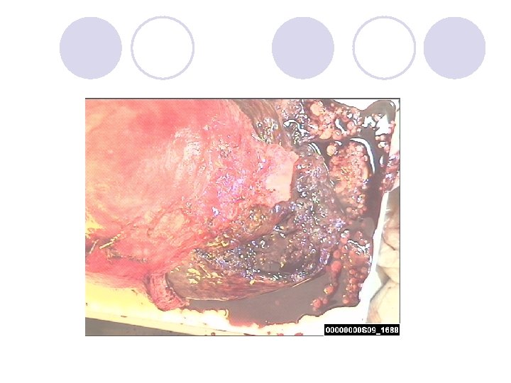
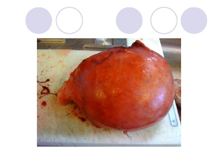
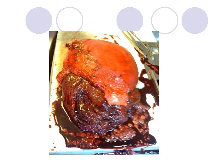
Invasive hydatidiform mole l It is clinically identified by the combination of an abnormal uterine ultrasound scan and a persistent or rising B-HCG level after uterine evacuation of a molar pregnancy l Pathologic confirmation of invasion is rarely required
Choriocarcinoma l Highly malignant l Greater risk of hemorrhage and metastases l May arise from any type of pregnancy
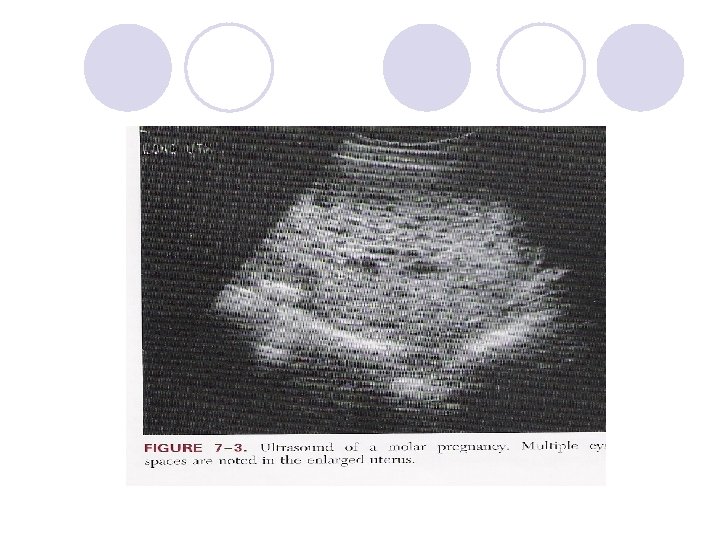
Diagnosis l In many patients the first evidence to suggest the presence of a hydatidiform mole is the passage of vesicular tissue l A quantative pregnancy test of greater than 100, 000 IU/L, an enlarged uterus, and vaginal bleeding suggest a diagnosis of a hydatidiform mole l Ultrasound (test of choice) will show multiple echoes (snow storm)
β-h. CG assays l The family of pituitary and placental glycoprotein hormones: HCG, FSH, LH and TSH, all have a common α-subunit and a distinct β-subunit l Many β-h. CG assays are available, some detect intact β-h. CG and others are selective for individual fragments l The competitive RIA using a polyclonal antibody recognizing all forms of β-h. CG remains a goldstandard assay for use in the management of GTD
β-h. CG assays l The amount of h. CG produced corresponds with tumor volume so that a serum h. CG of 5 IU/L corresponds to approximately 10, 000 to 100, 000 viable tumor cells
Symptoms and signs l Vaginal bleeding is the most common symptom l Patients with complete mole may have: first trimester pre-eclampsia, hyperthyroidism, hyperemesis, increased uterine size and theca-lutein cysts l Patients with partial moles are diagnosed clinically as missed or incomplete abortion
Management l Evacuation curettage: the method of evacuation l RH –ve patients should receive Rhogam l IV oxytocin should be administered after a moderate amount of the tissue has been removed l Complications may include: uterine perforation, hemorrhage, and trophblastic embolization l Hysterectomy may be selected as a method of evacuation in patients who desire sterilization
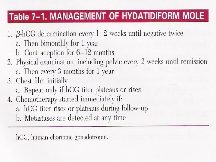
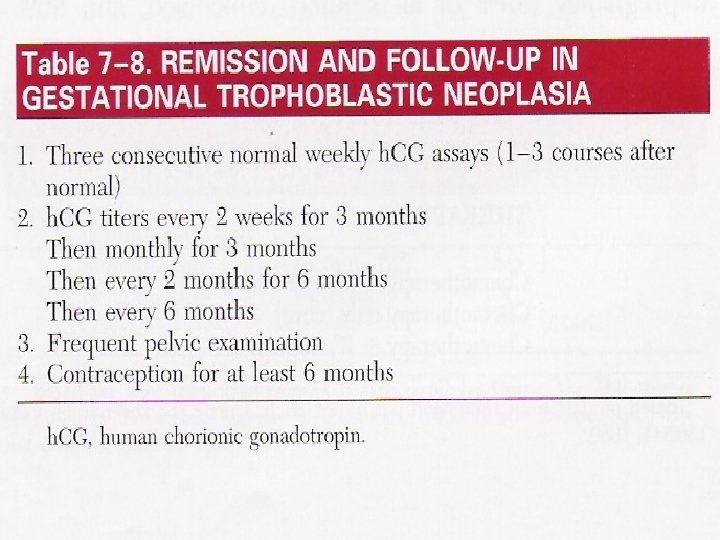
Follow-up of molar pregnancy
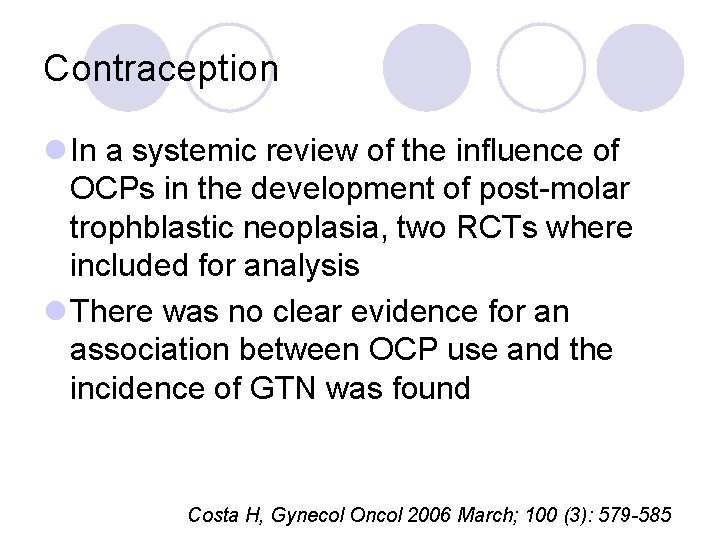
Contraception l In a systemic review of the influence of OCPs in the development of post-molar trophblastic neoplasia, two RCTs where included for analysis l There was no clear evidence for an association between OCP use and the incidence of GTN was found Costa H, Gynecol Oncol 2006 March; 100 (3): 579 -585
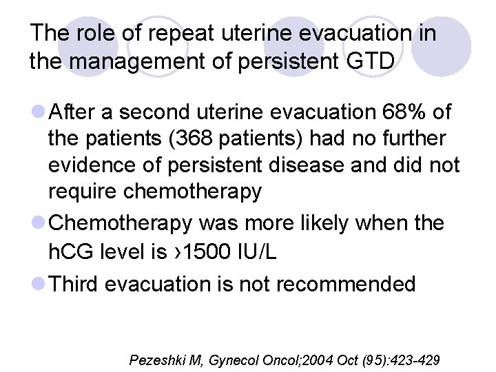
The role of repeat uterine evacuation in the management of persistent GTD l After a second uterine evacuation 68% of the patients (368 patients) had no further evidence of persistent disease and did not require chemotherapy l Chemotherapy was more likely when the h. CG level is › 1500 IU/L l Third evacuation is not recommended Pezeshki M, Gynecol Oncol; 2004 Oct (95): 423 -429
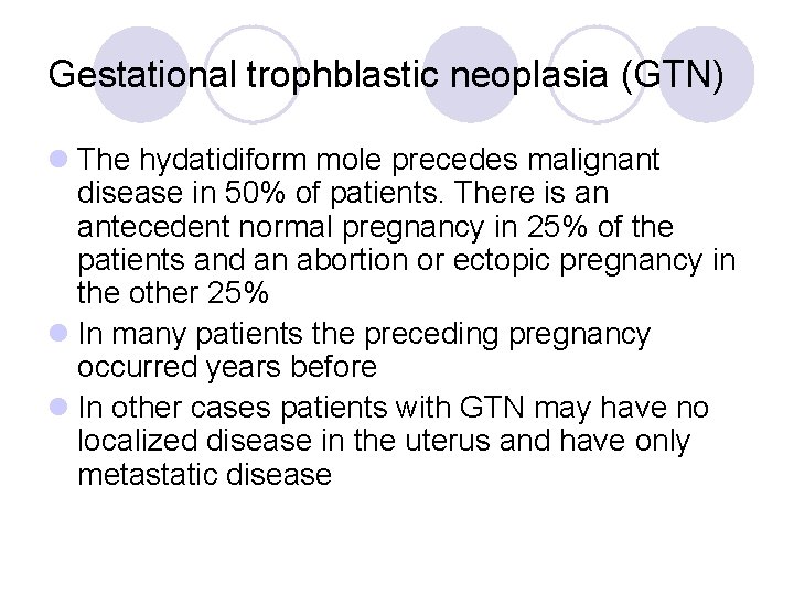
Gestational trophblastic neoplasia (GTN) l The hydatidiform mole precedes malignant disease in 50% of patients. There is an antecedent normal pregnancy in 25% of the patients and an abortion or ectopic pregnancy in the other 25% l In many patients the preceding pregnancy occurred years before l In other cases patients with GTN may have no localized disease in the uterus and have only metastatic disease
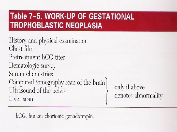
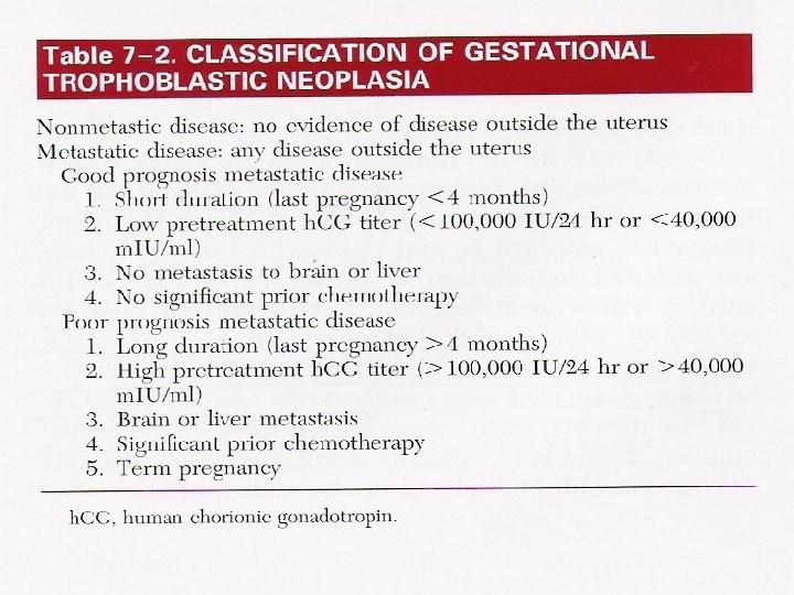
Classification of GTN
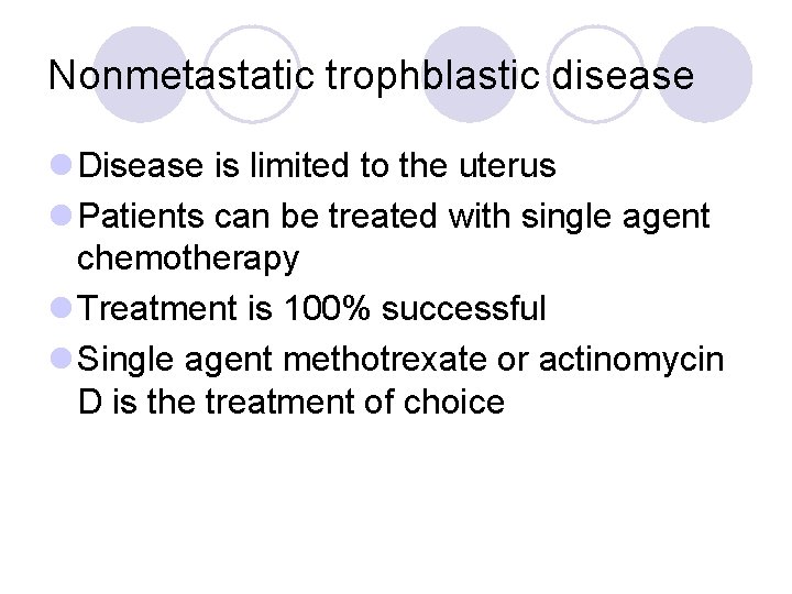
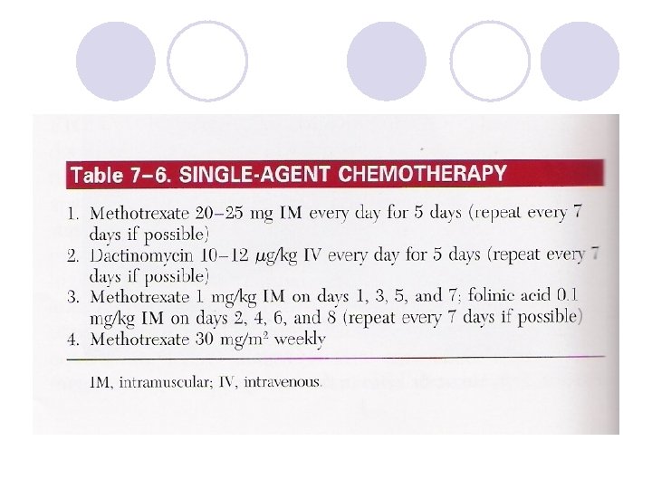
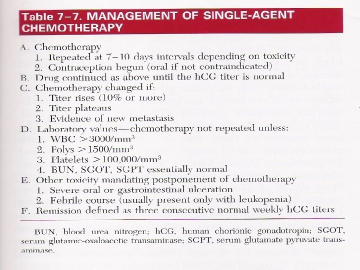
Nonmetastatic trophblastic disease l Disease is limited to the uterus l Patients can be treated with single agent chemotherapy l Treatment is 100% successful l Single agent methotrexate or actinomycin D is the treatment of choice
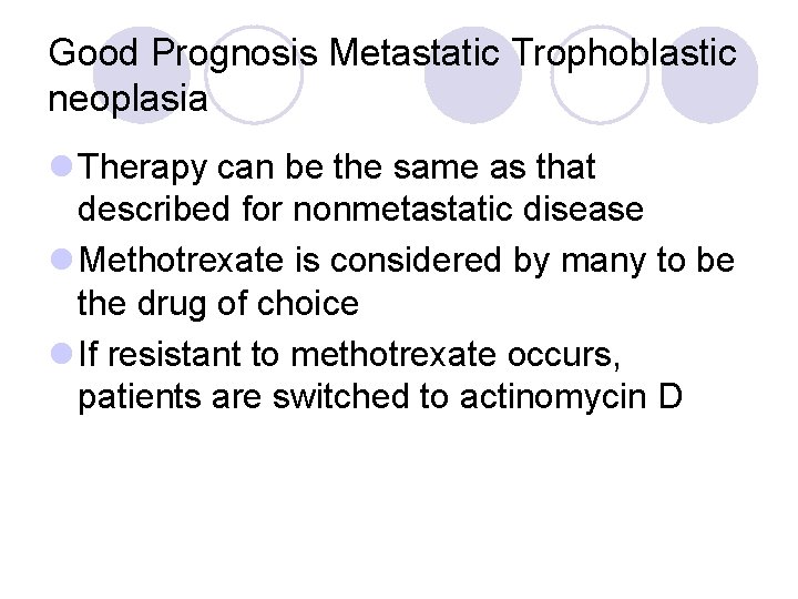
Good Prognosis Metastatic Trophoblastic neoplasia l Therapy can be the same as that described for nonmetastatic disease l Methotrexate is considered by many to be the drug of choice l If resistant to methotrexate occurs, patients are switched to actinomycin D
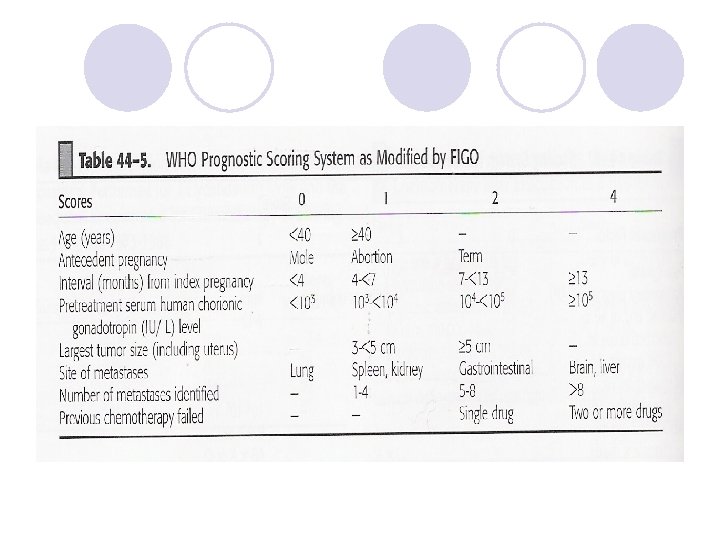
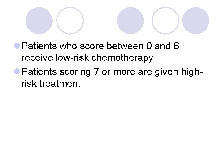
l Patients who score between 0 and 6 receive low-risk chemotherapy l Patients scoring 7 or more are given highrisk treatment
Drug-resistant disease l CT of the chest and abdomen together with MRI of the brain and pelvis is often helpful and can detect deposits not previously seen
Poor Prognosis Metastatic Trophoblastic neoplasia l Multiple agent chemotherapy is recommended in this disease l EMA-CO is considered the regimen of choice in most high-risk patients (Etoposide, Methotrexate, Actinomycin D, Cyclophosphamide, Vincristin) l The overall survival rate for these patients is 80 -85%
Poor Prognosis Metastatic Trophoblastic neoplasia l Patients with cerebral or hepatic metastases are treated concurrently with radiotherapy for the whole brain or liver (for hemostasis) l Surgery is not necessary in most patients, it may play a role in cases of tumor resistance to chemotherapy
Persistent Low HCG Levels l Pituitary HCG l False +ve HCG results l Quiescence GTD
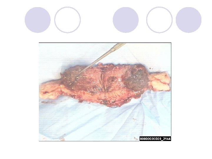
Placental Site Trophoblastic Tumor l Rare tumors (account for 0. 23% cases of GTD) l It has a variety of clinical features and its course is unpredictable l Can appear shortly after termination of pregnancy or years later l Hysterectomy is considered optimal therapy and is usually adequate in most situations l Chemotherapy can still play a major role Hassaida A, Gynecol Oncol, 2005 August; 99: 603 -607
Future Childbearing l After treatment of GTN, molar pregnancies occur in only about 1 -2% of subsequent pregnancies l These patients should be evaluated with a first trimester ultrasonography l Pregnancy outcome in women with history of molar gestation is similar to those with no such history l Standard chemotherapy protocols have minimal impact on the subsequent ability to reproduce

Thank you
- Slides: 42