Genus Clostridium v General characteristic of Clostridium group


 Clostridium botulinum v. It causes botulism which is intoxication not a bacterial infection")


 is the most")


 Lactose Sugar Casein Protein Litmus")


 It affects cattle mainly & causes black leg (black quarter).")


 bacillus. The spiral")
- Slides: 43
Genus Clostridium v General characteristic of Clostridium group: - Large Gram positive Straight or slightly curved rods with slightly rounded ends Anaerobic bacilli Spore bearing Motility: mostly are motile by peritrichous flagella except Cl. perfringens (formely named Cl. welchii), which is non motile. Capsule formation: All of them are non-capsulated except Cl. perfringens, which is capsulated Saprophytes Some are commensals of the animal & human gut which invade the blood and tissue when host die and initiate the decomposition of the carcass (dead body)
Culture characteristic: They grow under strict anaerobic condition that can be achieved by several commercial kits that use a chemical reaction to replace O 2 with CO 2 as the Anaerobic Gas. Pak System. They require enriched media for growth as (Cooked meat medium, liver-infusion media or thioglycolate broth). Biochemical reactions: Clostridia are classified into 4 groups according to their action on protein and carbohydrates found in cooked meat medium or liverinfusion broth. A- Proteolytic group: Ex. Cl. histolyticum. B- Saccharolytic group: - Ex. Cl. perfringens, Cl. chauvoei & Cl. septicum. C- Saccharolytic and proteolytic group: Ex. Cl. sporagenes and Cl. botulinum D- Non-Saccharolytic and Non-proteolytic group: Ex. Cl. tetani.
Pathogencity Clostridia are classified according to their ability to produce exotoxins & to invade tissue into two groups: Non invasive group Invasive group Neurotoxic Clostridia Cl. tetani Cl. botulinum Histotoxic clostridia Enterotoxaemia clostridia Clostridia associated with antibiotic inducing disease
Non invasive group v. Members of this group have no power to invade and multiply in living tissues & internal organs. v. They produce powerful exotoxins.
1 - Clostridium Causing Tetanus Cl. tetani Gram positive, straight, slender rod with rounded ends All species form endospore (drumstick with a large round end) Fermentative Obligate anaerobe Motile by peritrichous flagella Grows well in cooked meat broth and produces a thin spreading film when grown on enriched blood agar Spores are highly resistant to adverse conditions Iodine (1%) in water is able to kill the spores within a few hours
Toxins Cl. tetani produces two types of toxins: Tetanolysin, which causes lysis of RBCs Tetanospasmin is neurotoxin and essential pathogenic product – Tetanospasmin is toxic to humans and various animals when injected parenterally, but it is not toxic by the oral route – Tetanospasmin which causes increasing excitability of spinal cord neurons and muscle spasm
Laboratory Diagnosis of Tetanus The diagnosis of tetanus depends primarily upon the clinical manifestation of tetanus including muscle spasm and rigidity. Specimen: Wound exudates using capillary tube Culture: – On blood agar and incubated anaerobically – Growth appears as a fine spreading film. Gram stain is a good method for identifying Clostridium – Cl. tetani is Gram positive rod motile with a round terminal spore giving a drumstick appearance
(2) Clostridium botulinum v. It causes botulism which is intoxication not a bacterial infection that result from ingestion of foodstuffs containing toxin produced by Cl. botulinum growing in the food. v. Botulinum toxin is extremely potent & used as biological weapon in bioterrorism. v. Morphology: is a gram positive Large, straight rods , Motile, non-capsulated & spore-forming bacilli, Spores are oval and mainly subterminal (spoon-like in shape) v. Culture characters: Cl. Botulinum is strictly anaerobic, grow best at temperature around 30°C, isolated on cooked meat broth - Trypticase-peptoneglucose-yeast extract (TPGY) broth – Liver veal egg yolk agar in an anaerobic environment
On egg yolk agar produces iridescent layer on & around colonies due their lipase activity. On horse blood agar produces haemolysis Biochemical reaction All types ferment glucose and maltose only. All types produce H 2 S and are indole negative. Cl. botulinum posess seven types (A-G) vary in their saccharolytic and proteolytic activites. (a) Non- proteolytic type C, D, E, G. (b) Proteolytic type A, B, F.
Botulism Synonyms: Botulinus intoxication- Forage poisoning - Limber neck. Botulism gets it name from “botulus” which is a Latin word meaning sausage Definition: It is a rare but serious paralytic illness caused by botulinum toxin, which is produced by the bacterium Clostridium botulinum Mode of transmission: Ingestion of neurotoxin in contaminated feed stuffs is the main method of transmission in animals & man. Pathogenesis: Cl. botulinum produces a very potent neurotoxin
Following ingestion the toxin is absorbed from the gut blood reaches the neuromuscular junction then binds irreversibly to the presynaptic nerve ending of the peripheral nervous system & cranial nerves, where it blocks the release of acetylcholine no muscle contraction leading to flaccid paralysis.
Diagnosis: Diagnosis of botulism can be determined by obtaining a good history. Gram stained films shows Gram positive spoon-like bacilli. Isolation of the organism from the food by anaerobic cultivation, then identification which is based on culture characters and biochemical reactions. The definitive diagnosis comes from demonstration of the toxin in serum, gut contents or organs. ELISA test & PCR is available for determining neurotoxin types. Pathogenicity test: - Injection of botulinum toxin found in food remnants or serum in guinea pig or mice death with generalized flaccid paralysis. Mouse protection test: - Typing botulinum toxin can be done by neutralization with specific antitoxin in mice
Invasive group • Members of this group have a power to invade and multiply in tissues & internal organs. • The toxin is less potent than the non invasive group exotoxin. • These organisms are known as gangrene group in animals & man.
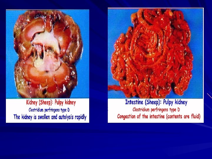
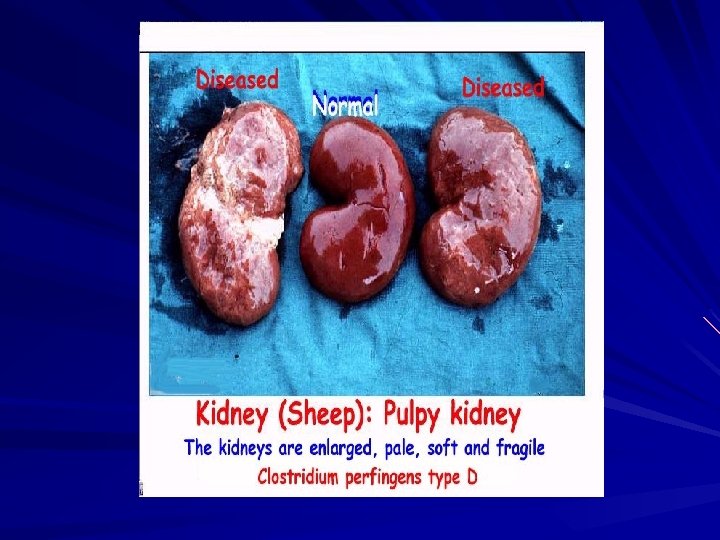
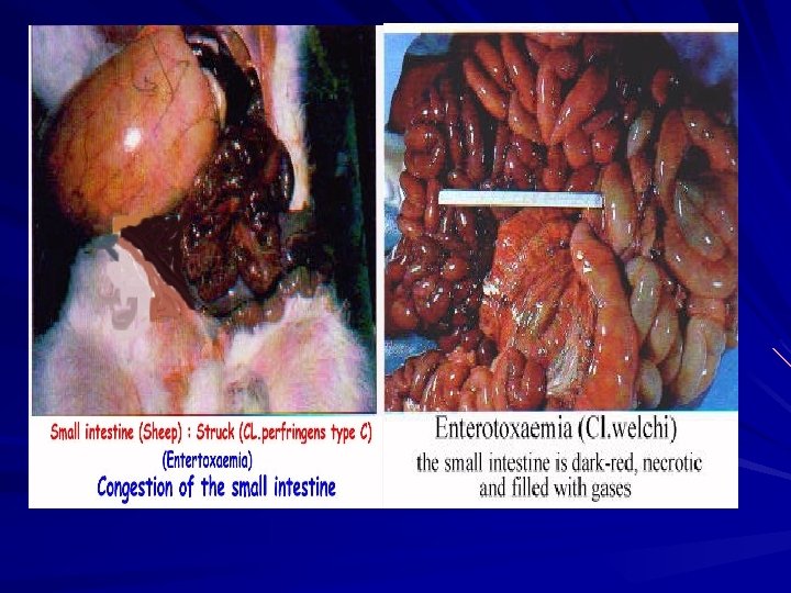
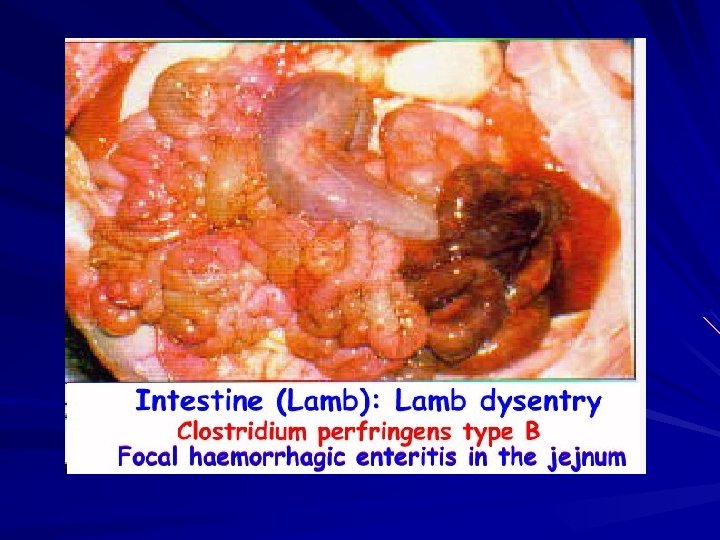
Enterotoxaemia Clostridial enterotoxaemias are acute, highly fatal intoxication caused by Closlridium perfringens types (A, B, C, D & E) exotoxins.
Clostridium perfringens Cl. perfringens type A Major toxins Enterotoxin α- toxin Hosts Disease Clinical Signs human Food poisoning human Calves & lambs Gas gangrene Enterotox-aemia Abdominal pain, nausea & diarrhea without vomiting Gas gangrene Haemorrhagic entritis, diarrhea & death. -Haemorrhagic diarrhea & rapidly fatal enterotoxaemia -Gas gangrene in muscles B α- toxin β- toxin ε- toxin Lamb dysentery C α- toxin Sheep & goat Struck β- toxin chicken Avian Necrotic entritis D α- toxin ε- toxin Adult sheep Pulpy kidney E α- toxin i- toxin Calves & lambs Enterotox-aemia Convulsions, enterotoxaemia & sudden death Diarrhea, enterotoxaemia Death within few hrs vascular damage, particularly of capillaries in the brain & decomposition of kidney & sudden death. Edema & enterotoxaemia
Clostridium perfringens Large Gram-positive bacilli with stubby ends Capsulated Non motile (Cl. tetani is motile) Anaerobic Grown quickly on selective media Can be identified by Nagler reaction
Toxins The toxins of Cl. perfringens – toxin (phospholipase C, lecithinase) is the most important toxin Lyses of RBCs, platelets, leucocytes and endothelial cells Increased vascular permeability with massive hemolysis and bleeding tissue destruction Hepatic toxicity and myocardial dysfunction – -toxin is responsible for necrotic lesions in necrotizing enterocolitis – Enterotoxin is heat labile toxin produced in colon → food poisoning
Laboratory Diagnosis Ø Specimen: Histological specimen or wound exudates Ø Histological specimen transferred aseptically into a sterile screwcapped bottle & used immediately for microscopical examination & culture Ø Specimens of exudates should be taken from the deeper areas of the wound where the infection seems to be most pronounced Ø Microscopical examination (Gram, Spore stain etc) Ø Gram-positive bacilli, non motile, capsulated & sporulated Ø The spore is oval, sub-terminal & non bulging Ø Spores are rarely observed Ø Culture: Anaerobically at 37 C Ø On Robertson's cooked meat medium → blackening of meat will observed with the production of H 2 S and NH 3 Ø On blood agar → β-hemolytic colonies
Biochemical Tests Cl. perfringnes characterized by: ØIt ferments many carbohydrates with acid & gas ØIt acidified litmus milk with stormy clot production ØNagler reaction is positive
Reaction on Litmus Milk Contains Skimmed Milk (Without Fat) Lactose Sugar Casein Protein Litmus indicator Acid Base and Redox indicator
Reaction on Litmus Milk 1 - Acidic Reaction Lactose Fermentation Acid Litmus Indicator Pink Color (Milk Sugar) 2 - Basic Reaction Litmus Indicator Digestion Casein (Milk Protein) Alkaline amines Blue Color
Reaction on Litmus Milk Stormy Clot Formation Lactose Fermentation Acid + Gas Stormy Clot Milk Sugar Coagulation Casein Milk Protein Clot
Reaction on Litmus Milk
Nagler’s Reaction This test is done to detect the lecithinase activity – The M. O is inoculated on the medium containing human serum or egg yolk (contains lecithin) – The plate is incubated an aerobically at 37 C for 24 h – Colonies of Cl. perfringens are surrounded by zones of turbidity due to lecithinase activity and the effect is specifically inhibited if Cl. perfringens antiserum containing antitoxin is present on the medium
Nagler’s Reaction Procedure of Nagler Reaction Positive Nagler Reaction
Toxin neutralization tests for identifying the types of C. perfringens implicated in enterotoxaemias Antitoxin (specificity) Test results Toxins identified in intestinal contents α α, β, ε α, β α, ε α, I Type A (anti α) - D D Type B (anti α, β, ε) - - - Type C (anti α, β) - D D Type D (anti α, ε) - D D - D Type E (anti α, I) - D D D - D: death of mouse or dermal necrosis of guinea-pig; toxins are not neutralized. -: mouse or guinea-pig unaffected; toxins are neutralized
Histotoxic Clostridia These are invasive group of clostridia that cause extensive destruction of muscle and connective tissue and are characterized by the formation of gas. Include C. chauvoei, C. colinum, C. hemolyticum, C. novyi, C. perfringens type A and C, C. septicum and C. sordellii
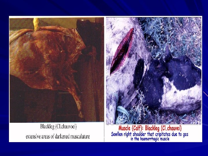
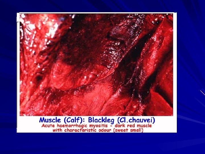
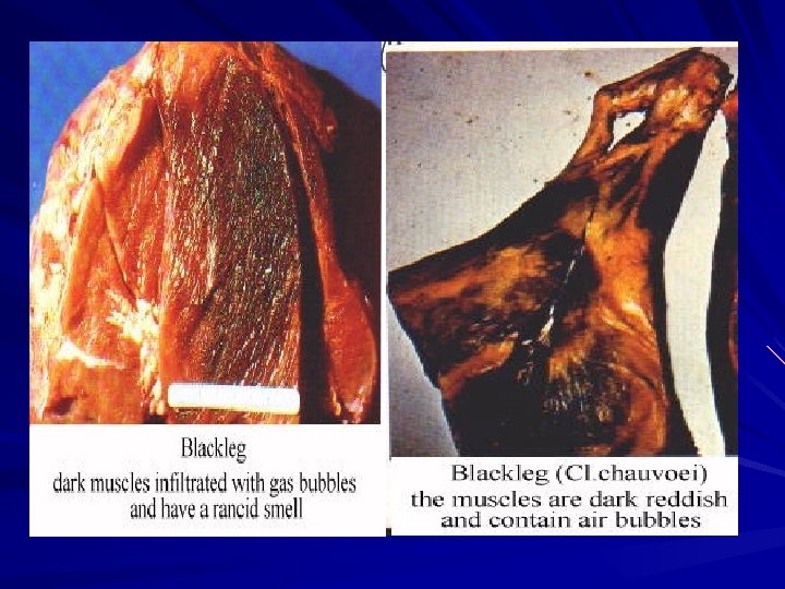
Clostridium chauvoei (Cl. Feseri) It affects cattle mainly & causes black leg (black quarter). In sheep it causes a symptomatic anthrax while equines are resistant The disease is characterized by swellings in the hip, shoulder, chest, back, neck or elsewhere. First the swelling is small, hot and painful. As the disease progresses, the swelling enlarges and becomes spongy and gaseous. If you press the swelling, gas can be felt under the skin. The animal usually dies in 12 to 48 hours.
Morphology: It is Gram positive straight long rod with rounded ends, arranged singly or in pairs. Motile with peritrichus flagella. Form central to subterminal ''lemon shape'' endospores. Culture characteristics: It is strict anaerobic, grows poorly after 3 -5 days at 37 C°. On ordinary media it requires glucose, blood or serum for good growth & produces minute (1 - 2 mm in diameter), greyish white transparent colonies. On liver sheep blood agar produces a narrow zone of haemolysis. On cooked meat medium produce poor growth after 3 days in the form of slight turbidity with some gases & foul odor. Biochemical reactions: Sacharolytic and non-proteolytic: - It ferments glucose, lactose, maltose and sucrose, but not salicin with slight gas production. Liquefaction of gelatin and H 2 S production positive. Nagler's-reaction & indole test negative. Litmus milk is unchanged or form acid with partial clotting. Antigenic structure & toxins: C. chauvoei has 3 types of antigens, one common somatic antigen and 2 flagellar antigens. It produces 2 types of toxins, lethal and haemolytic toxins
Diagnosis: 1 - Clinical signs and post-mortem examinations of no significance because the course of the disease is similar to that of malignant oedema in cattle and braxy disease in sheep caused by Cl. septicum. 2 - Direct staining with fluorescent labeled antibodies 3 - Isolation and identification see cultural characters and biochemical reactions. 4 - Protection Test: Inject two guinea pigs S/C with 0. 5 -1 ml of 72 hrs old culture or extract from infected muscles. One of them is protected by injection with specific C. chauvoei antiserum. Result: The first guinea pig dies within 72 hrs with oedema at the site of injection with bloody stained serous fluid and the muscles are very dark red in colour with foul odour. The protected one remains alive.
Items 1 - Disease 2 - On solid medium 3 -Colonial appearance 4 -Sucrose fermentation 5 -Salicin fermentation 6 -Indole production 7 - H 2 S 8 - Susceptibility to lab animals: Guinea pigs. Pigeons 9 - Gram’s stained impression smear of experimentally dead G. pig (liver& peritoneal surface). 10 - Toxins 11 - Antigenic structure C. chauvoei. Black quarter disease or black leg disease or symptomatic anthrax. Grows slowly within 48 -72 hrs Minute small delicate not more than 0. 5 mm with rounded edges. +ve -ve +ve C. septicum Malignant edema in cattle or braxy in sheep. Grows rapidly within 18 hrs. Large spreading filamentous colonies. -ve +ve after 72 hrs Resistant+ve after 18 -24 hrs highly susceptible 2436 hrs Short rods in short chains not more than 2 -3 bacilli. Long large bacilli in very long chains. 2 types of toxins: - lethal toxin - heamolytic toxin 8 types of toxins: lethal, necrotizing, haemolytic, leucocidin, fibrinolysin, hyalurinidase, gelatinase, proteinase. One somatic and 2 flagellar 2 somatic and 4 flagellar + spore antigen shared antigenically to that of C. chauvoei
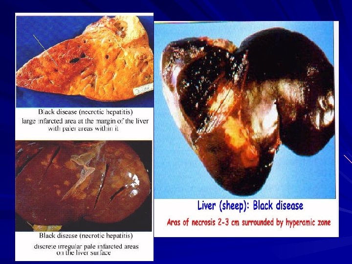
Clostridium novyi Cl. novyi is an anaerobic bacterium found in soil, manure, and plants Different types of Cl. Novyi: Type Toxin Animal susceptible Diseases Cl. novji A Alpha +++ Delta ++ Gamma + Man and animals Cl. novji B Beta +++ Alpha + Cattle & sheep Turkeys Sheep Cl. novji C No toxins Non pathogenic Beta ++ Red water disease (Infectious icterohaemoglobin urea) Cl. novji D (Cl. haemolyticum) Cattle & sheep Malignant oedema gas gangrene Black disease Infections necrotic hepatitis. Swelled head in rams
- Large, strongly Gram positive rods with rounded ends and peritrichous flagella. - Spores oval, central or subterminal spoon like in shape, non capsulated. Cultural characters: - Strict anaerobic & all types grown well in cooked meat media after 3 -5 days. yi type A & B produce raised opaque, dome-shaped colonies in (young cultures) but often flattened, large & irregular in older cultures. - On blood agar Type D (Cl. haemolyticum) formed pin point, dew drops colonies surrounded by a wide zone of β- haemolysis. - On lactose-eggs-yolk-milk agar all types of Cl. novyi fail to ferment lactose and opalescence will be produced ie: positive Nagler's reaction. Biochemical reaction: Cl. novyi type A is saccharolytic and mildly proteolytic. All types do not ferment lactose. Cl. novyi types A, B and D are Nagler's reaction positive which inhibited specifically with all antitoxins of Cl. novyi only. It liquefies Gelatin & produces H 2 S. Type A strains do not produce indole or reduce sulphite.
Clostridia associated with antibiotic induced disease Cl. difficile Clostridium difficile stands for Greek kloster = spindle & Latin difficile = difficult. It is a large, Gram positive rod that forms oval, subterminal spores. Onto blood agar the colonies are non haemolytic, raised with a rhizoid edge. It produces an enterotoxin (A) and cytotoxin (B). It causes human, rabbits, and guinea-pigs enterocolitis initiated by prolonged antibiotic therapy.
Cl. spiroforme It is a helically coiled, anaerobic, gram-positive sporeforming (terminal) bacillus. The spiral form occurs in smears from cultures on blood agar but in faeces or caecal contents, the spiral morphology is not so marked. On blood agar under anaerobic condition it is nonhaemolytic and produces convex, circular, shiny, and whitish to grey colonies. The organism produces a cytotoxin and an exotoxin that is identical to the iota toxin of C. perfringens type E. It causes diarrhoea in weaning rabbits & in adults after adminstration of antibiotics especially clindamycine