GENITOURINARY DISORDERS Islamic University Nursing College Genitourinary Tract


 is short")

 Glomerular filtration rate: measured by the creatinine")


 UTI is the presence of significant numbers of microorganisms anywhere")
 Escherichia coli (80% of cases) and other gram-negative enteric-organisms are")


: � � � low grade fever")


 is failure of one or both testes to descend normally through the")
 Cryptorchid testes are often accompanied by congenital hernias and abnormal testes, and")
 CM � Non-palpable testes � Affected hemiscrotum will appear smaller than the")
 Surgical repair is done to � prevent damage to the undescended testicle")
 Retrograde flow of urine from the bladder up the ureters and")
 Grading system depends on the extend of the VUR , dilatation")
 Primary reflux: congenital anomaly affects the ureterovesical junction Secondary reflux: occurs")
 Nursing Diagnosis � � � High risk for injury related to")

 Inflammation of the Glomeruli occurs as an immune complex disease after")
 Clinical manifestations � � � Tea-colored urine Anorexia Joint stiffness &")
 Management � Usually resolves spontaneously, treatment is focused on relief of")
 Complications �Acute/chronic renal failure �Hyperkalemia �Nephrotic syndrome �Chronic glomerulonephritis � Hypertension")
 Nursing Diagnosis � Fluid volume excess r/t decreased U. O. �")
 Unknown cause of high proteinuria as a result of damage to")
 Clinical Manifestations � � � � weight gain Puffiness of face,")
 Diagnostic test �Marked proteinuria +1 - +4 �Minimal hematuria � Reduce")
 Managements � Reduce urinary protein excretion � Maintain a protein-free urine")
 Nursing Diagnosis � Fluid volume excess related to fluid accumulation in")
 Interventions � Assess I&O � Assess changes in edema � Measure")

 Renal failure is the inability of the kidneys to excrete waste")
 ARF is an abrupt decline in glomerular and tubular function")
 Clinical manifestations � Azotemia: accumulation of nitrogenous waste (Blood Urea")
: Prevention recognize patients at risk (postoperative states, cardiac surgery, septic")
: Management Treat the underlying disease Management of the complications Provision")


- Slides: 47
GENITOURINARY DISORDERS Islamic University Nursing College
Genitourinary Tract Main function of GU is � Maintaining the composition and volume of the body fluids in equilibrium � Production of certain hormonal substance (e. g. , erythropoietin) � Remove wastes from bloodstream
Genitourinary Tract � � The nephrons increase in number throughout gestation and reach their full complement by birth but still immature and less effective Glomerular filtration and absorption are relatively low at birth and do not reach adult values until 12 years
Genitourinary Tract � Loop of Henle (site of the urine concentrating mechanism) is short in the newborn which reduces the ability of the newborn to reabsorb sodium and water. Concentrating ability reaches adult levels by around 3 rd month of age � Amount of urine excreted in 24 hours depends on : fluid intake, state of kidney health, and age
GU: Diagnostic tests: urine analysis
GU: Diagnostic tests Urine Culture (suprapubic aspiration) Glomerular filtration rate: measured by the creatinine clearance test (100 ml/min) BUN: � is used to measure the amount of urea nitrogen in the blood � tests glomerular function (N= 5 – 20 mg/ 100 ml) Serum creatinine: 0. 7 – 1. 5 mg/ 100 ml
GU: Diagnostic tests Sonography & MRI � To visualize the sizes of kidneys, ureters � differentiate between solid or cystic masses. X-ray: KUB IVP: intravenous pyelogram CT scan: size & density of kidneys, adequacy of urine flow Cystoscopy : evaluate stenosis Voiding Cystourethrogram (VCUG): evaluate reflux in ureters Renal biopsy
Genitourinary Tract: Assessment Chief concern: � � � � Burning or cries during urination Blood in urine/ Frequency of urination Abdominal pain/ Flank pain Enuresis Periorbital edema Poor appetite Strong urine odor Diaper rash Family history (Renal disease) Pregnancy history(Nephrotoxic drugs) Past illnesses (Recurrent UTI)
Urinary Tract Infection (UTI) UTI is the presence of significant numbers of microorganisms anywhere within the urinary tract � May present without clinical manifestations � Peak incidence between 2 -6 years of age � Female � The has greater risk of developing UTI likelihood of reoccurrence in female is 50% � Prevalence girls of UTI in infants is 2% in boys and 3. 7% in
Urinary Tract Infection (UTI) Escherichia coli (80% of cases) and other gram-negative enteric-organisms are most commonly causative agents A number of factors contribute to the development of UTI including: Anatomy of UT � Physical properties of UT � Chemical conditions properties of the host’s urinary tract �
Factors contributing to UTI Shorter urethra in females Uncircumcised males Incomplete bladder emptying (reflux, stenosis) Altered urine and bladder chemistry/ sterility: � Adequate fluid intake promote urine sterility � Use of cranberry juice increased urine acidity and so prevent UTI Extrinsic factors: � Poor hygiene, use of bubble bath, hot tubs � Bladder neck obstruction, chronic constipation, tight clothing/ diapers � Altered Normal. flora: antimicrobial agents � Catheters
UTI: Assessment Any child with fever, dysuria, urgency should be evaluated for UTI Clean – catch urine for culture & sensitivity UTI, urine is positive for proteinuria due to bacterial growth Hematuria due to mucosal irritation Increase WBC Urine p. H is more alkaline (>7)
Gastrointestinal Tract: clinical manifestation Cystitis (infection of bladder): � � � low grade fever (LGF) Mild abdominal pain Enuresis (preschooler) Pyelonephritis (kidneys): � � � Symptoms are more acute High fever Flank or abdominal pain Vomiting Malaise
UTI: Clinical Manifestations
UTI: Management Identify contributing factors to � eliminate the infection � reduce the risk of recurrence � Prevent urosepsis � Preserve renal function 7 -10 days antibiotics matching organism sensitivity (penicillins, sulfonamide, cephalosporins, tetracyclines) Mild analgesics/ antipyretics Increase fluid intake: flush out infection Clean – catch urine after 72 hrs to assess effectiveness For recurrent UTI, prophylactic antibiotics for 6 months
UTI: Nursing Care Education regarding prevention & treatment Instruct parents to observe for clues that suggest UTI: � � � Incontinence in a toilettrained child Strongsmelling urine Frequency
Cryptorchidism (Crptorchism) is failure of one or both testes to descend normally through the inguinal canal into the scrotum Absence of testes within the scrotum can be a result of �Undescended (cryptorchid) testes, �Retractile testes (withdrawal of the testes) �Anorchia (absence of testes) �Actopic : emerges outside the inguinal ring
Cryptorchidism (Crptorchism) Cryptorchid testes are often accompanied by congenital hernias and abnormal testes, and they are at risk for subsequent torsion Unknown cause, but this problem is believed to be partly inherited Risk Factors � Prematurity; Low birth weight; Twin � Down syndrome (fetus); Hormonal abnormalities (fetus) � Toxic exposures in the mother � Mother younger than 20 or older than 35 years of age � A family history of undescended testes
Cryptorchidism (Crptorchism) CM � Non-palpable testes � Affected hemiscrotum will appear smaller than the other � In retractile testes : Intermittently observing the testes in the scrotum , thus hands should be warm when examining the baby in a warm room Management � Retractile testis can be manipulated into the scrotum. � By 1 year of age, cryptorchid testes will descend spontaneously in approximately 75% of cases in both fullterm and preterm infants � In true undescended testes rarely descend spontaneously after 1 year of age and need a surgery
Cryptorchidism (Crptorchism) Surgical repair is done to � prevent damage to the undescended testicle & decrease the incidence of tumor formation, � avoid trauma and torsion & prevent the cosmetic and psychologic handicap of an empty scrotum Postoperative care: � prevention of infection � instructing parents in home care of the child about: pain control; carefully cleansing the operative site of stool and urine Observation of the wound for complications; Activity restriction
Vesicoureteral Reflux (VUR) Retrograde flow of urine from the bladder up the ureters and possibly to the kidneys during micturation The cause may be � � a defective bladder valve (UTI) incorrect placement of ureters Severity of VUR depends on the degree/grade of VUR
Vesicoureteral Reflux (VUR) Grading system depends on the extend of the VUR , dilatation of ureter and calyces (part of the kidney where urine collects)
Vesicoureteral Reflux (VUR) Primary reflux: congenital anomaly affects the ureterovesical junction Secondary reflux: occurs as a result of an acquired condition, UTI, neuropathic bladder dysfunction Radiological Tests � � Renal/Bladder Ultrasound Voiding Cystourethrogram (VCUG) Management � � Spontaneous resolution over time 20 -30% Continuous low-dose antibacterial therapy (prophylactic antibiotics) Frequent urine cultures Surgical correction for grades IV & V, anatomical abnormalities, recurrent UTI
Vesicoureteral Reflux (VUR) Nursing Diagnosis � � � High risk for injury related to possibility of kidney damage from chronic infection (pyelonephritis) Anxiety related to unfamiliar procedures Altered family processes related to illness of a child Nursing Interventions � � � Administration of antibiotics Education Prevention Perineal hygiene; Complete bladder emptying; Frequent voiding
Hypospadias/Epispadias Is a condition in which the urethral opening is located below the glans penis or anywhere along the ventral surface of the penile shaft mild cases the meatus is just below the tip of the penis. � severe malformations the meatus is located on the perineum between the halves of the scrotum Management � � Surgical repair Circumcision delayed to save the foreskin for repair Surgical correction by 1 year old, before toilet training
Acute Glomerulonephritis (AGN) Inflammation of the Glomeruli occurs as an immune complex disease after infection Common in school age children 1 -2 weeks After Streptococcal Infection (sore throat) antibodies are formed, an immune complex reaction is then occurs after a period of time which become trapped in the glomerular capillary loop
Acute Glomerulonephritis (AGN) Clinical manifestations � � � Tea-colored urine Anorexia Joint stiffness & pain Lab Results Urine analysis: ↑ WBC, epithelial cells, RBC casts Proteinuria Serum: ↑ BUN, creatinine, ESR, � decreased Hgb Hypoalbuminemia Serum ASO titers may be elevated
Acute Glomerulonephritis (AGN) Management � Usually resolves spontaneously, treatment is focused on relief of symptoms. � Antibiotics, such as penicillin to destroy any streptococcal bacteria that remain in the body. � Antihypertensive medications and diuretic medications to control swelling and high BP � Dietary salt restriction may be necessary to control swelling and high blood pressure � > 90% recover from AGN
Acute Glomerulonephritis (AGN) Complications �Acute/chronic renal failure �Hyperkalemia �Nephrotic syndrome �Chronic glomerulonephritis � Hypertension �Congestive heart failure or pulmonary edema (inspiratory crackles)
Acute Glomerulonephritis (AGN) Nursing Diagnosis � Fluid volume excess r/t decreased U. O. � Risk for impaired skin integrity r/t edema and decreased activity � Anxiety r/t hospitalization, knowledge deficit of disease Management � No � � added salt diet & Fluid restriction Q 4 h BP & Daily weights I&O
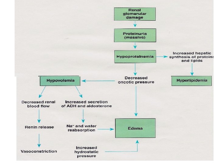
Nephrotic Syndrome (NS) Unknown cause of high proteinuria as a result of damage to the Glomerular Capillary Wall leading to low serum albumin and edema NS is a sign of a disease that damages the glomeruli in the kidney Forms of NS � Primary: � � Minimal Change Nephrotic Syndrome (MCNS) Idiopathic 80% of all cases Good prognosis Secondary to another disorder Congenital: autosomal recessive gene
Nephrotic Syndrome (NS) Clinical Manifestations � � � � weight gain Puffiness of face, periorbital at morning which subsides during the day swelling of abdomen, scrotum & lower extremities is more prominent Respiratory difficulty (pleural effusion) Edema of intestinal mucosa cause diarrhea, loss of appetite, poor intestinal absorption Decrease urine volume/dark, frothy Irritable, easily fatigued
Nephrotic Syndrome (NS) Diagnostic test �Marked proteinuria +1 - +4 �Minimal hematuria � Reduce serum albumin < 2 g/dl �Increase serum cholesterol: > 450 -1500 mg/dl �Increase SG �Elevated ESR
Nephrotic Syndrome (NS) Managements � Reduce urinary protein excretion � Maintain a protein-free urine � Reduce edema & Prevent infection � Minimize complications � General measures: Daily weight & bed rest during edema, change position Q 2 hrs to decrease pressure on body and reduce edema Antibiotics with infections Diet: restricted salt during massive edema, high protein diet Corticosteroids: prednisone (side effect ↑ chance for infection) Immunosuppressants (do not administer immunization) Albumin (plasma expander) and lasix
Nephrotic Syndrome (NS) Nursing Diagnosis � Fluid volume excess related to fluid accumulation in tissues � Risk for fluid volume deficit (intravascular) r/t proteinuria, edema, and effects of diuretics � Risk for impaired skin integrity r/t edema and decreased circulation � Risk for infections r/t urinary loss of gammaglobulins � Anxiety (parental) r/t caring for child with chronic disease and hospitalization
Nephrotic Syndrome (NS) Interventions � Assess I&O � Assess changes in edema � Measure abd girth � Measure edema around eyes / & dependent areas � Weigh daily note degree of pitting � Test urine for specific gravity and albumin (hyperalbuminuria ) � Administer corticosteroids (to reduce excretions of urinary protein) � Administer diuretics (relieve edema) � Limit fluids as indicated
CM APGN MCNS Strep. antibody titers Elevated Normal BP Elevated Normal or decreased Edema Primarily periorbital and Generalized & severe peripheral Circulatory congestion Common absent Proteinuria Mild to moderate Massive Hematuria Gross or microscopic Microscopic or none Serum protein levels Minimal reduction Marked decreased Serum lipid levels Normal Elevated Peak age onset 5 -7 years 2 -3 years
Renal Failure (RF) Renal failure is the inability of the kidneys to excrete waste material, concentrate urine, and conserve electrolytes Could be acute or chronic renal failure
Acute Renal Failure (ARF) ARF is an abrupt decline in glomerular and tubular function Could be caused by Escherichia coli (which is usually contracted from eating improperly cooked meat or contaminated dairy products) Classic sign is Elevated blood urea nitrogen level
Acute Renal Failure (ARF) Clinical manifestations � Azotemia: accumulation of nitrogenous waste (Blood Urea Nitrogen (BUN)) within the blood � circulatory congestion/ hypervolemia electrolytes abnormalities: Increased K(potassium level > 7 m. Eq/L) & phosphate Decreased Na+ (seizures) & calcium � � metabolic acidosis, hypertension oliguria: output < 1 ml/kg/hr; Anuria: no urinary output in 24 hours Nausea, Vomiting, Drowsiness
Acute Renal Failure (ARF): Prevention recognize patients at risk (postoperative states, cardiac surgery, septic shock) prevent progression from pre-renal to renal preserve renal perfusion � isovolemia, � cardiac output, � normal blood pressure avoid nephrotoxins (aminoglycosides, NSAIDS)
Acute Renal Failure (ARF): Management Treat the underlying disease Management of the complications Provision of supportive therapy Strictly monitor intake and output (weight, urine output, insensible losses, IVF) & monitor serum electrolytes Adjust medication dosages according to GFR Nutrition � provide adequate caloric intake � limit protein intake to control increases in BUN � minimize potassium and phosphorus intake � limit fluid intake If adequate caloric intake can not be achieved due to fluid limitations, some form of dialysis should be considered
Chronic Renal Failure Progressive deterioration of kidneys functions over months or years produces a variety of clinical and biochemical disturbances that eventually culminate in the clinical syndrome known as uremia Uremia is a retention of nitrogenous products that produce toxic symptoms Renal damage is judged by elevated serum creatinine (Normal 0. 4 - 1. 2 d/L) Renal function is compromised when creatinine is above 1. 2 The end-stage renal disease (ESRD), is irreversible
Chronic Renal Failure Uremia � Retention of waste products � Water and sodium retention � Hyperkalemia � Metabolic acidosis � Anemia � Calcium & phosphorus disturbances � Growth disturbance
Chronic Renal Failure Uremic symptoms can affect every organ system, � Neurological system: cognitive impairment, personality change, asterixis (motor disturbance that affects groups of muscles), seizures � GI: nausea, vomiting, food distaste � Blood-forming system–anemia due to erythropoetin deficiency, easy bruising and bleeding due to abnormal platelets � Pulmonary system–fluid in the lungs, with breathing difficulties � Cardio: chest pain due to pericarditis & pericardial effusion � Skin –generalized itching
Chronic Renal Failure: Management Peritoneal Dialysis/Hemodialysis � is required when the glomerular filtration rate decreases below 10% to 15% of normal Restrict protein intake Calcium and Vitamin D, Antihypertensives, Diuretics, Bicarbonate, Antiepileptics, Antihistamines Transplantation