GENETICS PPT DNA Protein Synthesis Punnett Squares Human


 � To get DNA blueprint for protein synthesis")


")


 Cell division � The growing of")
 The time a cell spends performing its function and preparing for")


 1. 2. 3. 4. Prophase Metaphase Anaphase Telophase")
 DNA has already been replicated and is coiled tightly enough to")
 Chromatids move to central zone called metaphase plate Chromatids line up")
 Centromere of each chromatid splits creating daughter chromosomes Daughter chromosomes are")
 Nuclear membranes form � DNA uncoils Cells are preparing to enter")
 Cytoplasmic division that forms two daughter cells � Usually begins in")
")

 Are a result of abnormal cell growth and division � Benign")
 The result of very active malignant cells Stimulates additional blood vessel")


 Sister chromatids Chiasmata Spindle")


- Slides: 47
GENETICS PPT DNA, Protein Synthesis, Punnett Squares, Human Genetics
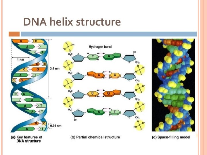
DNA - REVIEW Made up of nucleotides � 5 carbon sugar, nitrogenous base (A, T, C, G), and phosphate group Sugar and phosphate backbone with nitrogenous bases pointing out Two strands connected in the middle with hydrogen bonds between the nitrogenous bases Pairing is A-T and C-G
DNA Located in the nucleus in eukaryotes and cytoplasm in prokaryotes Made up of genes which code for proteins Generally tightly coiled so the histones block the start site of the gene. When the cell is triggered to make a protein, the DNA uncoils a bit and the weak hydrogen bonds are broken to allow access to the start site for the protein. - allows transcription
Figure 3 -17 DNA Organization and Chromosome Structure. Nucleus Supercoiled region Cell prepared for division Chromosome Nondividing cell Chromatin in nucleus DNA double helix © 2013 Pearson Education, Inc. Nucleosome Histones
QUICK REMINDERS OF SOME DEFINITIONS DNA strands � Contain the genetic code that dictates an organism's inherited traits � Genetic code is arranged in triplets Genes � Are the functional units of heredity � Contain all the triplets needed to produce specific proteins Protein Synthesis – process used to make proteins � Transcription + Translation = Protein
TRANSCRIPTION Forms messenger RNA (m. RNA) � To get DNA blueprint for protein synthesis from the nucleus out to the cytosol � Remember that DNA cannot leave the nucleus but the ribosomes are outside of the nucleus Transcription is used to take the message in one gene of DNA and copy it into m. RNA so it can be taken to the ribosome
RNA polymerase binds to promoter gene and "unzips" the DNA Gene G C A T T A G C A T G C T A A T C G G C C G C T A T C G G C A T T A G C A T A G Promoter RNA polymerase T C A T C G A T Triplet 1 A 1 C G Triplet 2 G Triplet 3 C 2 3 T 1 Complementary triplets TRANSCRIPTION – STEP 1 DNA G C 2 G A 3 G C G T T 4 A Triplet 4 C 4 A A T KEY © 2013 Pearson Education, Inc. A Adenine U Uracil (RNA) G Guanine T C Cytosine Thymine (DNA)
TRANSCRIPTION – STEP 2 G C A T A New RNA codon triplets are formed, using uracil instead of thymine Remember in DNA, A-T, and C-G but in RNA, A-U and C-G © 2013 Pearson Education, Inc. T T A G C A T A U C G G C A T Codon 1 G C C G C T C G RNA nucleotide A G C T A A T KEY A Adenine U Uracil (RNA) G Guanine T C Cytosine Thymine (DNA)
TRANSCRIPTION – STEP 3 A Codon 1 At DNA "stop" signal, m. RNA detaches and "unzipped" DNA reattaches m. RNA strand U G C Codon 2 C G A G Codon 3 C Codon 4 (stop codon) U A A KEY © 2013 Pearson Education, Inc. A Adenine U Uracil (RNA) G Guanine T C Cytosine Thymine (DNA)
Figure 3 -18 Transcription. DNA C A T T A G C A T G C T A A T C G G C C G T A C G G C A T T A G C A T A G Promoter RNA polymerase T A T C C G Triplet 2 G 2 C T Triplet 3 C 3 1 3 Triplet 4 T T T 4 A A C A G C G C A T G C Codon 1 G A Codon 3 T C Codon 4 (stop codon) U A A C G A RNA G nucleotide C T A T G Codon 2 T U m. RNA strand C C A C U A T G C 4 Codon 1 T T 2 G A T A G A 1 A A A T A C G Triplet 1 G T Complementary triplets Gene G A A T KEY After transcription, the two DNA strands reattach. © 2013 Pearson Education, Inc. A Adenine U Uracil (RNA) G Guanine T C Cytosine Thymine (DNA)
TRANSCRIPTION WRAP-UP RNA polymerase is the enzyme that adds RNA nucleotides to the growing RNA strand Read in triplets of three bases called codons which will be decoded in translation The end result of this process is a strand of m. RNA that has been made as a copy of a DNA gene. NOTE – while we talk about this process making m. RNA, it is used to make the other forms we will talk about as well – t. RNA, m. RNA
TRANSLATION Definition – synthesis of a protein using the information provided by the sequence of codons along the m. RNA strand Uses new m. RNA codons (triplet of three bases) to signal the assembly of specific amino acids in series m. RNA leaves the nucleus and binds to a ribosome in cytoplasm Transfer RNA (t. RNA) delivers amino acids to ribosomes Each t. RNA has a complement to the codon called an anticodon
RIBOSOME STRUCTURE Remember the ribosome is composed of r. RNA (ribosomal)
STEPS TO TRANSLATION "Start" codon of m. RNA combines with small ribosomal subunit and first t. RNA � Coding for methionine with the base sequence AUG Small and large ribosomal subunits enclose the m. RNA 1. 2. NUCLEUS m. RNA The m. RNA strand binds to the small ribosomal subunit and is joined at the start codon by the first t. RNA, which carries the amino acid methionine. Binding occurs between complementary base pairs of the codon and anticodon. The small and large ribosomal subunits interlock around the m. RNA strand. 2 Amino acid 1 KEY A Adenine Small ribosomal subunit U G Guanine C Cytosine U Uracil 1 t. RNA Anticodon A C G G C t. RNA binding sites U A C A U G C C Start codon G A G C A U A m. RNA strand C C G C A G U A A Large ribosomal subunit
STEPS TO TRANSLATION 3. 2 nd t. RNA brings another amino acid � Its anticodon binds to 2 nd codon of m. RNA A second t. RNA arrives at the adjacent binding site of the ribosome. The anticodon of the second t. RNA binds to the next m. RNA codon. 1 2 1 KEY The chain elongates until the stop codon is reached; the components then separate. The first amino acid is detached from its t. RNA and is joined to the second amino acid by a peptide bond. The ribosome moves one codon farther along the m. RNA strand; the first t. RNA detaches as another t. RNA arrives. U A 2 Peptide bond Small ribosomal subunit 3 C A Adenine 2 U G Guanine C Cytosine U Uracil Completed polypeptide 1 U A C A U G G G C C C G A U A A G C Stop codon A U G 3 C G G G C C C G A G C U A A Large ribosomal subunit G C C G A C G U A A U A
STEPS TO TRANSLATION Ribosomal enzymes remove 1 st amino acid and attach it to the 2 nd with a peptide bond 4. � Ribosome moves along the codons repeating these steps A second t. RNA arrives at the adjacent binding site of the ribosome. The anticodon of the second t. RNA binds to the next m. RNA codon. 1 2 1 KEY The chain elongates until the stop codon is reached; the components then separate. The first amino acid is detached from its t. RNA and is joined to the second amino acid by a peptide bond. The ribosome moves one codon farther along the m. RNA strand; the first t. RNA detaches as another t. RNA arrives. U A 2 Peptide bond Small ribosomal subunit 3 C A Adenine 2 U G Guanine C Cytosine U Uracil Completed polypeptide 1 U A C A U G G G C C C G A U A A G C Stop codon A U G 3 C G G G C C C G A G C U A A Large ribosomal subunit G C C G A C G U A A U A
STEPS TO TRANSLATION Amino acids continue to be added until ribosome reaches the "stop" codon at end of m. RNA 5. � Ribosome detaches leaving the strand of A second t. RNA arrives at The chain elongates until the m. RNA and a site newly completed polypeptide stop codon is reached; the adjacent binding of the ribosome. The anticodon of the second t. RNA binds to the next m. RNA codon. 1 The first amino acid is detached from its t. RNA and is joined to the second amino acid by a peptide bond. The ribosome moves one codon farther along the m. RNA strand; the first t. RNA detaches as another t. RNA arrives. 2 1 KEY U A 2 Peptide bond components then separate. Small ribosomal subunit 3 C A Adenine 2 U G Guanine C Cytosine U Uracil Completed polypeptide 1 U A C A U G G G C C C G A U A A G C Stop codon A U G 3 C G G G C C C G A G C U A A Large ribosomal subunit G C C G A C G U A A U A
THE GENETIC CODE How do we figure out which amino acid/t. RNA molecule will attach to the m. RNA?
Entire Protein Synthesis
STAGES OF A CELL'S LIFE CYCLE (3 -8) Cell division � The growing of new somatic cells and replacing old � Mitosis Is the DNA replication of the genetic material in nucleus � Meiosis Is the production of sex cells—the sperm and ova � Apoptosis Is genetically controlled cell death for cells that do not divide © 2013 Pearson Education, Inc.
INTERPHASE (3 -8) The time a cell spends performing its function and preparing for mitosis � G 1 phase is when the cell duplicates organelles and adds cytosol � S phase is when DNA is replicated in the nucleus � G 2 phase is when centrioles are replicated © 2013 Pearson Education, Inc.
FIGURE 3 -20 STAGES OF A CELL’S LIFE CYCLE. G 2 Protein synthesis rs hou S IS Proph ase Me T MI se s ha ho ap e se Telopha An tap ha ur s O 8 or more hours o 5 THE CELL CYCLE 2 t G 1 Normal cell functions plus cell growth, duplication of organelles, protein synthesis S DNA replication, synthesis of histones 1 to 3 CYTO SIS E KIN MITOSIS AND CYTOKINESIS (see Fig. 3 -22) © 2013 Pearson Education, Inc. INTERPHASE 6 to 8 hours
FIGURE 3 -21 DNA REPLICATION. 2 7 8 9 6 5 4 3 © 2013 Pearson Education, Inc. 1 DNA polymerase Segment 2 DNA nucleotide KEY Adenine Guanine Cytosine Thymine 6 7 Segment 1 8 1 2 3 4 DNA polymerase 5
FOUR STAGES OF MITOSIS (3 -8) 1. 2. 3. 4. Prophase Metaphase Anaphase Telophase PLAY A&P FLIX™ Mitosis © 2013 Pearson Education, Inc.
PROPHASE (3 -8) DNA has already been replicated and is coiled tightly enough to be seen under a microscope- chromosomes are condensed � Each of the two copies of DNA is called a chromatid, and they are connected to each other at a point called the centromere � Nucleoli then disappear � Two pairs of centrioles move to opposite poles � Spindle fibers appear INTERPHASE STAGE 1 a EARLY PROPHASE Spindle fibers STAGE 1 b LATE PROPHASE Centriole Chromosome with two sister chromatids MITOSIS BEGINS Centrioles (two pairs) Centromeres
METAPHASE (3 -8) Chromatids move to central zone called metaphase plate Chromatids line up parallel to the plate STAGE 2 Metaphase plate © 2013 Pearson Education, Inc. METAPHASE
ANAPHASE (3 -8) Centromere of each chromatid splits creating daughter chromosomes Daughter chromosomes are pulled apart and move toward the centrioles STAGE 3 ANAPHASE Daughter chromosomes © 2013 Pearson Education, Inc.
TELOPHASE (3 -8) Nuclear membranes form � DNA uncoils Cells are preparing to enter interphase again STAGE 4 Cleavage furrow © 2013 Pearson Education, Inc. TELOPHASE
CYTOKINESIS (3 -8) Cytoplasmic division that forms two daughter cells � Usually begins in late anaphase � Continues throughout telophase � Is usually completed after a nuclear membrane has re-formed around each daughter nucleus INTERPHASE Daughter cells CYTOKINESIS © 2013 Pearson Education, Inc.
FIGURE 3 -22 A INTERPHASE, MITOSIS, AND CYTOKINESIS. Spindle fibers Nucleus (contains replicated DNA) STAGE 1 b Centriole MITOSIS BEGINS Centrioles (two pairs) Centromeres LATE PROPHASE Chromosome with two sister chromatids © 2013 Pearson Education, Inc. STAGE 1 a EARLY PROPHASE INTERPHASE
FIGURE 3 -22 B INTERPHASE, MITOSIS, AND CYTOKINESIS. 2 METAPHASE STAGE 3 ANAPHASE STAGE 4 TELOPHASE © 2013 Pearson Education, Inc. STAGE INTERPHASE Daughter cells Daughter chromosomes Metaphase plate Cleavage furrow CYTOKINESIS
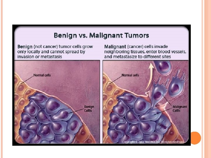
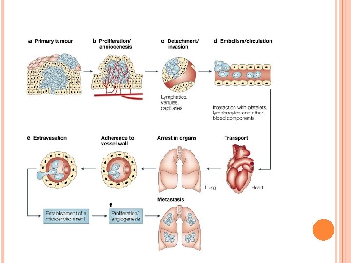
TUMORS (3 -9) Are a result of abnormal cell growth and division � Benign tumors Are usually encapsulated and rarely life threatening � Malignant tumors Spread from original tissue capsule through a process called invasion Having spread to other tissues and organs, the tumor has metastasized © 2013 Pearson Education, Inc.
CANCER (3 -9) The result of very active malignant cells Stimulates additional blood vessel growth Triggers a positive feedback mechanism of further growth and metastasis © 2013 Pearson Education, Inc.
TREATMENTS Surgery. Radiation Therapy. Chemotherapy. Immunotherapy - Helps your immune system fight cancer. Targeted Therapy - Targets the changes in cancer cells that help them grow, divide, and spread Hormone Therapy - Slows or stops the growth of cancer that uses hormones to grow Stem Cell Transplant - Procedures that restore bloodforming stem cells in people who have had theirs destroyed by high doses of cancer treatments, such as chemotherapy and radiation therapy.
MEIOSIS Reduction division Used to make gametes – sperm/egg Will take the 2 n cells and make them n Goes through Meiosis I and Meiosis II Only in sex cells – mitosis is done in all body cells and then after fertilization which produces a 2 n cell again Results in variation from � Crossing over � Independent Assortment � Segregation
Figure 13. 8 a Prophase I Centrosome (with centriole pair) Sister chromatids Chiasmata Spindle Telophase I and Cytokinesis Anaphase I Metaphase I Sister chromatids remain attached Centromere (with kinetochore) Metaphase plate Fragments Homologous chromosomes of nuclear envelope Homologous chromosomes separate Microtubule attached to kinetochore Cleavage furrow Each pair of homologous chromosomes separates. Chromosomes line up Duplicated homologous chromosomes (red and blue) by homologous pairs. pair and exchange segments; 2 n 6 in this example. Two haploid cells form; each chromosome still consists of two sister chromatids.
Figure 13. 8 b Prophase II Metaphase II Anaphase II Telophase II and Cytokinesis During another round of cell division, the sister chromatids finally separate; four haploid daughter cells result, containing unduplicated chromosomes. Sister chromatids separate Haploid daughter cells forming
Figure 13. 9 a MEIOSIS MITOSIS Parent cell MEIOSIS I Chiasma Prophase I Duplicated chromosome Chromosome duplication 2 n 6 Chromosome duplication Homologous chromosome pair Metaphase I Anaphase Telophase Anaphase I Telophase I Daughter cells of meiosis I 2 n Daughter cells of mitosis 2 n Haploid n 3 MEIOSIS II n n Daughter cells of meiosis II
SUMMARY Property Mitosis Meiosis DNA replication Occurs during interphase before mitosis begins Occurs during interphase before meiosis I begins Number of divisions One, including prophase, metaphase, and telophase Two, each including prophase, metaphase, and telophase Synapsis of homologous chromosomes Does not occur Occurs during prophase I along with crossing over between nonsister chromatids; resulting chiasmata hold pairs together due to sister chromatid cohesion Number of daughter cells and genetic composition Two, each diploid (2 n) and genetically identical to the parent cell Four, each haploid (n), containing half as many chromosomes as the parent cell; genetically different from the parent cell and from each other Role in the animal body Enables multicellular adult to arise from zygote; produces cells for growth, repair, and, in some species, asexual reproduction Produces gametes; reduces number of chromosomes by half and introduces genetic variability among the gametes
gure 13. 9 MITOSIS MEIOSIS Parent cell MEIOSIS I Chiasma Prophase I Duplicated chromosome Chromosome duplication 2 n 6 Chromosome duplication Homologous chromosome pair Metaphase I Anaphase Telophase Anaphase I Telophase I Daughter cells of meiosis I 2 n Haploid n 3 MEIOSIS II 2 n Daughter cells of mitosis n n Daughter cells of meiosis II SUMMARY Property Mitosis Meiosis DNA replication Occurs during interphase before mitosis begins Occurs during interphase before meiosis I begins Number of divisions One, including prophase, metaphase, and telophase Two, each including prophase, metaphase, and telophase Synapsis of Does not occur homologous chromosomes Occurs during prophase I along with crossing over between nonsister chromatids; resulting chiasmata hold pairs together due to sister chromatid cohesion Two, each diploid (2 n) and genetically Number of daughter cells identical to the parent cell and genetic composition Four, each haploid (n), containing half as many chromosomes as the parent cell; genetically different from the parent cell and from each other Role in the animal body Enables multicellular adult to arise from zygote; produces cells for growth, repair, and, in some species, asexual reproduction Produces gametes; reduces number of chromosomes by half and introduces genetic variability among the gametes
SOURCES OF VARIATION – CROSSINGOVER
SOURCES OF VARIATION – INDEPENDENT ASSORTMENT
SOURCES OF VARIATION – SEGREGATION
OOGENESIS AND SPERMATOGENESIS