Genetic testing DR HEBA M KAREEM ASSISTANT PROFESSOR

Genetic testing DR. HEBA M. KAREEM ASSISTANT PROFESSOR OF MEDICAL BIOCHEMISTRY AND MOLECULAR BIOLOGY

WHAT IS GENETIC TESTING The analysis of human DNA in any of its forms or related products (chromosomes, RNA, proteins). The ultimate goal is to recognize the potential for a genetic condition at an early stage. Definitions: Genotype vs. Phenotype • The genetic make-up, as distinguished from the physical appearance Mutation • A genetic change, usually one that is associated with a disease Karyotype • A visual presentation of chromosomes

TYPES OF GENETIC TESTING 1. DIAGNOSTIC TESTING used to confirm or rule out a known or suspected genetic disorder in a person with disease symptoms. Confirming a diagnosis may alter medical management for the individual (PKU). 2. Predictive testing: offered to individuals who do not have symptoms at the time of testing but have a family history of a genetic disorder. 3. Carrier Testing 3 Test to identify individuals who have a gene mutation for a disorder inherited in an autosomal recessive or X-linked recessive manner

4. 1. Prenatal genetic testing Detect genetic disorders and birth defects. > 200 single gene disorders can be diagnosed. Testing done only when a family history or other risk a. Ultrasound: Noninvasive, uses reflected sound waves converted to an image. Transducer placed on abdomen See physical features of fetus, not chromosomes. May identify some chromosomal abnormalities by physical features b. Amniocentesis • Diagnose > 100 disorders, cells analyzed for chromosomal and biochemical disorders. • Risk of infection and spontaneous abortion. • 4 • Normally only used when: - Advanced maternal age. - History of chromosomal disorder. - Parent with chromosomal abnormality. Mother carrier of X-linked disorder

C. PREIMPLANTATION GENETIC DIAGNOSIS Eggs collected, fertilized, allowed to develop. ~ 3 rd day of fertilization, embryo has 6– 8 cells. For PGD, one cell, a blastomere, is removed. DNA extracted and tested (DNA analysis, karyotyping, biochemical analysis for PKU). Embryo without genetic disorder are implanted into mother. 5. Newborn Screening identifies individuals who have an increased chance of having a specific genetic disorder so that treatment can be started as soon as possible. performed on a small blood sample, which is taken by pricking the baby’s heel. a parent will usually only receive the result if it is positive. if the test result is positive, additional testing is needed to determine whether the baby has a genetic disorder. performed routinely at birth 5 •
 • Tests for mutations that")
TYPES OF GENETIC TESTS: I. Constitutional (affect germ cells) • Tests for mutations that affect ALL CELLS in the body, and have been there since conception II. Acquired (affect somatic cells) • Tests for changes that affect only certain cells or cell types in the body, and that occurred later in life. Genetic testing includes: 1. Direct genetic testing (Molecular): examination of DNA (or RNA) to determine if mutations are present. 2. Cytogenetic testing: examination of the chromosomes for visible alterations that indicate a genetic defect. (karyotyping) 6 3. Biochemical genetic testing: assay for specific metabolites that indicate a genetic disease

1. Molecular Test: Example Analysis of DNA sequence in patient with a rare inherited disease (Muscular Dystrophy ). --Gene: DMD • Clinical Picture: - progressive muscle weakness starting in early childhood. wheelchair by age 12. Obtain blood sample from child Read the DNA sequence of the DMD gene. Identify the mutation that caused the disease 2. Cytogenetic Test: Karyotype – to examine the chromosomal complement of an individual including number, form, and size of the chromosomes 7 a trisomy has 47 chromosomes instead of 46. e. g Down syndrome

3. BIOCHEMICAL TEST Analyzes the quantity of a downstream product of a gene (e. g. not looking directly at the gene, or the chromosome). Example: Newborn Screening. To determine if enzymes in the body are abnormal in some way. • performed on a blood, urine, spinal fluid, or other tissue sample. • the disease is usually the result of a mutation that causes an enzyme to be absent, unstable, or to have altered activity. 8 • diseases often called "inborn errors of metabolism" because they are present at birth and affect how the body's metabolism works

Congenital Hypothyroidism: Inadequate or absent production of thyroid hormone. Thyroid hormone replacement therapy begun by 1 month of age can prevent mental and growth retardation. II. Acquired genetic diseases: Cancer is a heterogeneous disease. It is not a single disease. Cancer is a genetic disease: All cancers involve genetic changes in somatic cells, the germ line, or both. Most gene mutations in cancer occur in somatic cells and are acquired (multifactorial etiology). (single tumors, late-onset, unilateral). However, some mutations do occur in the germline and may be inherited and passed on to future generations. multiple tumors, earlyonset, bilateral). 9 (

PROCEDURE: GENETIC TESTING & PROFILING • Take a sample of cells (blood, hair root, amniotic fluid, mouth swab). Ø Use staining of chromosomes to locate any chromosome abnormalities. Ø Extract the DNA from cells – Cut up the DNA – Separate the DNA fragments – Analyse the DNA fragments. 10 Molecular testing: I. RFLP. II. DNA sequencing. III. Blotting techniques.
 v v v polyms that alter the length")
I. RFLPs (Restriction Fragment Length Polymorphisms) v v v polyms that alter the length of restriction fragments. Result from changes (e. g. SNPs) that introduce or delete a restriction enzyme site. Genotyping by Southern or PCR-RFLP Principle: Ø Isolation of DNA. 11 Ø DNA is amplified by PCR. Ø Amplified DNA is incubated with restriction endonucleases. Ø Then electrophoresis. Ø Visualization of different bands.
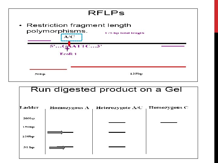

RFLP v. Description of previous figure: veg. , if A gene of length 175 bp. is cut by Eco. R 1 which specifically hydrolyses bond between G & A. so that 2 fragments are produced of length 50 & 125 bp. a mutation occurs converting normal A to C. This R. endonuclease will not act. So, the DNA fragment will be only of 175 bp length. v. By electrophoresis, we can detect : 1. the normal subject which has 2 fragments (50 & 125), called homozygote for A. 2. The heterozygote containing one normal (A) and one mutated base (C). 3. The homozygote having mutation of both alleles of this gene (C & C). 13 v. If

II. DNA SEQUENCING: DEFINITION DNA sequencing is the process of determining the precise order of nucleotides within a DNA molecule. It is used to determine the order of the four bases in a strand of DNA isolated from cells of animals, plants, bacteria, or virtually any other source of genetic information. Overall process: First, DNA has to be extracted from the cells of the organism being studied. The sequencing reaction is then performed on the DNA, and the sequenced DNA strands are sorted by size using capillary electrophoresis. Finally, the DNA code is read by a computer and analysed. Uses of sequencing: Detect the presence of known 14 genes for medical purposes: - genetic testing (ex. diagnostic). Forensic identification. - Parental testing.
 sequencing A modified DNA replication reaction. Growing chains are terminated")
1. Chain termination (Sanger) sequencing A modified DNA replication reaction. Growing chains are terminated by dideoxynucleotides. Limitations: The dideoxy method is good only for 5001000 bp reactions. Expensive & Takes time. Brief Bio Background as regards DNA Polymerase • DNA polymerase can add free nucleotides and forms phosphodiester bonds. • No known DNA polymerase is able to begin a new chain, so needs primer. • It requires DNA template (ss DNA). • It requires presence of d. NTPs ( d. ATP, d. GTP, d. CTP, TTP ) 15

DNA POLYMERASE ACTION d. ATP 16 d. GTP
 SEQUENCING Principle: Uses DNA polymerase to synthesize a second DNA")
1. CHAIN TERMINATION (SANGER) SEQUENCING Principle: Uses DNA polymerase to synthesize a second DNA strand that is labeled. DNA polymerase always adds new bases to the 3’ end of a primer that is base-paired to the template DNA. Also uses chain terminator nucleotides: dideoxy nucleotides (dd. NTPs), which lack the -OH group on the 3' carbon of the deoxyribose. When DNA polymerase inserts one of these dd. NTPs into the growing DNA chain, the chain terminates, as nothing can be added to its 3' end. 17

DIDEOXIES BLOCK ELONGATION dd. GTP 18 H
 Primer annealing. (primer)")
Steps: 1. 2. 3. 4. 5. 6. Strand separation. (denaturation; heating) Primer annealing. (primer) Primer extension. (DNA polymerase, d. NTPs) Chain termination. (dd. NTPs) Electrophoresis. (capillary) Detection & analysis. ( computer; electropherogram). ………………………………… Steps 1 (strand separation) Double-stranded DNA needs to be denatured, or separated into single strands, before it can be sequenced. 19 This process is accomplished by heating the DNA.
 Next, a small single-stranded DNA piece of about 20 bases,")
Steps 2 (primer annealing) Next, a small single-stranded DNA piece of about 20 bases, called an oligonucleotide, is annealed to the denatured template strand. In addition, a large excess of primers is used to again ensure that the primers will out-compete the complementary DNA strand for annealing to the template. The oligonucleotide primer must be of complementary sequence to the template strand in order to bind by base-pair interactions. Steps 3 (primer extension): During the extension phase, a 20 bacterial DNA polymerase enzyme begins assembling a new DNA chain from the d. NTPs, provided in the reaction mixture. The nucleotides are added in the order specified by the complementary bases in the template strand.
 sequencing Steps 4 (chain termination) The reaction mixture also contains")
1. Chain termination (Sanger) sequencing Steps 4 (chain termination) The reaction mixture also contains small amounts of each of the 4 dideoxynucleotides, or “dd. NTPs, ” which lack the 3'-hydroxyl group necessary for chain extension. Whenever a dd. NTPs is incorporated into a growing DNA chain, it terminates chain growth. When DNA polymerase reaches a base for which some dd. NTP is present, the chain will either: d. NTPs >>> dd. NTPs 21 • terminate if a dd. NTP is added, or: • continue if the corresponding d. NTP is added. • which one happens is random, based on ratio of d. NTP to dd. NTP in the tube.

Template 3 T C A dd. A A dd. G A G dd. T A G G A Four different labels: Each of the four nucleotide chains has a different dye. For example: Red for dd. ATP. Yellow for dd. GTP. Blue for dd. TTP. Green for dd. CTP. T dd. C T 5 C dd. T
 The newly synthesized DNA strands, each labeled with one of")
Steps 5 (capillary electrophoresis) The newly synthesized DNA strands, each labeled with one of four dyes, are now sorted by length using capillary electrophoresis. An electrical current pulls the negatively charged DNA strands through the capillary. This tube is used to separate strands that differ in length by only one base. Shorter DNA strands migrate through the gel material more quickly, and come out the bottom of the capillary first. A G T C dd. T A G T dd. C A G dd. T A dd. G dd. A Size & color 23 The fragments are separated by

Genetic techniques. II. DNA sequencing: 1. Chain termination sequencing Capillary electrophoresis 5- A G T C T -3 24 Electropherogram
 sequencing Steps 6 (detection) Fluorescent")
Genetic techniques. II. DNA sequencing: 1. Chain termination (Sanger) sequencing Steps 6 (detection) Fluorescent dyes can absorb and emit fluorescent light at specific wavelengths. As the strands emerge out the bottom of the capillary they pass through a laser beam that excites the fluorescent dye attached to the dd. NTPs at the end of each strand. This causes the dye to fluoresce, or glow, at a specific wavelength, or color. This color is then detected by a photocell, which feeds the information to the computer. 5- A G T C T -3 ØThe template strand sequence is: 3- T C A G A -5 25 Reading the sequence ØThe sequenced strand is:

Automated Sequencing: detecting a G/G mutation red A/A Homozygos mutation yellow A ATCT G/A A GTCT Yellow + red Heterozygos mutation Normal A G/A T C T
 Computer analysis: The computer displays the information received from the photocell")
Steps 6 (detection) Computer analysis: The computer displays the information received from the photocell as an electropherogram; Chromatogram. It also prints the letter of the appropriate base below each of the signal peaks. 27 successive peaks correspond to DNA segments differing in length by one nucleotide.

Genetic techniques. III. Blotting technique: History: Professor Sir Edwin Southern, Professor of Biochemistry developed this method in 1975. The technique is known as DNA transfer or 'Southern blotting. Common types: • Southern blot: it is used to detect DNA using DNA probe. • Northern blot : it is used to detect RNA using DNA probe. • Western blot : it is used to detect protein using antibody as a probe. Hybridization • The binding between ss labeled probe to a complementary nucleotide sequence on the target DNA. Making a probe: • 28 • A probe is a small length of DNA (20 -30 nucleotides) or RNA. Complementary to the sequence (gene) of interest.
. Denaturation (alkali). Transfer")
1. SOUTHERN HYBRIDIZATION Steps: DNA extraction & digestion. Separation (gel electrophoresis). Denaturation (alkali). Transfer to nitrocellulose membrane (blotted). Hybridize with labeled probe. Band visualization. The labeled probes detect specific DNA sequences. Transfer buffer 29 1. 2. 3. 4. 5. 6.

30 Genetic techniques. III. Blotting technique: 1. Southern hybridization
. Digest")
Steps 1: DNA separation & digestion: DNA is extracted from cells (ex. leukocytes). Digest the DNA with an appropriate restriction enzyme. Steps 2: electrophoresis: The complex mixture of fragments is subjected to gel electrophoresis to separate the fragments according to size. 31 The lengths of the fragments are compared with band of relative standard fragments of known size (Marker).

Steps 3: denaturation & blot: The restriction fragments present in the gel are denatured with alkali. The denatured DNA fragments are transferred to nitrocellulose membrane for analysis. The gene of interest is on only one of these pieces of DNA. 32 Blot This procedure preserves the distribution of the fragments in the gel, creating a replica of the gel on the filter.

Not Hybridize Steps 4: Hybridization: The filter is incubated with a specific labeled DNA probe. Hybridize The probe hybridizes to the complementary DNA restriction fragment. Steps 5: Detection: 33 Excess probe is washed away and the probe bound to the filter is detected by autoradiography, which reveals the DNA fragment to which the probe hybridized.

Genetic techniques. III. Blotting technique: 2. NORTHERN Blot Definition: Northern blotting is a technique for detection of specific RNA sequences. RNA is isolated from several biological samples (e. g. various tissues, various developmental stages of same tissue etc. ) RNA is more susceptible to degradation than DNA. Application: • Study of gene expression at the level of m. RNA in eukaryotic cells. • To measure the amount & size of RNAs transcribed from eukaryotic genes.

3. Western Blot Definition: Western blotting is an Immunoblotting technique which rely on the specificity of binding between a protein of interest and a probe (antibody) raised against that particular protein). Applications: • To determine the molecular weight of a protein (identification). • To measure relative amounts (quantitation) of the protein present in complex mixtures of proteins. It is used as confirmatory test for HIV. Advantages: • WB is highly sensitive technique. As little as 1 -5 ng of an average-sized protein can be detected. • Detection and interpretation: A prestained MW standard is included during electrophoresis to allow the identification of the MW of the target protein.

Applications of some genetic tests: ** Duchenne muscular dystrophy l l l Xp 21; gene is 2. 3 Mb long; 79 exons 2/3 of cases due to deletions of some exons Diagnosis: -Southern analysis. - PCR of exons 17 C 18 P 1 19 P 2 44 P 3 45 46 47 48 49 P 4 Patient 1: exon 44 deleted 17 19 44 45 48 Patient 2: exon 17 deleted Patient 3: exon 45 deleted Patient 4: exon 19 deleted
 To determine the genetic father")
Applications of some genetic tests: ** Forensic (Paternity testing) To determine the genetic father of a specific child
- Slides: 37