Genetic Counseling and Genetic Testing Genetics Genetic Testing


 can be performed earlier (between the 10 th and 12")


- Slides: 17
Genetic Counseling and Genetic Testing Genetics
Genetic Testing § The ultimate goal of genetic testing is to recognize the potential for a genetic condition at an early stage. § In some cases, genetic testing allows people to make informed choices about reproduction. § In other cases, genetic testing allows early intervention that may lessen or even prevent the development of the condition. § For those who know that they are at risk for a genetic condition, genetic testing may help alleviate anxiety associated with the uncertainty of their situation.
§ Genetic testing includes prenatal testing and postnatal testing. § Prenatal genetic tests are those that are conducted before birth and now include procedures for diagnosing several hundred genetic diseases and disorders. § The major purpose of prenatal tests is to provide families with the information that they need to make choices during pregnancies and, in some cases, to prepare for the birth of a child with a genetic condition. § The Human Genome Project has accelerated the rate at which new genes are being isolated and new genetic tests are being developed.
§ In spite of these advances, prenatal tests are still not available for many common genetic diseases, and no test can guarantee that a “perfect” child will be born.
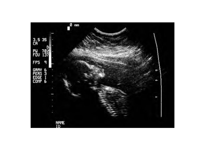
prenatal diagnosis: Ultrasonography Some genetic conditions can be detected through direct visualization of the fetus. § Such visualization is most commonly done with the use of ultrasonography— usually referred to as ultrasound. § In this technique, high-frequency sound is beamed into the uterus; when the sound waves encounter dense tissue, they bounce back and are transformed into a picture. § The size of the fetus can be determined, as can genetic conditions such as neural-tube defects (defects in the development of the spinal column and the skull) and skeletal abnormalities.
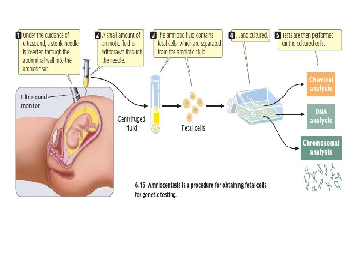
Amniocentesis Most prenatal testing requires fetal tissue, which can be obtained in several ways. The most widely used method is amniocentesis, a procedure for obtaining a sample of amniotic fluid from a pregnant woman. Amniotic fluid—the substance that fills the amniotic, sac and surrounds the developing fetus—contains fetal cells that can be used for genetic testing. Amniocentesis is routinely performed as an outpatient procedure either with or without the use of a local anesthetic.
1. First, ultrasonography is used to locate the position of the fetus in the uterus. 2. Next, a long, sterile needle is inserted through the abdominal wall into the amniotic sac, and a small amount of amniotic fluid is withdrawn through the needle. 3. Fetal cells are separated from the amniotic fluid and placed in a culture medium that stimulates them to grow and divide. 4. Genetic tests are then performed on the cultured cells. Complications with amniocentesis (mostly miscarriage) are uncommon, arising in only about 1 in 400 procedures.
§ A major disadvantage of amniocentesis is that it is routinely performed at about the 15 th to 18 th week of a pregnancy. § The cells obtained by amniocentesis must then be cultured before genetic tests can be performed, requiring yet more time. § For these reasons, genetic information about the fetus may not be available until the 17 th or 18 th week of pregnancy. § By this stage, abortion carries a risk of complications and is even more stressful for the parents.
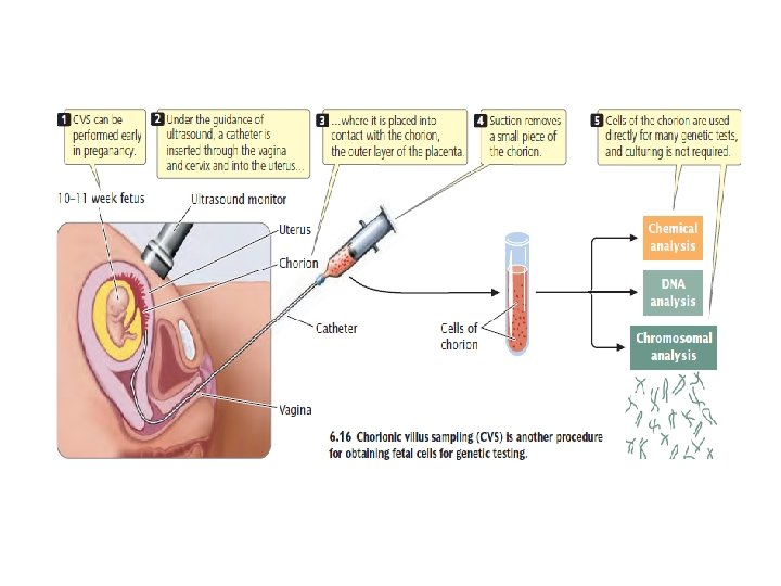
Chorionic villus sampling (CVS) can be performed earlier (between the 10 th and 12 th weeks of pregnancy) and collects a larger amount of fetal tissue, which eliminates the necessity of culturing the cells. 1. In CVS, a catheter—a soft plastic tube—is inserted into the vagina 2. and, with the use of ultrasonography for guidance, is pushed through the cervix into the uterus. 3. The tip of the tube is placed into contact with the chorion, the outer layer of the placenta. 4. Suction is then applied, and a small piece of the chorion is removed.
Although the chorion is composed of fetal cells, it is a part of the placenta that is expelled from the uterus after birth; so the tissue that is removed is not actually from the fetus. This tissue contains millions of actively dividing cells that can be used directly in many genetic tests. Chorionic villus sampling has a somewhat higher risk of complication that of amniocentesis; the results of several studies suggest that this procedure may increase the incidence of limb defects in the fetus when performed earlier than 10 weeks of gestation.
Fetal cells obtained by amniocentesis or by CVS can be used to prepare a karyotype, which is a picture of a complete set of metaphase chromosomes. Ø Karyotypes can be studied for chromosome abnormalities Ø Biochemical analyses can be conducted on fetal cells to determine the presence of particular metabolic products of genes. Ø For genetic diseases in which the DNA sequence of the causative gene has been determined, the DNA sequence can be examined for defective alleles.
Maternal blood screening tests Increased risk of some genetic conditions can be detected by examining levels of certain substances in the blood of the mother. However, these tests do not determine the presence of a genetic problem; rather, they simply indicate that the fetus is at increased risk and hence are referred to as screening tests. When increased risk is detected, follow-up tests (additional blood-screening tests, ultrasound, amniocentesis, or all three ) are usually conducted.
One substance examined in maternal screening tests is α-fetoprotein, a protein that is normally produced by the fetus during development and is present in fetal blood, amniotic fluid, and the mother’s blood during pregnancy. The level of α-fetoprotein is significantly higher than normal when the fetus has a neural-tube defect or one of several other disorders. Some chromosome abnormalities produce lower-than-normal levels of α-fetoprotein. Measuring the amount of α-fetoprotein in the mother’s blood gives an indication of these conditions.
Reference § Benjamin A. Pierce, 2010. Genetics: A Conceptual Approach, 4 th Edition. W. H. Freeman.