General sensation Composed of sensory receptors throughout the


 Ora serrata Sclera Ciliary body Choroid Ciliary")


")


 Thalamic nucleus (ventral posterior Pons medial nucleus)")
. –")
 ear Middle ear Internal (inner) ear (labryinth) Auricle")
")


 Round window Helicotrema Human Anatomy and")
 Vestibular membrane Scala vestibuli")
 Afferent nerve fibers")
 middle ear")
 Air pressure Wavelength Area of")


 (Bouins, H&E, Bar = 36. 4 µm). 1.")
 Fibers")

- Slides: 47
General sensation • Composed of sensory receptors throughout the body. – General senses • – Special senses • • Exteroceptors Interoceptors Propioceptors (sketetal muscle). Conscious. General structure (histology, activity 1) – Modified dendrites. • • • Vision, hearing, equilibrium, olfaction, taste. Relative to the source of the stimuli. – – – • Touch, pressure, pain, heat, cold, stretch, vibration. Free nerve endings Merkel discs in epidermis Hair follicle receptors Meissner corpuscles: light touch Ruffini corpuscles: deep pressure and stretching. Pacinian corpuscles: pressure recptor. Muscle spindles Golgi tendon organs Receptor physiology – Act as signal transducers. Direct relationship between importance and number of clustering of the receptors; punctuate distribution.
Special Senses 1. 2. 3. 4. Vision Olfaction Taste Hearing and equilibrium
1. Vision • • Organ: Eye ball Accessory structures – Lacrimal apparatus. • Lacrimal gland: secretes tears; solution of salts and lyzozyme. • Lacrimal canals • Nasolacrimal canal – – – – • Eyelids (papebrae) Medial and lateral canthus Caruncle Conjunctiva. Mucus membrane that lines inner surface of eyelids. Stratified squamous and stratified cilindrical epithelium. Eyelashes Ciliary glands: lubricate eyeball Tarsal glands: lubricate eyeball 6 extrinsic eye muscles Activity 1
The eye and associated accessory structures Site where conjunctiva merges with cornea Palpebral fissure Eyebrow Eyelid Eyelashes Pupil Lacrimal caruncle Lateral commissure (canthus) Medial commissure (canthus) Iris Sclera (covered by conjunctiva) Eyelid (a) Human Anatomy and Physiology, 7 e by Elaine Marieb & Katja Hoehn Copyright © 2007 Pearson Education, Inc. , publishing as Benjamin Cummings.
The lacrimal apparatus Lacrimal gland Lacrimal sac Excretory ducts of lacrimal gland Lacrimal punctum Lacrimal canaliculus Nasolacrimal duct Inferior meatus of nasal cavity Nostril Human Anatomy and Physiology, 7 e by Elaine Marieb & Katja Hoehn Copyright © 2007 Pearson Education, Inc. , publishing as Benjamin Cummings.
The eye and associated accessory structures Levator palpebrae superioris muscle Orbicularis oculi muscle Eyebrow Tarsal plate Palpebral conjunctiva Tarsal glands Cornea Palpebral fissure Eyelashes Bulbar conjunctiva Conjunctival sac Orbicularis oculi muscle (b) Human Anatomy and Physiology, 7 e by Elaine Marieb & Katja Hoehn Copyright © 2007 Pearson Education, Inc. , publishing as Benjamin Cummings.
Extrinsic eye muscles Trochlea Superior oblique muscle Superior oblique tendon Superior rectus muscle Axis at center of eye Inferior rectus muscle Medial rectus muscle Lateral rectus muscle Conjunctiva Optic nerve (a) Inferior rectus oblique muscle Name Lateral rectus Medial rectus Superior rectus Inferior oblique Superior oblique Annular ring (b) Controlling cranial nerve Moves eye laterally VI (abducens) Moves eye medially III (oculomotor) Elevates eye and turns it medially III (oculomotor) Depresses eye and turns it medially III (oculomotor) Elevates eye and turns it laterally III (oculomotor) Depresses eye and turns it laterally IV (trochlear) Action (c) Human Anatomy and Physiology, 7 e by Elaine Marieb & Katja Hoehn Copyright © 2007 Pearson Education, Inc. , publishing as Benjamin Cummings.
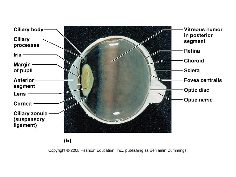
Internal structure of the eye • Composed of 3 layers or tunics – External • – Cornea (transparent) and Sclera (white). Middle • Anterior – – • Posterior – – Retina: sensory region Additional internal structures – – – • Choroid: dark pigment. Inner • • Iris: radial smooth muscle (III ocoulomotor) Uvea: vasculariazed. Cilliar body(muscles) : control the curvature of the lens (cristaline). Cilliary processes: secrete aqueous humor. Humor vitreous Macula lutea (yellow spot): high cone density Fovea centralis: area of maximum (very high amount of cones) sight acuity. Sclera venous sins( Schlemm canal) Ciliary zonule Activity 2 (eye dissection next class)
Internal structure of the eye (sagittal section) Ora serrata Sclera Ciliary body Choroid Ciliary zonule (suspensory ligament) Cornea Iris Pupil Anterior pole Retina Macula lutea Fovea centralis Posterior pole Optic nerve Anterior segment (cavity) Lens Scleral venous sinus (Canal of Schlemm) Posterior segment (cavity) (contains vitreous humor) Central artery and vein of the retina Optic disc (blind spot) (a) Human Anatomy and Physiology, 7 e by Elaine Marieb & Katja Hoehn Copyright © 2007 Pearson Education, Inc. , publishing as Benjamin Cummings.
Circulation of aqueous humor Cornea Lens Iris Lens epithelium Cornea Posterior segment containing vitreous humor Lens Corneal epithelium ANTERIOR Corneal endothelium Aqueous humor Anterior chamber Anterior segment Ciliary zonule (suspensory ligament) Posterior chamber Scleral venous sinus Ciliary processes Limbus (cornealscleral junction) Ciliary body Anterior ciliary vein Bulbar conjunctiva Ciliary muscle Sclera Human Anatomy and Physiology, 7 e by Elaine Marieb & Katja Hoehn Copyright © 2007 Pearson Education, Inc. , publishing as Benjamin Cummings.
Focusing for distant and close vision Sympathetic + Nearly parallel rays from distant object Lens Ciliary zonule Inverted image Ciliary muscle Lens (a) Lens is flattened for distant vision Ciliary zonule (suspensory ligaments) Parasympathetic + Divergent rays from close object Ciliary muscle Inverted image (c) Anterior segment viewed from behind (b) Lens bulges for close vision Human Anatomy and Physiology, 7 e by Elaine Marieb & Katja Hoehn Copyright © 2007 Pearson Education, Inc. , publishing as Benjamin Cummings.
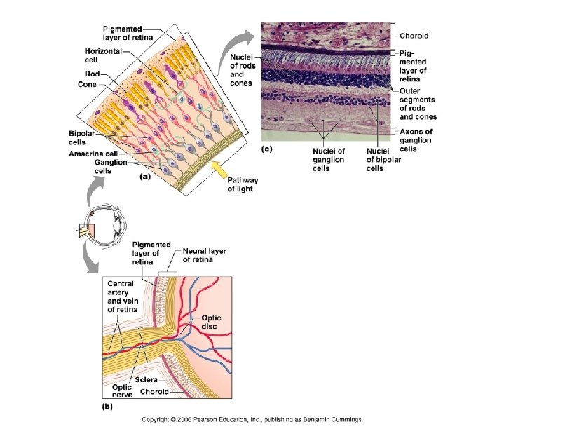
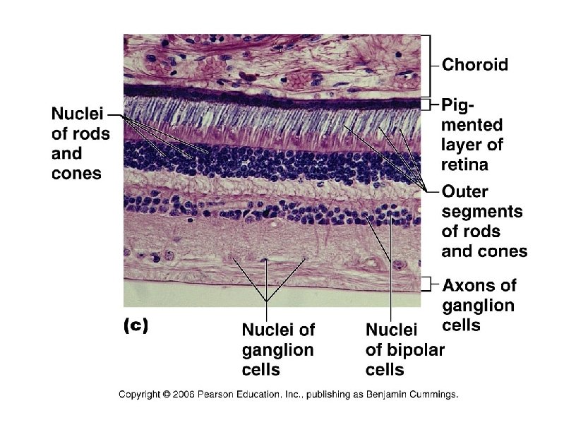
Histology of the retina • Sensory tunic – Outer pigmented epithelial layer – Inner neural layer. • • Photoreceptors: cones or rods. Bipolar neurons Ganglion Axons of ganglions • Activity 3
Synaptic terminals Rod cell body Inner fibers Rod cell body Cone cell body (a) Human Anatomy and Physiology, 7 e by Elaine Marieb & Katja Hoehn Nuclei Mitochondria Retinal Outer segment Inner segment Pigmented layer Outer fiber Light Li gh t Process of bipolar cell Light Photoreceptors of the retina (b) Opsin Connecting cilia Apical microvillus Discs being phagocytized Discs containing visual pigments Melanin granules Pigment cell nucleus Basal lamina (border with choroid) Copyright © 2007 Pearson Education, Inc. , publishing as Benjamin Cummings.
Visual fields of the eyes and visual pathway to the brain, inferior view Fixation point Right eye Left eye Optic nerve Suprachiasmatic nucleus Pretectal nucleus Optic chiasma Optic tract Lateral geniculate body Superior colliculus (sectioned) Uncrossed (ipsilateral) fiber Crossed (contralateral) fiber Optic radiation Lateral geniculate body of thalamus Superior colliculus (a) Human Anatomy and Physiology, 7 e by Elaine Marieb & Katja Hoehn Occipital lobe (visual cortex) (b) Corpus callosum Copyright © 2007 Pearson Education, Inc. , publishing as Benjamin Cummings.
2. Olfaction • Sensory structure: – Olfactory epithelium. Chemoreceptor. • Olfactory receptor cell: bipolar neuron (I) • Support cells: pseudo stratified ciliated epithelium. • Basal cells. • Activity 1
Olfactory receptors Olfactory epithelium Frontal lobe of cerebrum Olfactory tract Olfactory bulb Mitral cell Olfactory tract Glomeruli Nasal conchae Route of inhaled air Cribriform plate of ethmoid bone Olfactory epithelium Filaments of olfactory nerve Lamina propria connective tissue Axon Basal cell Olfactory receptor cell Supporting cell Mucus Dendrite Olfactory cilia Olfactory gland Route of inhaled air containing odor molecules Human Anatomy and Physiology, 7 e by Elaine Marieb & Katja Hoehn Copyright © 2007 Pearson Education, Inc. , publishing as Benjamin Cummings.
Red: support cell Blue: bipolar neuron Green: basal cell Green: Bowman’s gland cell
3. Taste • Sensory structure – Taste bud. Chemoreceptor. Mechanoreceptor. Thermo receptor. • Basic tastes: sweet, sour, salt, bitter. • Types (location in figure) – Fungiform papillae (mushrooms) – Circunvallate papilla (globes) – Filiform papilla • Cell types – Gustatory (taste) cells: bipolar neurons. – Support cells • Activity 2
Fg: fungiform papillae Fl: filliform papillae Weather’s Fucntional Histology
Location and structure of taste buds on the tongue Taste fibers of cranial nerve Gustatory hair Epiglottis Taste pore Palatine tonsil Lingual tonsil Basal cell Circumvallate papilla Foliate papillae Stratified squamous epithelium of tongue Gustatory (taste) cells Taste pore Connective Gustatory tissue receptor cells (c) Connective tissue (a) Fungiform papillae (b) Taste bud Basal cells (d) Human Anatomy and Physiology, 7 e by Elaine Marieb & Katja Hoehn Copyright © 2007 Pearson Education, Inc. , publishing as Benjamin Cummings.
The gustatory pathway Gustatory cortex (in insula) Thalamic nucleus (ventral posterior Pons medial nucleus) Solitary nucleus in medulla oblongata Facial nerve (VII) Glossopharyngeal nerve (IX) Human Anatomy and Physiology, 7 e by Elaine Marieb & Katja Hoehn Vagus (nerve X) Copyright © 2007 Pearson Education, Inc. , publishing as Benjamin Cummings.
4. Hearing and balance • Organ: Ear (sensory receptor for hearing and equilibrium). – Outer ear. Hearing • • – Auricle External acoustic meatus Tympanic membrane Ceruminous glands Middle ear. Hearing • Tympanic cavity – • – Osicles » Malleus (hammer) » Incus (anvil) » Staples (stirrup). Connects with the oval window. Pharingotimpanic tube. Pressure equalization of middle ear and environment. Inner ear • Bony and membranous labyrinth – – – Cochlea: hearing Vestible: equilibrium 3 perpendicular semicircular canals: equilibrium
Structure of the ear External (outer) ear Middle ear Internal (inner) ear (labryinth) Auricle (pinna) Helix Lobule External acoustic meatus Tympanic membrane Pharyngotympanic (auditory) tube (a) Human Anatomy and Physiology, 7 e by Elaine Marieb & Katja Hoehn Copyright © 2007 Pearson Education, Inc. , publishing as Benjamin Cummings.
Middle ear • Middle ear. Hearing – Tympanic cavity • Osicles – Malleus (hammer) – Incus (anvil) – Staples (stirrup). Connects with the oval window. • Pharingotimpanic tube. Pressure equalization of middle ear and environment.
Structure of the middle ear Entrance to mastoid antrum in the epitympanic recess Auditory ossicles Malleus (hammer) Incus (anvil) Stapes (stirrup) Semicircular canals Vestibule Vestibular nerve External acoustic meatus Cochlear nerve Cochlea Tympanic membrane Oval window (deep to stapes) (b) Human Anatomy and Physiology, 7 e by Elaine Marieb & Katja Hoehn Internal jugular vein Pharyngotympanic (auditory) tube Round window Copyright © 2007 Pearson Education, Inc. , publishing as Benjamin Cummings.
The three auditory ossicles in the right middle ear Malleus Incus Epitympanic recess Superior Anterior Pharyngotym- Tensor panic tube tympani muscle Human Anatomy and Physiology, 7 e by Elaine Marieb & Katja Hoehn Tympanic Stapes membrane (medial view) Stapedius muscle Copyright © 2007 Pearson Education, Inc. , publishing as Benjamin Cummings.
Inner ear • Inner ear – Bony labyrinth • Contains perilymph; similar composition to CSF since it is connected to the arachnoid space. – Membranous labyrinth • Suspended in perilymph, contains endolymph. • Divisions: – Cochlea: organ of Corti, hearing VIII. – Vestible: » utricle and sacule. Macula; vertcial acceleration (equilibrium). – 3 perpendicular semicircular canals » Semicircular duct. Ampula; angular acceleration (equilibrium).
Membranous labyrinth of the internal ear Temporal bone Facial nerve Semicircular ducts in semicircular canals: Vestibular nerve • Anterior Superior vestibular ganglion • Posterior Inferior vestibular ganglion • Lateral Cochlear nerve Cristae ampullares in the ampullae Maculae Spiral organ (of Corti) Utricle in vestibule Cochlear duct in cochlea Saccule in vestibule Stapes in oval window Human Anatomy and Physiology, 7 e by Elaine Marieb & Katja Hoehn Round window Copyright © 2007 Pearson Education, Inc. , publishing as Benjamin Cummings.
Anatomy of the cochlea Modiolus Oval window (a) Round window Helicotrema Human Anatomy and Physiology, 7 e by Elaine Marieb & Katja Hoehn Cochlear nerve, division of the vestibulocochlear nerve (VIII) Spiral ganglion Osseous spiral lamina Vestibular membrane Cochlear duct (scala media) Copyright © 2007 Pearson Education, Inc. , publishing as Benjamin Cummings.
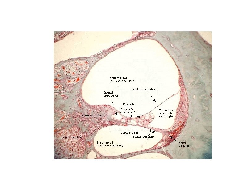
Anatomy of the cochlea Tectorial membrane Cochlear duct (scala media) Vestibular membrane Scala vestibuli Stria vascularis Spiral organ (of Corti) Basilar membrane Scala tympani Spiral ganglion Osseous spiral lamina (b) Human Anatomy and Physiology, 7 e by Elaine Marieb & Katja Hoehn Copyright © 2007 Pearson Education, Inc. , publishing as Benjamin Cummings.
Anatomy of the cochlea Tectorial membrane Inner hair cell Hairs (stereocilia) Afferent nerve fibers Outer hair cells Supporting cells (c) Human Anatomy and Physiology, 7 e by Elaine Marieb & Katja Hoehn Fibers of cochlear nerve Basilar membrane Copyright © 2007 Pearson Education, Inc. , publishing as Benjamin Cummings.
Hearing physiology • Sound waves travel from the outer ear (timpanic membrane) middle ear (Malleus, Incus, staples, oval window) inner ear (scala tympani)/scala vestibuli stimulates the stereocillia of the organ of corti VIII • The round window serves as a pressure relief valve.
Sound: source and propagation Area of compressed molecules (a) Air pressure Wavelength Area of rarefaction Crest Trough Time (b) Amplitude (c) Human Anatomy and Physiology, 7 e by Elaine Marieb & Katja Hoehn Copyright © 2007 Pearson Education, Inc. , publishing as Benjamin Cummings.
Route of sound waves through the ear External ear Tympanic membrane Malleus, incus, stapes (ossicles) Internal ear Oval window Fluids in cochlear canals Upper and middle Lower Pressure Pinna Air External acoustic meatus Middle ear One vibration Amplitude Human Anatomy and Physiology, 7 e by Elaine Marieb & Katja Hoehn Amplification in middle ear Spiral organ (of Corti) stimulated Time Copyright © 2007 Pearson Education, Inc. , publishing as Benjamin Cummings.
Resonance of the basilar membrane and activation of the cochlear hair cells Stapes Scala Cochlear vestibuli nerve Perilymph Oval window Round window Scala tympani Basilar membrane Cochlear duct (a) Base Relative lengths of basilar fibers within different regions of basilar membrane (b) Hz 20, 000 (High notes) Human Anatomy and Physiology, 7 e by Elaine Marieb & Katja Hoehn Apex Basilar membrane 500 Hz 4000 Hz Hz 1500 Hz 20 (Low notes) (c) 24, 000 Hz Copyright © 2007 Pearson Education, Inc. , publishing as Benjamin Cummings.
Simplified diagram of the auditory pathway to the auditory cortex of the brain Medial geniculate body of thalamus Primary auditory cortex in temporal lobe Inferior colliculus Lateral lemniscus Superior olivary nucleus (ponsmedulla junction) Midbrain Cochlear nuclei Vibrations Medulla Vestibulocochlear nerve Spiral ganglion of cochlear nerve Bipolar cell Spiral organ (of Corti) Human Anatomy and Physiology, 7 e by Elaine Marieb & Katja Hoehn Copyright © 2007 Pearson Education, Inc. , publishing as Benjamin Cummings.
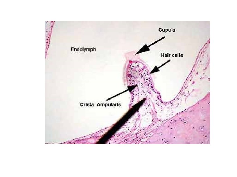
Equilibrium physiology • Located in the vestibular apparatus – Vestible • Utricle and saccule (sacs) within vesitble. – Receptor: Macculae (static equilibrium) » Hair cells (stereocillia) are embedded in the otholitic membrane which contain Ca. CO 3 (otoliths). Respond to vertical acceleration. – Membranous semicircular ducts • Ampulla (located at the base of each perpendicular duct). Mechism of dynamic equilibrium (angular acceleration). – Receptor: Crista ampullaris. Stereocilia covered by a gelatinous cupula. Endolymph stimulates the recptor.
Structure of a macula Macula of saccule Macula of utricle Kinocilium Stereocilia Otoliths Otolithic membrane Hair bundle Hair cells Vestibular nerve fibers Human Anatomy and Physiology, 7 e by Elaine Marieb & Katja Hoehn Supporting cells Copyright © 2007 Pearson Education, Inc. , publishing as Benjamin Cummings.
The effect of gravitational pull on a macula receptor cell in the utricle Otolithic membrane Kinocilium Stereocilia Depolarization Receptor potential (Hairs bent toward kinocilium) Nerve impulses generated in vestibular fiber Increased impulse frequency Human Anatomy and Physiology, 7 e by Elaine Marieb & Katja Hoehn Excitation Hyperpolarization (Hairs bent away from kinocilium) Decreased impulse frequency Inhibition Copyright © 2007 Pearson Education, Inc. , publishing as Benjamin Cummings.
Macula within the utriculus (otolith chamber) (Bouins, H&E, Bar = 36. 4 µm). 1. ciliated sensory cells; 2. sustenticular cells; 3. connective tissue; 4. cupula (gelatinous matrix); 5. globular sensory epithelium; 6. collagenous connective tissue; 7. otolith chamber; 8. cranium.
Location and sturcture of a crista ampullaris Flow of endolymph Crista ampullaris (a) Fibers of vestibular nerve Cupula (b) Turning motion Cupula Position of cupula during turn (c) Human Anatomy and Physiology, 7 e by Elaine Marieb & Katja Hoehn Increased firing (d) Ampulla of left ear Cupula at rest Ampulla of right ear Position of cupula during turn Fluid motion in ducts Horizontal ducts Decreased firing Afferent fibers of vestibular nerve Copyright © 2007 Pearson Education, Inc. , publishing as Benjamin Cummings.
Pathways of the balance and orientation system Vestibular receptors Visual receptors Somatic receptors Vestibular nuclear complex Reticular nuclei Input Cerebellum Central nervous system processing Oculomotor control (cranial nerve nuclei III, IV, VI) (eye movements) Spinal motor control (cranial nerve nuclei XI and vestibulospinal tracts) (neck movements) Output Human Anatomy and Physiology, 7 e by Elaine Marieb & Katja Hoehn Copyright © 2007 Pearson Education, Inc. , publishing as Benjamin Cummings.