General Principles of Positioning Chapter 12 Terminology Caudal


 Right (R) Dorsal (D) Medial (M) Lateral (L)")


- Slides: 26
General Principles of Positioning Chapter 12
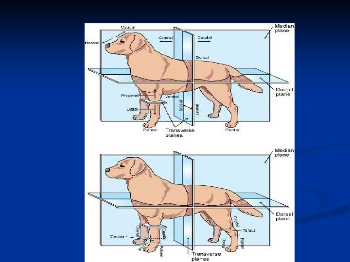
Terminology Caudal: Parts of the head, neck and trunk positioned towards the tail from any given point. Also aspect of limbs above the carpal and tarsal joints that face the rear of the animal. n Cranial- Describes parts of the neck, trunk, and tail positioned toward the head from any given point. Cranial also describes those aspects of the limb above the carpal and tarsal joints that face toward the head. n
Terminology, cont. n n n Distal- Farther away from the point of origin of a structure. Dorsal- Upper aspect of the head, neck, trunk, and tail. The term also means toward the upper aspect of the animal. Dorsal also describes the aspects of the legs from the carpus and tarsus joints distally that face toward the head. Lateral- the x-ray beam enters through either the left or right side of the body and emerges on the opposite side, where the cassette is positioned.
Terminology, cont Mediolateral- the x-ray beam enters a limb through the medial side and exits on the lateral side. Most lateral radiographs of the limbs are taken in lateromedial projection in large animal radiography. n Palmar- Used instead of caudal when describing the forelimb from the carpal joint distally. n Plantar- Used instead of caudal when describing the hindlimb from the tarsal joint distally. n
And yet more Terminology Proximal- Nearer to the point of origin of a structure. n Recumbent- The animal is lying down when the radiograph is made. Most radiographs of the dog and cat are made with the animal in the recumbent position, and this position should be presumed unless otherwise stated on the radiograph. n
Almost Done Rostral- parts of the head positioned toward the nares from any given point on the head. n Superior and Inferior- Used to describe the upper and lower dental arcades, respectively. n Ventral- Lower aspect of the head, neck, trunk, and tail. The term also means toward the lower aspect of the animal. n
Abbreviations n n n Left (L) Right (R) Dorsal (D) Medial (M) Lateral (L) Cranial (Cr) Rostral (R) Caudal (Cd) Palmar (Pa) Oblique (O) Plantar (Pl)
Abbreviations Abbreviated term used for the position designates the direction of the x-ray beam. n First letter states where the x-ray beam enters the body, the second designates where it exits. n V/D – enters ventrally and exits dorsally n DMPa. LO- indicates that carpus is rotated to a selected -degree angle and the central x-ray enters the dorsal/medial surface and exits the palmar/lateral surface. n
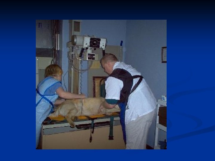
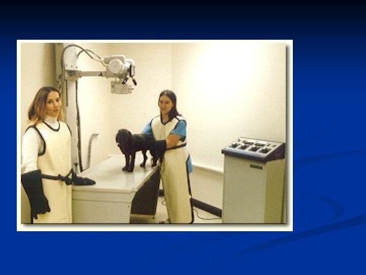
Basic Criteria of Positioning Refrain from overt physical restraint n Primary goal is to produce a good quality radiograph of the area being examined. n Factors to consider: n 1. Welfare of the patient. n 2. Restraint and immobilization of the patient. n 3. Minimal trauma to area of interest. n The least risk of exposing those assisting with the examination to radiaton. n
Patient factors The comfort and welfare of the patient should be considered at all times. n PATIENCE!!!!!! n Radiography can be frightening to animal. n Noises of prep and of x-ray can be very disconcerting. Make sure take into consideration when taking radiographs. n
How to handle animals Handle in a slow, quiet, manner. n Use a calm, soft voice and reassure animals through touch. n Avoid quick, loud movements. n Avoid and severe restraint. n
Prep sounds n When depressing prep button, machine will make noise. It is good idea to let patients hear this sound as you are positioning so as to avoid the frightening newness of the sound once the radiograph is taken.
General Positioning May require sedation, general anesthesia, and positional devices n All essential anatomical regions should be included in the primary beam when taking x-rays n PRIMARY GOAL- to find the most comfortable posture/position for the animal to produce an accurate reproduction of the area of interest n
PREPARATION!!! n Prepare, prepare n Prior to radiograph being taken make sure: n Correct patient is present n All chemicals and processor are in working order. n That cassette is positioned appropriately. n That identifying markers are in place.
Measurement Caliper- measuring device for anatomic area of interest. n Measures area in centimeters n If unsure, always measure thickest spot. If large differences in sizes exist, may have to take two separate radiographs. n
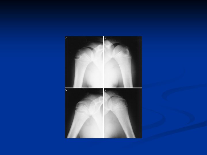
Required Views n Radiograph is a two dimensional picture of a three-dimensional structure. n n n 2 views will help you see something that you might miss on 1 view Area of interest closest to film n n Therefore two views must be taken at right angles to one another in order to get a good idea of structure of anatomy. Minimizes distortion and magnification Comparison n To compare to other area to see if there any pathological changes
Splitting the Cassette n n Taking more than 1 picture on a cassette You need to place lead over the “non-used” portion of the cassette, take the exposure, remove the lead, cover the “exposed” side and expose the “non-exposed” side n n Lead gloves can be used – inexpensive, handy Not practical when using bucky tray Split as many times as you have room The 2 views need to be in the same direction
Collimation, Collimation Very important n Decreases the amount of scatter n n n Which increases the amount of contrast Have you heard this before? ? ?
Positioning Guidelines n n Should be Taken over Thickest area General Rule: n The center of the primary beam (+) should be directly in the center of the area of interest. n n CASSETTE SIZE IS IMPERATIVE Specific anatomy must be included for each anatomic area. Long bones should include the shaft of the bone, as well as the joint above and the joint below n Joints should have the beam centered over the joint space, and the small portion of long bones above and below the joint. n
Patient Preparation n Clean & dry hair coat Removal of splints, bandage material, collars, leashes, etc. Chemical restraint is preferred but not always allowed n n No matter how good an animal seems, always expect the worse Use of positioning devices n n Sandbags, foam blocks, wood blocks, trough, tape, gauze, rope Positioning devices should not be placed directly above or below area of interest – not completely radiopaque
Film Identification Proper labeling of a radiograph is mandatory for legal and practical reasons. n Should include: n Appropriate patient identification n Appropriate markers (R ) (L) and view if necessary. n Should mark side that is down on the patient. n Lateral projections of the leg should have the marker placed cranially to the leg. n
Views n Standard is a lateral view and v/d or d/v view. n Oblique view n n n Patient is rotated – not the tube head Degree of angulations varies on area of interest – usually pretty minimal = 10 to 15 degree’s They allow for a more dimensional view n n Eliminates superimposition Comparison Views n n n Compare R w/ L Young animals, extremities – most common Helpful to do both projections on 1 film