General Information about viruses A virus is a

General Information about viruses § A virus is a small infectious agent that can infect all types of living organisms such as including animals, plants, bacteria and archaea. But viruses are not live organisms (do not belong to any of three domains and six kingdoms of life) § Lack cell structures for growth and reproduction § Must replicate inside a living host § Smaller than bacteria; need electron microscope to see
")
Structure § All viruses contain nucleic acids (either DNA or RNA, but not both) and proteins. Some may have lipids. § Usually the nucleic acids are wrapped in protein coat called capsid. Some have an envelope (taken from host cell membrane) Classification (the most useful and widely used is based on nucleic acids) § DNA viruses (double-strand single-strand DNA viruses) Such as Hepatitis B virus, Epstein–Barr virus, Papillomavirus. § RNA viruses (double-strand single-strand RNA viruses) Such as Ebola virus, , Hepatitis C virus § Reverse transcribing virus: Hepatitis B virus and HIV

Tobacco mosaic virus HIV Bacteriophage Ebola virus

Transcription and Translation 1. DNA viruses DNA RNA Proteins

2. RNA viruses Single-strand RNA positive-sense RNA. Viral RNA with the same base sequence as m. RNA; during replication it functions as m. RNA, serving as a template for protein synthesis. negative-sense RNA. Viral RNA with a base sequence complementary to that of m. RNA; during replication it must be converted to positive-sense RNA before translation. Double-strand RNA
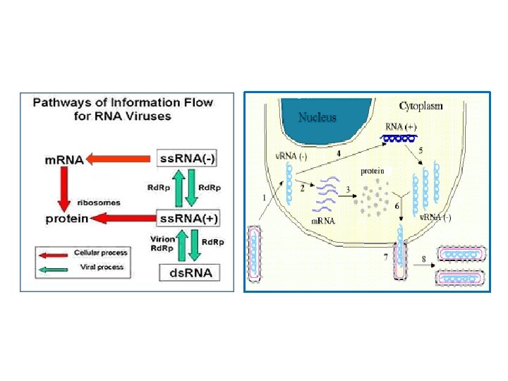
 RNA DNA Infection and Reproduction m. RNA protein")
3. Retrovirus (HIV) RNA DNA Infection and Reproduction m. RNA protein
")
Lytic Growth (all virus)
")
Lysogenic Growth (some virus)

Viruses and Cancer § The Epstein-Barr virus has been linked to Burkitt's lymphoma. This virus infects B lymphocytes of the immune system and epithelial cells § The hepatitis B virus has been linked to liver cancer in people with chronic infections. § Human papilloma viruses have been linked to cervical cancer. § Human herpes virus-8 (HHV-8) and HIV have been linked to the development of Kaposi sarcoma causes patches of abnormal tissue to develop in various area of the body including in the skin, in the lining of the mouth, nose, and throat or in other organs.

Discovery of EBV In 1958 an Irish surgeon, Denis Burkitt discovered an aggressive form of lymphoma, now called Burkitt’s lymphoma. In 1964 two British virologists, Epstein and Barr, analyzed the Burkitt’s lymphoma cells from Africa and found that there was an infectious agent lodged inside of the lymphoma cells, which was a new virus named Epstein-Barr virus or EBV. In 1970, the virus was shown to be able to immortalize B lymphocytes.

Discovery of HBV In 1964, after a brief tenure at the NIH , Baruch Blumberg moved to the Institute for Cancer Research in Philadelphia (the Fox Chase Cancer Center) to study blood antigen. One antigen was found frequently in Asian (including Australians) and African populations, but not in Europeans and Americans. He called this antigen as Australia antigen ( Au). He noticed that individuals carrying the Au often suffered from chronic hepatitis, whereas the Au negative individuals has much less this disease. In 1966 one of his patients was negative for Au with a mental-disability but later suddenly changed to Au positive and hepatitis was developed. How could this happened? Soon he found that Au was a foreign protein floating in the patient blood, which was produced by a new virus called hepatitis B virus. Further research showed that chronic infection with HBV had great risk for cancer development. Thus, HBV was/is a carcinogen!

Blumberg received Nobel Prize in Physiology or Medicine in 1976 for his work on HBV.

Mechanisms of some virus induced cancer 1. Carrying oncogenes 2. Activate human oncogens 3. Inhibits tumor suppressor genes
")
Human Immunodeficiency virus (HIV)

Lymphocytes are immune cells that allow the body to remember and recognize previous invaders and help the body destroy them. NK (Nature killer) Cells: bigger than T and B lymphocytes, activated by cytokines. Nature killer cells can release cytotoxic factors. B lymphocytes: response to pathogens and produce antibodies to neutralize foreign objects. T Lymphocytes: § § Cytotoxic Suppressor T cells are CD 8 positive cells that release cytotoxic factors. Helper T lymphocytes are CD 4 positive cells that can bind to antigen and release cytokines. The cytokines act as growth factors and help to active B cells and NK cells.

Structure of HIV: HIV has a viral capsid that contain RNA. The RNA has two identical strands. Inside the core are three enzymes required for HIV replication called reverse transcriptase, integrase and protease. The capsid is surrounded by an envelop of host-cell origin. As HIV buds out of the host cell during replication, it acquires a phospholipid envelope. The envelope includes glycoproteins, gp 120 and gp 41.
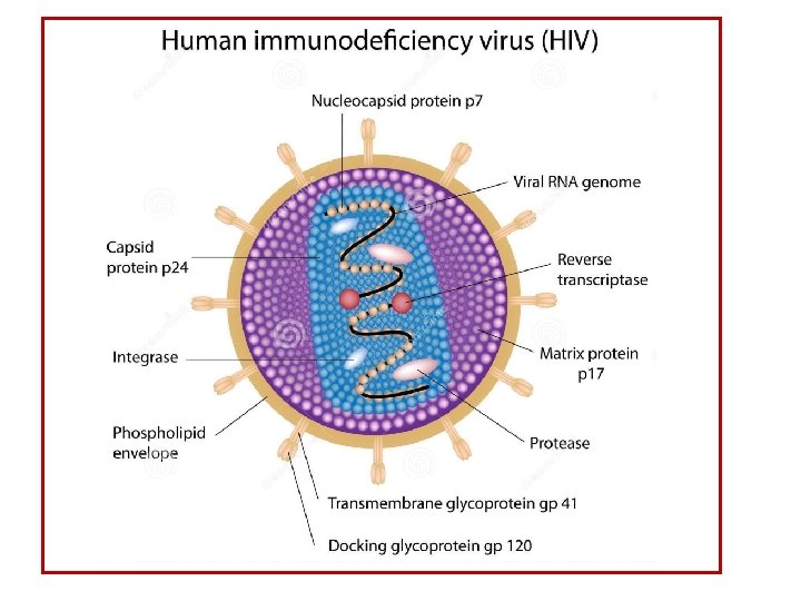
 § GP 120 of HIV recognizes and binds to CD")
Infection Pathway (signal pathway) § GP 120 of HIV recognizes and binds to CD 4 of CD 4+ T cells. § After HIV has bound to the target cell, the HIV RNA and various enzymes, including reverse transcriptase, integrase, ribonuclease, and protease, are injected into the cell. § Shortly after the viral capsid enters the cell, the reverse transcriptase and the single-stranded (+)RNA are released from the capsid and into cytosol where the RNA are reverse transcribed into a complementary DNA (c. DNA) molecule.

§ The integration of the viral DNA into the host cell's genome is carried out by another viral enzyme called integrase. § During viral replication, the integrated DNA is transcribed into m. RNA which is then spliced into smaller pieces. These small pieces are exported from the nucleus into the cytoplasm, where they are translated into proteins § The final step of the viral cycle, assembly of new HIV-1 viruses begins at the plasma membrane of the host cell.

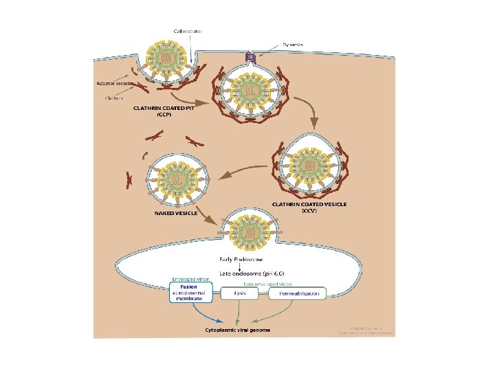
Diagram of the process that HIV entering human cells

Ebola Virus

Ebola, also called Ebola hemorrhagic fever, which is caused by Ebola virus Incubation period: 2 to 21 days. Main early symptoms: fever, headache, joint and muscle aches, sore throat, and weakness. Late symptoms: diarrhea, vomiting, stomach pain, rashes, bleeding, and organ failure. Main death causes: internal bleeding, organ failure, severe dehydration Infection pathways: Ebola virus can passes between animals and humans, by physical contact (blood and secretions) or eating (in animals) Main ebolar virus carriers: fruit bats, gorillas, monkeys, forest antelope, and chimpanzees. Mortality in human: 50 -90% depending on what type of Ebola viruses.
 Sudan Ebola virus (1998) Bundibugyo (BDBV) (2007)")
Types of Ebola Viruses Tai Forest (1995) Sudan Ebola virus (1998) Bundibugyo (BDBV) (2007) Zaire Ebola virus (1976) Reston (RESTV) (1990) Discovery of Ebola In 1976 by a international scientist team in a village in Zaire (Democratic Republic of Congo)

Structure of Ebola Virus tubular structure with diameter about 80 nm and long from 800 nm to 1000 nm Possible receptors for Ebola virus (1) cholesterol transporter protein, NPC-1, (2) TIM-1

Fig. Endocytosis imports extracellular molecules by forming vesicles from the plasma membrane
 Ebola virus protein recognize and attach to the receptors of host")
Infection Pathway (1) Ebola virus protein recognize and attach to the receptors of host cells. (2) The Ebola virus RNA into host cells and stay inside of vesicle through the mechanism called Endocytosis. (3) Once the virus is fused with the vesicle membrane, the negative RNA is released into cytoplasm, where it is converted to positive RNA using RNA polymerase. (4) The host cells use positive RNA to make virus proteins (5) The RNA and the proteins are assembled together, becoming new viruses.

HIV vs. Ebola Virus § Both Ebola virus and HIV use endocytosis to into host cells § HIV is retrovirus, which uses reverse transcriptase to make virus DNA even it has a positive RNA. § Ebola is not retrovirus, using RNA polymerase to make protein § HIV RNA is positive RNA, whereas Ebola virus has negative RNA.
- Slides: 29