General Histology Specialized Connective Tissues 1 Bone Bone

General Histology

Specialized Connective Tissues: 1 - Bone Ø Bone is the primary constituent of the adult skeleton. Ø It is a specialized type of connective tissue with a calcified extracellular matrix in which characteristic cells are embedded. Ø Bone functions: 1. Protect vital organs. 2. Support fleshy structures. 3. Provide a calcium reserve (bone contains about 99% of the body’s calcium).
 portion of the bone")
Structure of the bone: 1. Bone matrix: a. Inorganic (calcified) portion of the bone matrix (about 65% of the dry weight) is composed of calcium, phosphate, bicarbonate, citrate, magnesium, potassium, and sodium. b. Organic portion of the bone matrix (about 35% of the dry weight) consists primarily of type I collagen (95%). 2. Periosteum : is a layer of noncalcified connective tissue covering bone on its external surfaces. It is composed of an outer dense fibrous collagenous layer and an inner cellular osteogenic layer. 3. The endosteum is a thin specialized connective tissue that lines the marrow cavities and supplies osteoprogenitor cells and osteoblasts for bone growth and repair.

Bone cells 1. Osteoprogenitor cells: These spindle-shaped cells are located in the periosteum and the endosteum. 2. Osteoblasts: These are the cells that form new bone. They also come from the bone marrow and are related to structural cells. They have only one nucleus. 3. Osteocyte : a cell that lies within the substance of fully formed bone. It occupies a small chamber called a lacuna, which is contained in the calcified matrix of bone. 4. Osteoclasts: are large, motile, multinucleated cells (up to 50 nuclei) that resorb bone.

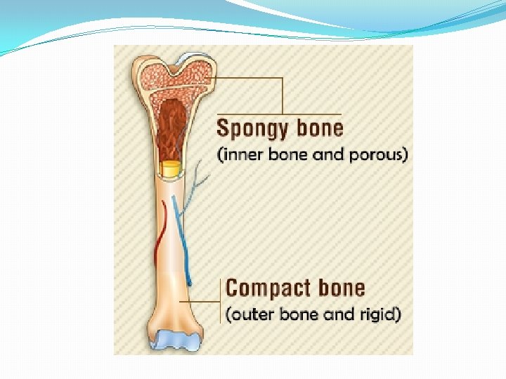
Classification of bone : 1. Gross observation of cross-sections of bone reveals two types: a. Spongy (cancellous) bone, which is composed of interconnected trabeculae. Bony trabeculae surround cavities filled with bone marrow. The trabeculae contain osteocytes and are lined on both surfaces by a single layer of osteoblasts. Spongy bone is always surrounded by compact bone. b. Compact (dense) bone has no trabeculae or bone marrow cavities. 2. Microscopic observation of bone reveals two types: a. Primary bone: also known as immature or woven bone that contains many osteocytes and large, irregularly arranged type I collagen bundles. It has a low mineral content. b. Secondary bone: is also known as mature or lamellar bone. It has a calcified matrix arranged in regular layers, or lamellae. It contains osteocytes in lacunae between, and occasionally within,

Organization of lamellae in compact bone is characteristic and consists of the following elements : 1. Haversian systems (osteons) are long cylindrical structures that run approximately parallel to the long axis of the diaphysis. This system are composed of 4 to 20 lamellae surrounding a central haversian canal, which contains blood vessels, nerves, and loose connective tissue. They are interconnected by Volkmann canals, which also connect to the periosteum and endosteum and carry the neurovascular supply. 2. Interstitial lamellae are irregularly shaped lamellae between haversian systems. They are remnants of remodeled haversian systems.

Structure of the bone:

2 - Cartilage: • Cartilage is an avascular specialized fibrous connective tissue. • It has a firm extracellular matrix that is less pliable than that of connective tissue proper, and it contains chondrocytes embedded in the matrix. Ø Functions: • Cartilage functions primarily to support soft tissues and assist in the development and growth of long bones.

Types: • The three types of cartilage; 1. Hyaline cartilage. 2. Elastic cartilage. 3. Fibrocartilage.

Hyaline cartilage : 1. Structure a. Matrix: The matrix is composed of an amorphous ground substance containing proteoglycan aggregates and chondronectin, in which type II collagen is embedded. b. Perichondrium is a layer of dense, irregular connective tissue that surrounds hyaline cartilage except at articular surfaces. It consists of an outer fibrous layer containing type I collagen, fibroblasts, and blood vessels and an inner cellular layer containing chondrogenic cells and chondroblasts. d. Chondrocytes are mature cartilage cells that are embedded within lacunae in the matrix.

B. Elastic cartilage: • It possesses a perichondrium and is nearly identical to hyaline cartilage except for a network of elastic fibers, which impart a yellowish color. C. Fibrocartilage • It lacks an identifiable perichondrium. It is characterized by alternating rows of fibroblastderived chondrocytes surrounded by scant matrix and thick parallel bundles of type I collagen fibers. Fibrocartilage is located in areas where support and tensile strength are required

3 - Blood • Blood is a specialized connective tissue that consists of formed elements erythrocytes, leukocytes, and platelets and a fluid component called plasma. • The volume of blood in an average human adult is approximately 5 L. • Functions: 1. Blood circulates in a closed system of vessels and transports nutrients, waste products, hormones, proteins, ions, oxygen (O 2), carbon dioxide (CO 2), and formed elements. 2. It also regulates body temperature. 3. It assists in regulation of osmotic and acid–base balance. 4. Blood cells have short life spans and are continuously replaced by a process called hemopoiesis.

BLOOD CONSTITUENTS A. Plasma consists of 90% water; 9% organic compounds (such as proteins, amino acids, and hormones); and 1% inorganic salts, dissolved gases, and nutrients. B. Formed elements of blood 1. Erythrocytes (red blood cells [RBCs]) 2. Leukocytes, or white blood cells (WBCs)

- Slides: 15