General Histology Practical Lab 8 Nervous System Central

General Histology Practical Lab 8

Nervous System Central nervous system • • • The brain and the spinal cord consists of white matter and gray matter. White matter is composed of myelinated nerve fibers, some unmyelinated nerve fibers and neuroglial cells. Gray matter consist of aggregations of neurons cell bodies, dendrites, unmyelinated portions of axons, and neuroglial cells. The three connective tissue covering of the brain and spinal cord are the meninges. The outer most layer of the meninges is the dura mater, the intermediate layer is the arachnoids, and the inner most intimate layer of the meninges is the pia mater.

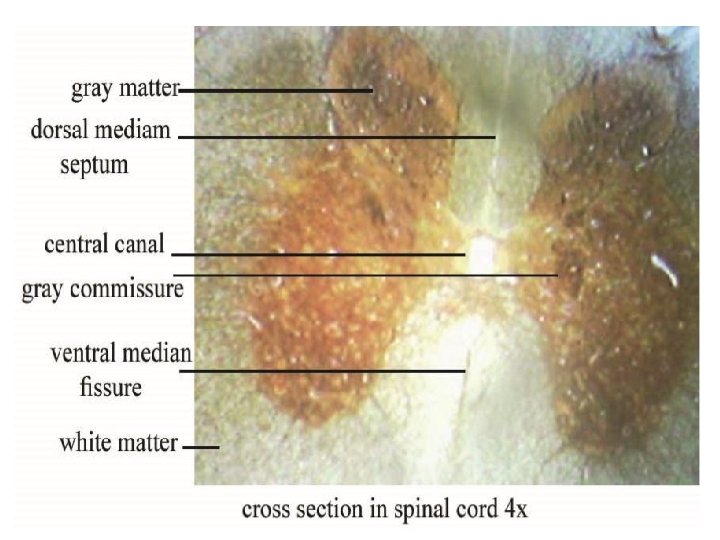
1 - Spinal cord: White matter is located in the periphery of the spinal cord whereas gray matter lies deep in the spinal cord, where it forms the shape of an H in cross section. • A small central canal , lined by ependymal cells and representing the lumen of the original neural tube, lies in the center of the cross bar of the H. • the upper vertical bars of the H represent the dorsal horns of the spinal cord. • The lower vertical bars of the H represent the ventral horns of the spinal cord, which house cell bodies of large multipolar motor neurons, whose axons exit the spinal cord via the ventral roots. •

2 - Cerebrum: • Gray matter in the brain is located at the periphery (cortex) of the cerebrum and cerebellum. • White matter lies deep to the cortex. • The gray matter at the periphery of the cerebral hemispheres is folded into many gyri sulci called the cerebral cortex. • This portion of the brain is responsible for: 1. motor response. 2. integration of sensory signals. • The cerebral cortex is divided into six layers composed of neuronal cells that is:

1 - Molecular layer: is composed of nerve terminals originating in other areas of the brain. 2 - External granular layer: contain mostly granule (stellate) cells and neuroglial cells. 3 - External pyramidal layer: contains neuroglial cells and large pyramidal cells, which become increasingly larger from the external to the internal border of this layer.

4 - Internal granular layer: is a thin layer and is characterized by closely arranged, small granule (stellate) cells, pyramidal cells, and neuroglial. This layer has the greatest cell density of the cerebral cortex. 5 - Internal pyramidal layer: contains the largest pyramidal cells and neuroglial. This layer has the lowest cell density of the cerebral cortex. 6 - Multiform layer: consists of various multiform cells called martinotti cells and neuroglia.

3 - Cerebellum: • The layer of gray matter located in the periphery of the cerebellum is called cerebellar cortex. • This portion of the brain is responsible for : ü 1. maintaining balance and equilibrium. ü 2. muscle tone. ü 3. coordination of skeletal muscles.

The cerebellar cortex is divided into three layers: 1 - Molecular layer: contains superficially located stellate cells, dendrites of purkinje cells, basket cells and unmyelinated axons from the granular layer. 2 - Purkinje cell layer: contains the large flask shaped purkinje cells, which are present only in the cerebellum. 3 - Granular layer: consists of small granule cells and glomeruli (cerebellar islands). Granuli are regions of the cerebellar cortex where synapsis are taking place between axons entering the cerebellum and the granule cells.

Granular Molecular Perpendicular

• Home Works q Q 1: What the typical draw for the central canal of spinal cord? q Q 2: what is the type of Purkinje cell?

. R O F U O Y G K N I N N A I T H T LIS
- Slides: 12