GENERAL EXAMINATION ITEMS OF GENERAL EXAMINATION Mental state


 �Under nails �Palmer crease")


")


- Slides: 55
GENERAL EXAMINATION
ITEMS OF GENERAL EXAMINATION Mental state Body built Posture Facial expression and general look Complexion (pallor, jaundice , cyanosis) Vital signs � Pulse � Blood pressure � Respiratory rate � temperature
ITEMS OF GENERAL EXAMINATION Head&neck Upper &lower limbs Lymphatic system Skin Breast genitalia
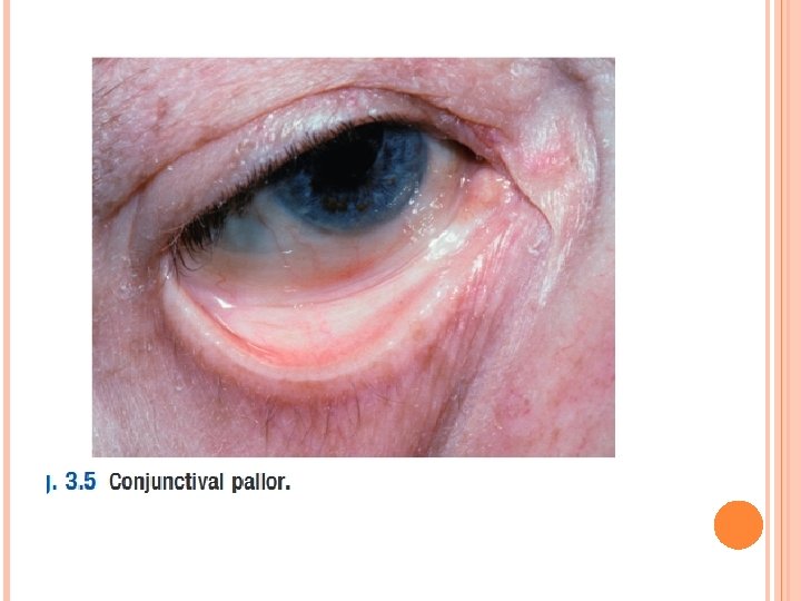
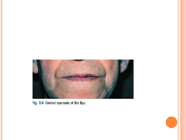
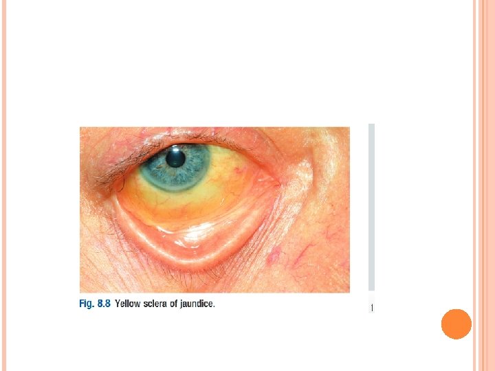
COMPLEXION Items �Pallor �Jaundice �cyanosis
PALLOR Site �Skin �Mucous membrane (conjunctiva, lips) �Under nails �Palmer crease
CAUSES OF PALLOR Anaemia Acute hemorrhage Cardiac Shock Acute nephritis
CYANOSIS Appears when vascular capillaries bed contains more than 5 gm reduced hemoglobin
TYPES Central � Blood pumbed into aorta > 5 gm reduced hemoglobin � Hypoxic hypoxia Peripheral � Due to stagnation of blood in peripheral circulation � stagnant hypoxia
CAUSES OF CENTRAL CYNOSIS Congenital cyanotic heart disease Lung diseases � Fibrosis � Collapse � Pulmonary empolism Abnormal hemoglobin � Met hemoglobin(congenital) � nitrites
CAUSES OF PERIPHERAL CYANOSIS � Venous thrombosis � Arterial embolism � Exposure to cold � Raynoud, s phenomena � Congestive heart failure
Central peripheral Site Tongue , inner side of lips Fingers , outer side of lips , tip of nose, ear lobule Hand Warm Cold Oxygenation Improve No effect Warming Increase Decrease Clubbing Present Absent
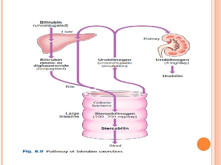
JAUNDICE Yellow discolouration of sclera&skin Bilirubin>2. 5 mg/dl Types � Hemolytic � Hepatocellular � obstructive
HEMOLYTIC JAUNDICE Appears with hemolytic anemia Increase unconjugated bilirubin Pallor Lemon yellow Dark stool Normal urine
HEPATOCELLULAR JAUNDICE Appears with hepatic cell failure Increase conjugated & unconjugated bilirubin Orange yellow Pale stool Dark urine
OBSTRUCTIVE JAUNDICE Appears with obstruction of bile ducts by stone & tumour Increase conjugated bilirubin Olive green Pale stool Dark urine
VITAL SIGNS
PULSE • Rate • Rhythm • Volume • Character • Equality on both hands • Vessel wall
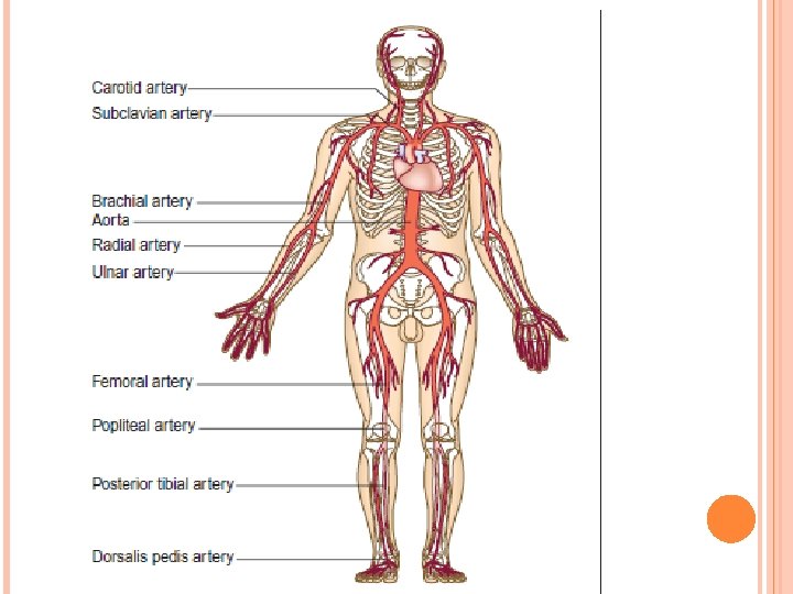
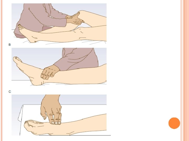
PERIPHERAL PULSATION § § § § Radial carotid bracial Femoral Popliteal Posterior tebial Dorsalis pedis
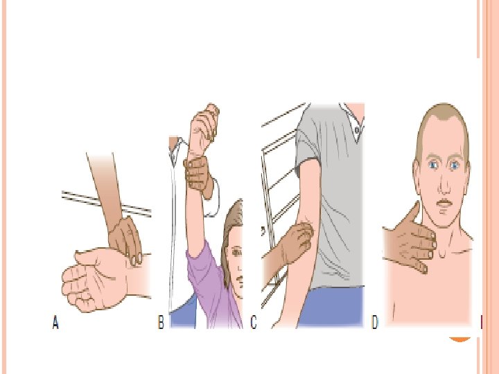
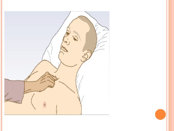
RADIAL ARTERY Place the pads of your index and middle fingers over the right radial artery. ■ Assess rate, and rhythm. ■ Count the pulse rate over 30 seconds; multiply by 2 to obtain the beats per minute (bpm). ■ To detect a collapsing pulse: first, check that the patient has no shoulder or arm pain or restriction on movement. Feel the pulse with the base of your fingers, then raise the patient’s arm vertically above the patient’s head. ■ Palpate both radial pulses simultaneously, assessing any difference in pulse volume between the two. §
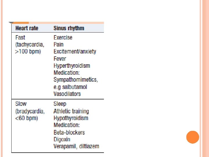
RATE Ø Resting heart rate is normally 60– 90 bpm. Ø Bradycardia is a pulse rate <60 bpm; Tachycardia is a rate of >100 bpm. Ø
RHYTHM Rhythm Sinus rhythm originates from the sinoatrial node and produces a regular rhythm. the pulse can be either regular or irregular During inspiration, parasympathetic tone falls and the heart rate increases; on expiration, the heart rate decreases.
VOLUME Volume refers to the perceived degree of pulsation and reflects the pulse pressure. Big volume � Exercise � Fever � Hyperthyrodism Small volume � Hypovolemic skock � Cardiac failure
CHARACTER Character refers to the waveform or shape of the arterial pulse. Water hammer pulse(big volume) � Exertion � Fever � Thyrotoxicosis � Anemia Plateau pulse(small volume) Aortic stenosis
EQUALITY OF PULSE Unequal pulse � Thromposis � Empolism � tumour
VESSEL WALL Palpable vessel wall atheroscelerosis
THE RESPIRATORY RATE Rate and pattern Rate : in a relaxed adult is about 14 -16 breaths per minute Tachypnea is an increased respiratory rate. I. � Exertion � Fever � Hypoxia � Nervous excitation bradypnea is an decreased respiratory rate � Increase in intracranial tension � Opium poisons
II. RESPIRATORY RHYTHM Normal rhythm is regular Abnormal rhythm � Hyperpnea increase in deapth of respiration Psychogenic condition Diabetic ketosis � Oligopnea decrease in deapth of respiration Pnemonia Depression of respiratory rate
TEMPERA TURE
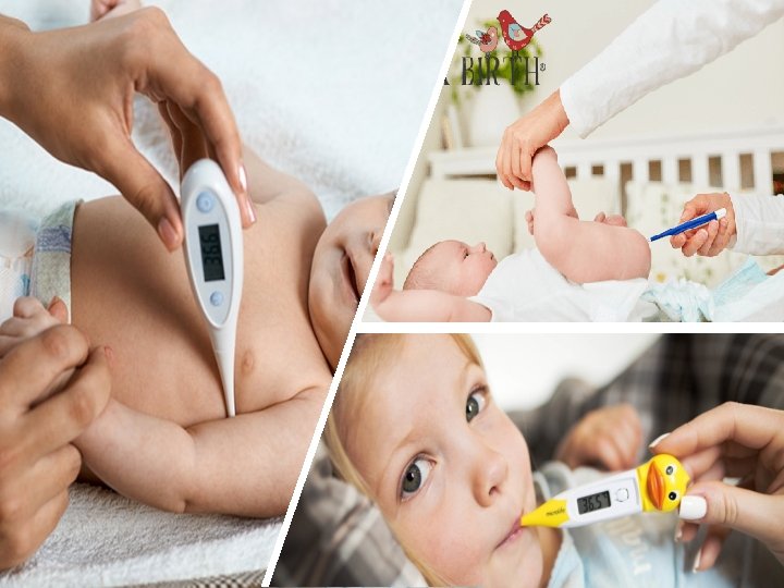
Temperature Body temperature may be recorded in the • mouth, axilla, ear or rectum. A ‘normal’ mouth temperature is 35. 8 -37°C. Those in the ear and rectum are 0. 5°C higher • and in the axilla 0. 5°C lower. • In women, ovulation is associated with a 0. 5°C • rise in temperature. In hospitalized patients, regular temperature measurements may identify certain characteristic patterns of disturbance.
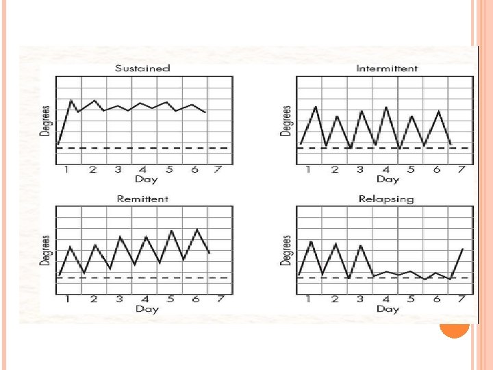
TYPES OF FEVERThe pattern of temperature changes mayoccasionally hint at the diagnosis: • Continuous/sustained fever: Temperature remains above normal throughoutthe day and does not fluctuate more than 1 °C in 24 hours, e. g. lobarpneumonia, typhoid fever, urinary tract infection, brucellosis. • Intermittent fever: The temperature elevation is present only for a certainperiod, later cycling back to
• Relapsing fever: temperature returns to normal for days before rising -Tertianfever (48 hour periodicity), typical of Plasmodium vivax or Plasmodium ovalemalaria • Remittent fever: Temperature remains above normal throughout the day and fluctuates more than 1 °C in 24 hours, e. g. , infective endocarditis. • Pel-Ebstein fever: A specific kind of fever associated with Hodgkins lymphoma, being high for one week and low for the next week and so on. However, there is some debate as to whether this
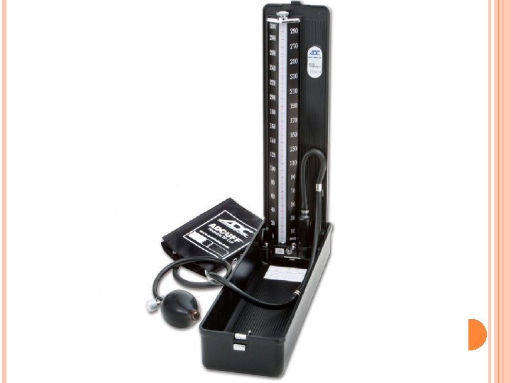
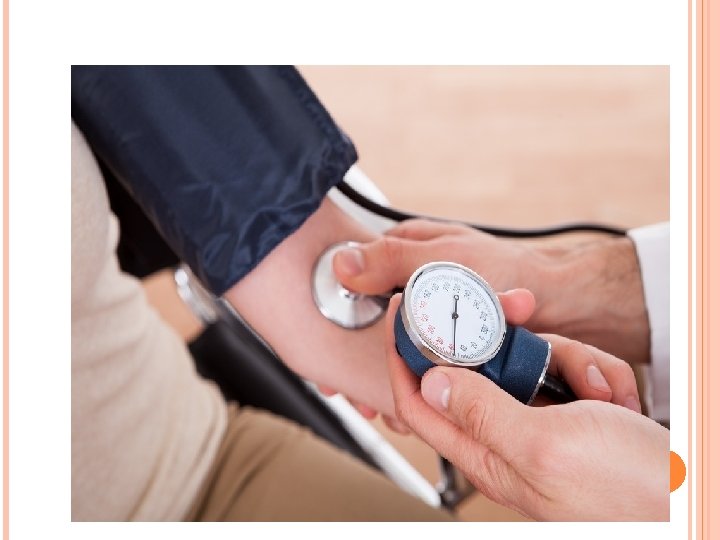
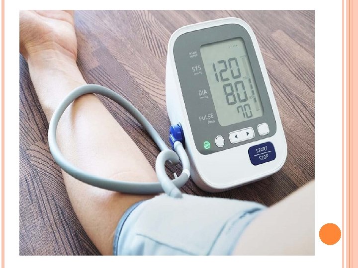
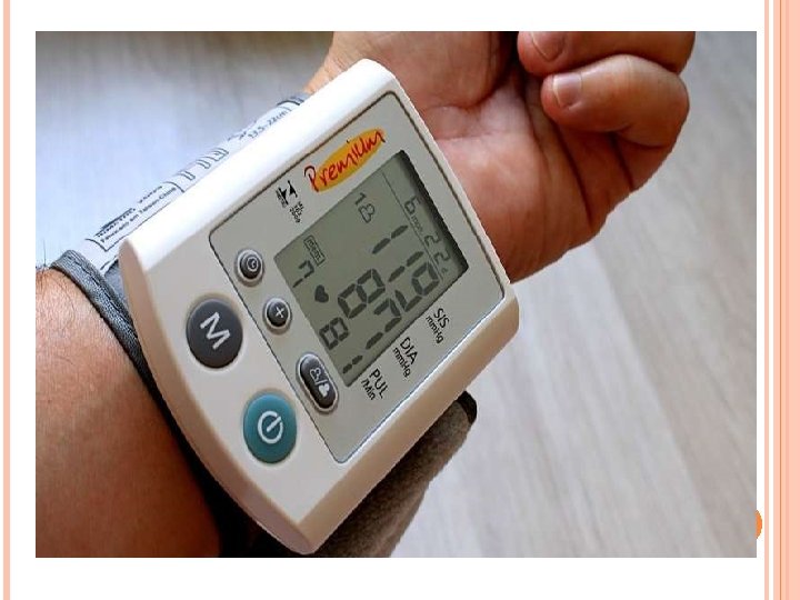
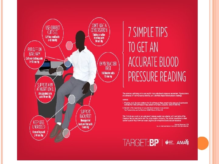
BLOOD PRESSURE
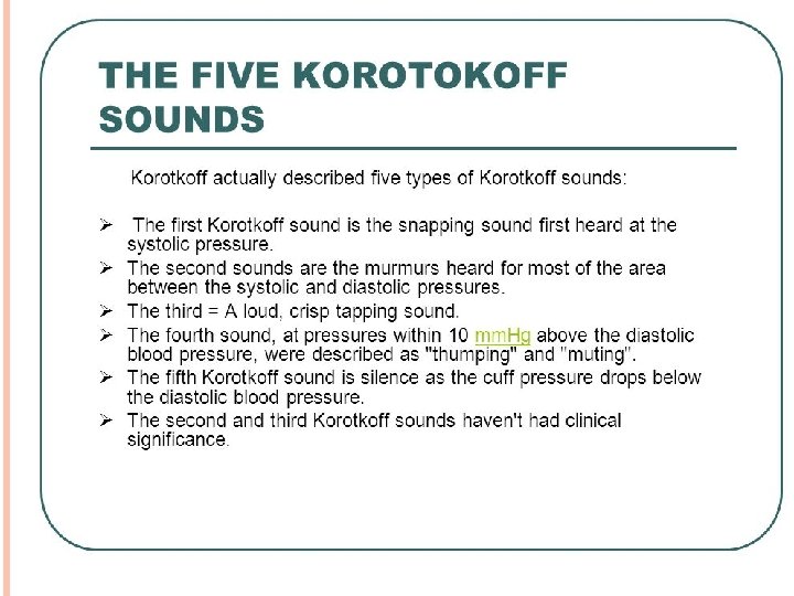
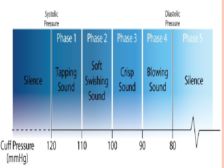
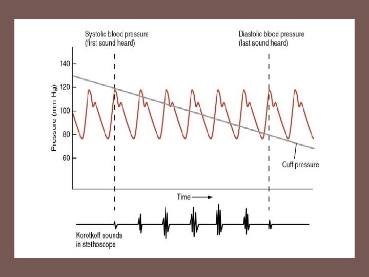
The physiology of blood pressure measurement (including a description of the Korotkoff sounds)
High blood pressure effects