
General Embryology Spermatogenesis Dr Ruqaya Alsmak Senior Embryologist
General Embryology Spermatogenesis Dr. Ruqaya Alsmak Senior Embryologist Ibn-Sina University of Medical and Pharmaceutical Sciences

Spermatogenesis • Maturation of Sperm Begins at Puberty Spermatogenesis, which begins at puberty, includes all of the events by which spermatogonia are transformed into spermatozoa. At birth, germ cells in the male infant can be recognized in the sex cords of the testis as large, pale cells surrounded by supporting cells, which are derived from the surface epithelium of the testis in the same manner as follicular cells, become Sertoli cells.

Structure of the Testis

Spermatogenesis • Shortly before puberty, the sex cords acquire a lumen and become the seminiferous tubules. At about the same time, PGCs give rise to spermatogonial stem cells, which then divide to form primary spermatocytes

Spermatogenesis • Primary spermatocytes then enter a prolonged prophase (22 days) followed by rapid completion of meiosis I and formation of secondary spermatocytes. During the second meiotic division, these cells immediately begin to form haploid spermatids Furthermore, spermatogonia and spermatids remain embedded in deep recesses of Sertoli cells throughout their development. In this manner, Sertoli cells support and protect the germ cells, participate in their nutrition, and assist in the release of mature spermatozoa.
Spermatogenesis • Spermatogenesis is regulated by luteinizing hormone (LH) production by the pituitary gland. LH binds to receptors on Leydig cells and stimulates testosterone production, which in turn binds to Sertoli cells to promote spermatogenesis. Follicle-stimulating hormone (FSH) is also essential because its binding to Sertoli cells stimulates testicular fluid production and synthesis of intracellular androgen receptor proteins.
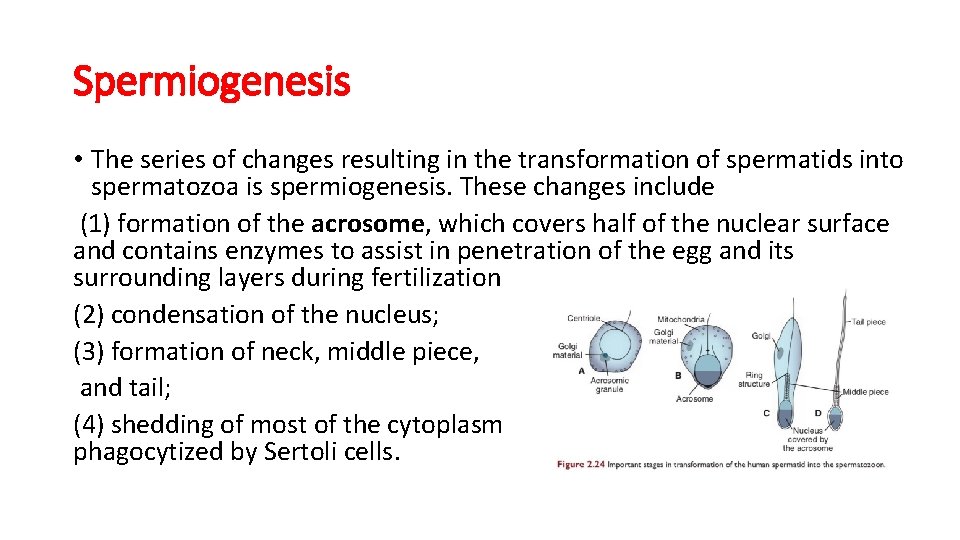
Spermiogenesis • The series of changes resulting in the transformation of spermatids into spermatozoa is spermiogenesis. These changes include (1) formation of the acrosome, which covers half of the nuclear surface and contains enzymes to assist in penetration of the egg and its surrounding layers during fertilization (2) condensation of the nucleus; (3) formation of neck, middle piece, and tail; (4) shedding of most of the cytoplasm as residual bodies that are phagocytized by Sertoli cells.
In humans, the time required for a spermatogonium to develop into a mature spermatozoon is approximately 74 days, and approximately 300 million sperm cells are produced daily.
First week of Development: Ovulation to implantation

Ovarian Cycle
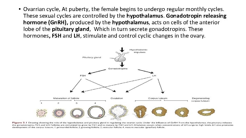
• Ovarrian cycle, At puberty, the female begins to undergo regular monthly cycles. These sexual cycles are controlled by the hypothalamus. Gonadotropin releasing hormone (Gn. RH), produced by the hypothalamus, acts on cells of the anterior lobe of the pituitary gland, Which in turn secrete gonadotropins. These hormones, FSH and LH, stimulate and control cyclic changes in the ovary.
• With each ovarian cycle, a number of primary follicles begin to grow under the influence of FSH, but usually one reaches full maturity, and only one oocyte is discharged at ovulation. Fsh also stimulates maturation follicular (granulosa) cells. In cooperation theca interna and granulosa cells produce estrogens. As a results of estrogen production • The uterine endometrium enters the follicular or proliferative phase • Thinning of the cervical mucosa allow passage of sperms • The anterior lobe of pituitary gland is stimulated to secrete LH
• At mid cycle, there is an LH surge that • The oocyte complete meiosis I and initiate meiosis II • Stimulate production of progestrone • and cause follicular rupture and ovulation
Ovulation • The ovulation under the influence of FSH and LH, the vesicular (Graafian follicle) grow to a diameter of 25 mm, there is an abrupt increase in LH that causes the primary oocyte to complete meiosis I and the follicle to enter the preovulatory mature vesicular stage. Meiosis II is also initiated, but the oocyte is arrested in metaphase approximately 3 hours before ovulation.
• . Prostaglandin levels also increase in response to the LH surge and cause local muscular contractions in the ovarian wall. Those contractions extrude the oocyte, which together with its surrounding granulosa cells from the region of the cumulus oophorus, breaks free (ovulation) and floats out of the ovary (Fig. 3. 3).
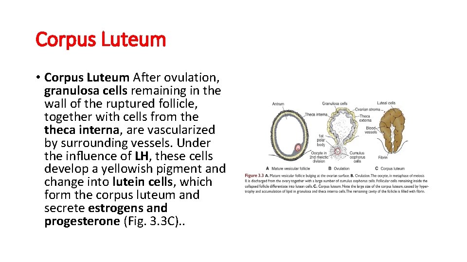
Corpus Luteum • Corpus Luteum After ovulation, granulosa cells remaining in the wall of the ruptured follicle, together with cells from theca interna, are vascularized by surrounding vessels. Under the influence of LH, these cells develop a yellowish pigment and change into lutein cells, which form the corpus luteum and secrete estrogens and progesterone (Fig. 3. 3 C). .
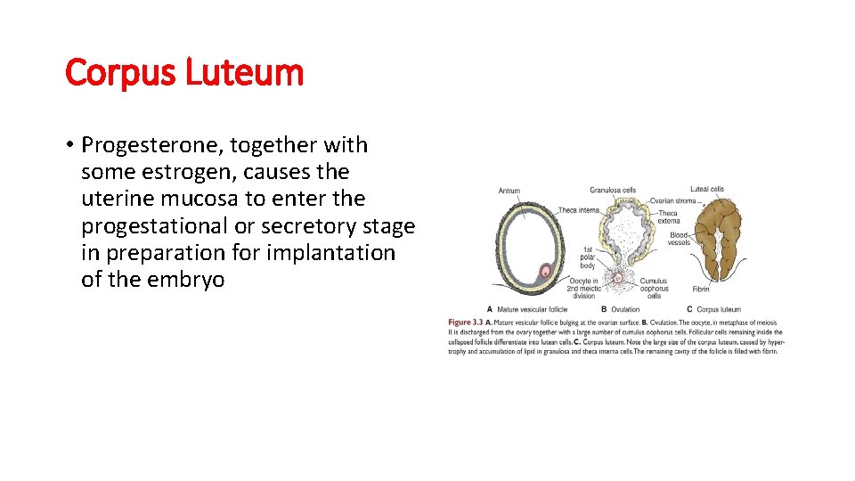
Corpus Luteum • Progesterone, together with some estrogen, causes the uterine mucosa to enter the progestational or secretory stage in preparation for implantation of the embryo

Oocyte transport • Shortly before ovulation, fimbriae of the uterine tube sweep over the surface of the ovary, and the tube itself begins to contract rhythmically. It is thought that the oocyte, is carried into the tube by these sweeping movements of the fimbriae and by motion of cilia on the epithelial lining. Once the oocyte is in the uterine tube, it is propelled by peristaltic muscular contractions of the tube and by cilia in the tubal mucosa with the rate of transport regulated by the endocrine status during and after ovulation.
Corpus Albicans • Corpus Albicans If fertilization does not occur, the corpus luteum reaches maximum development approximately 9 days after ovulation. It can easily be recognized as a yellowish projection on the surface of the ovary. Subsequently, the corpus luteum shrinks because of degeneration of lutean cells (luteolysis) and forms a mass of fibrotic scar tissue, the corpus albicans.
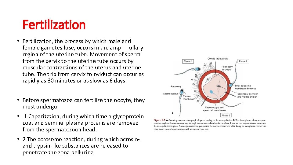
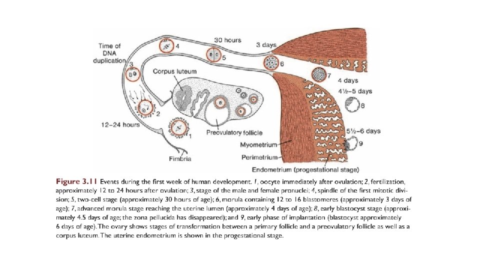
Fertilization • Fertilization, the process by which male and female gametes fuse, occurs in the amp ullary region of the uterine tube. Movement of sperm from the cervix to the uterine tube occurs by muscular contractions of the uterus and uterine tube. The trip from cervix to oviduct can occur as rapidly as 30 minutes or as slow as 6 days. • Before spermatozoa can fertilize the oocyte, they must undergo: • 1 Capacitation, during which time a glycoprotein coat and seminal plasma proteins are removed from the spermatozoon head. • 2 The acrosome reaction, during which acrosinand trypsin-like substances are released to penetrate the zona pellucida
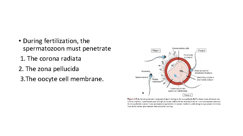
• During fertilization, the spermatozoon must penetrate 1. The corona radiata 2. The zona pellucida 3. The oocyte cell membrane.
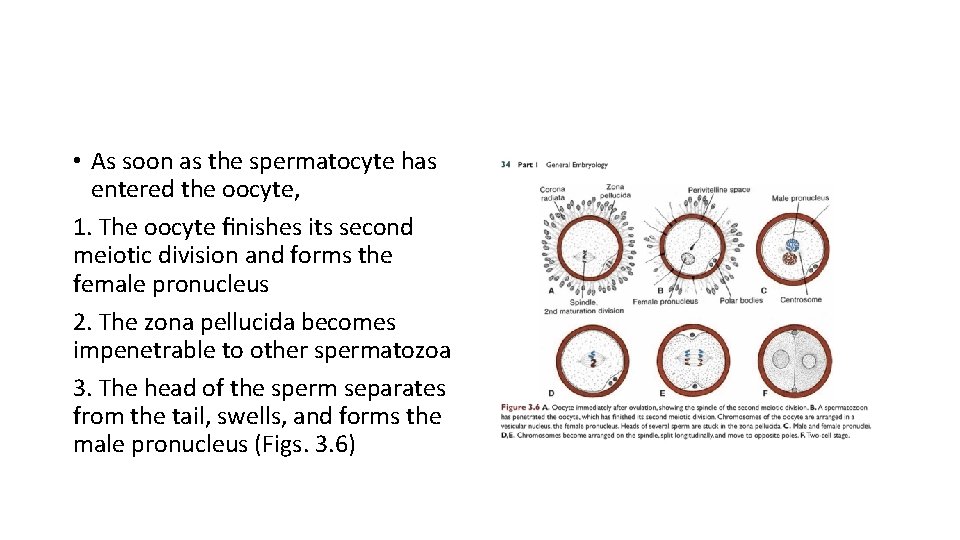
• As soon as the spermatocyte has entered the oocyte, 1. The oocyte finishes its second meiotic division and forms the female pronucleus 2. The zona pellucida becomes impenetrable to other spermatozoa 3. The head of the sperm separates from the tail, swells, and forms the male pronucleus (Figs. 3. 6)
• The main results of fertilization are as follows: ● Restoration of the diploid number of chromosomes, half from the father and half from the mother. Hence, the zygote contains a new combination of chromosomes different from both parents. ● Determination of the sex of the new individual. An X-carrying sperm produces a female (XX) embryo, and a Y-carrying sperm produces a male (XY) embryo. Therefore, the chromosomal sex of the embryo is determined at fertilization. ● Initiation of cleavage.
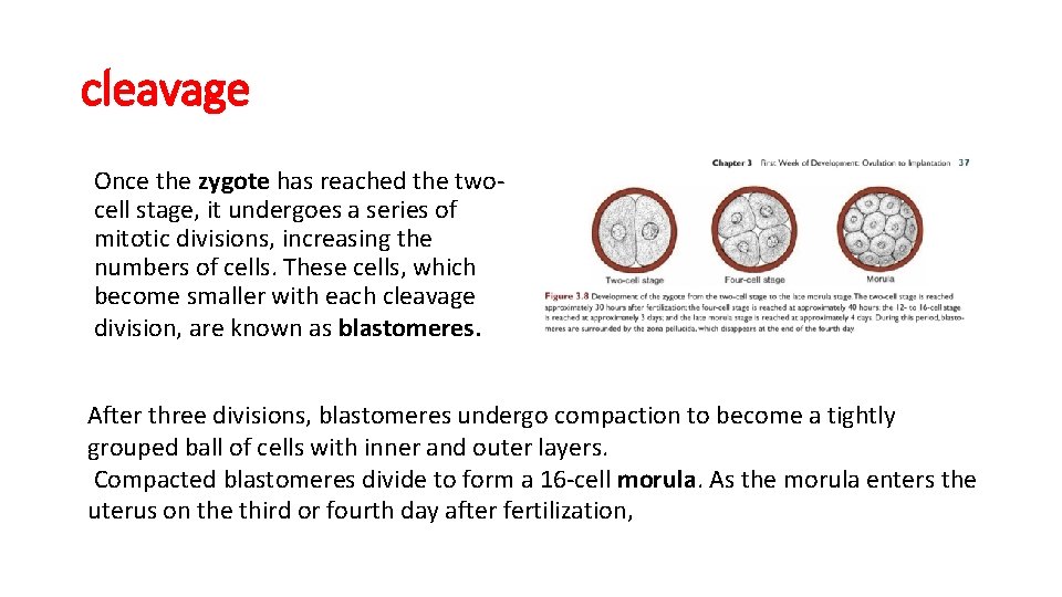
cleavage Once the zygote has reached the twocell stage, it undergoes a series of mitotic divisions, increasing the numbers of cells. These cells, which become smaller with each cleavage division, are known as blastomeres. After three divisions, blastomeres undergo compaction to become a tightly grouped ball of cells with inner and outer layers. Compacted blastomeres divide to form a 16 -cell morula. As the morula enters the uterus on the third or fourth day after fertilization,
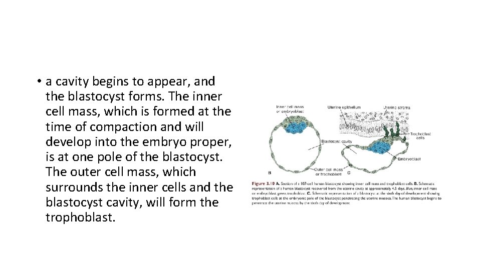
• a cavity begins to appear, and the blastocyst forms. The inner cell mass, which is formed at the time of compaction and will develop into the embryo proper, is at one pole of the blastocyst. The outer cell mass, which surrounds the inner cells and the blastocyst cavity, will form the trophoblast.
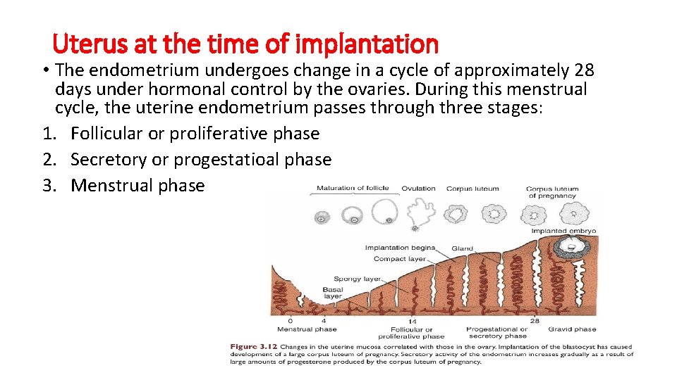
Uterus at the time of implantation • The endometrium undergoes change in a cycle of approximately 28 days under hormonal control by the ovaries. During this menstrual cycle, the uterine endometrium passes through three stages: 1. Follicular or proliferative phase 2. Secretory or progestatioal phase 3. Menstrual phase
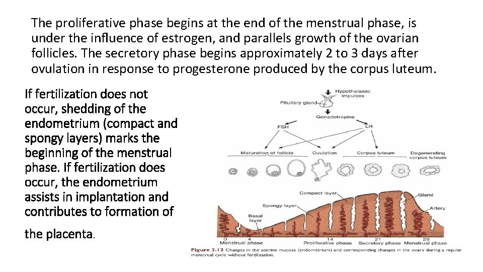
The proliferative phase begins at the end of the menstrual phase, is under the influence of estrogen, and parallels growth of the ovarian follicles. The secretory phase begins approximately 2 to 3 days after ovulation in response to progesterone produced by the corpus luteum. If fertilization does not occur, shedding of the endometrium (compact and spongy layers) marks the beginning of the menstrual phase. If fertilization does occur, the endometrium assists in implantation and contributes to formation of the placenta.
- Slides: 28