General Embryology Presented by Dr Amjad Al Shatarat

General Embryology Presented by Dr. Amjad Al- Shatarat Assistant professor of Anatomy and Embryology

What is embryology ? Is the science that deals with the development of the embryo from single cell to a baby in 9 months Development begins with FERTALIZATION What is fertilization? Fertilization is the process by which the male gamete the sperm and the female gamete the oocyte unite to form the zygote

Why do we need the union of two cells to form the zygote ? According to the number of chromosomes in the nucleus of the human cells we Have two types : 1 - Somatic cells 2 - Reproductive cells (also called sex cells)
 is any cell of the body other than a")
A somatic cell (soma body) is any cell of the body other than a germ cell. A germ cell is a gamete (sperm or oocyte) or any precursor cell destined to become a gamete Somatic cells : contain two sets of chromosomes: first set contains 23 chromosomes coming from the mother called maternal The second set contains 23 chromosomes coming from the father called paternal Therefore, Somatic cells called diploid cells (dipl- double; -oid form), symbolized 2 n. The two chromosomes that make up each pair are called homologous chromosomes (homo- same) they contain similar genes arranged in the same (or almost the same) order

HOMOLOGOUS CHROMOSOMES When examined under a light microscope generally look very similar. The exception to this rule is one pair of chromosomes called the sex chromosomes, designated X and Y. In females the homologous pair of sex chromosomes consists of two large X chromosomes; in males the pair consists of an X and a much smaller Y chromosome Note : If the sex pair is XX the individual is genetically female If the sex pair is XY the individual is genetically male

Where can we find somatic cells? All the cells in the human body are somatic except the sperm and the oocyte How they divide? Somatic cells divide by mitosis for growth and to replace cells that die from tear and wear Mitosis 46 Somatic cell (2 n) 46 + 46 (2 n) Daughter cells (2 n) Mitosis conserves chromosomes number t r n ta im po
 Reproductive cells develop in gonads (ovaries")
2 - Reproductive cells (also called sex cells) Reproductive cells develop in gonads (ovaries in female and testes in male) They contain only 23 chromosomes that is why they called haploid cells (1 n) Reproductive cells divide by meiosis 46 2 n Reproductive cell Diploid 46 chromosomes precursor cell destined to become a gamete 23 1 n + 23 1 n Meiosis does not conserve Chromosomes number instead It reduces it by half Gametes haploid cells 23 chromosomes im p an t r o t

Thus, It is impossible for a female to reproduce here self simply because here sex cells (the oocyts are haploid (23, 1 n) It is impossible for a male to reproduce him self simply because his sex cells (the sperms are haploid (23, 1 n) What to do ? Union + Oocyte 23 chromosomes 1 n, haploid Fertilization Sperm 23 chromosomes 1 n, haploid Zygote 46 chromosomes 2 n, diploid

o f tw o s e Th sist s: on iod c er cle , E S As H P ll i e c R en a iding, E T whot divd the M) N ( ll is n an I C a ce I T hen O T E, wing I M AS divid PH l cel rp o j ma cy
 all its chromosomes to")
Cell Division When a cell reproduces, it must replicate (duplicate) all its chromosomes to pass its genes to the next generation of cells 9/16/2021 ﻛﻠﻴﺔ ﺍﻟﻄﺐ - ﺍﻻﺭﺩﻧﻴﺔ ﺍﻣﺠﺪ ﺍﻟﺸﻄﺮﺍﺕ. ﺩ 10

The Interphase is a state of high metabolic activity; it is during this time that the cell does most of its growing. During interphase 1 - The cell replicates its DNA 2 -Produces additional organelles and cytosolic components Interphase consists of three phases: 1 -G 1 phase 2 -S phase 3 -G 2 phase 9/16/2021 ﻛﻠﻴﺔ ﺍﻟﻄﺐ - ﺍﻻﺭﺩﻧﻴﺔ ﺍﻣﺠﺪ ﺍﻟﺸﻄﺮﺍﺕ. ﺩ 11

a. shatarat@ju. edu. jo facebook amjadshatarat

all cells before division undergo DNA synthesis during the Interphase Chromosome's structure a + proteins called histones b Can c cond ense d to d 9/16/2021 form chro mos ome e ﻛﻠﻴﺔ ﺍﻟﻄﺐ - ﺍﻻﺭﺩﻧﻴﺔ e ﺍﻣﺠﺪ ﺍﻟﺸﻄﺮﺍﺕ. ﺩ 13

s si e th n sy Th Can condensed to form chromosome Interphase DNA replicates (duplicates) for s A. d N n D ta of Ss e 9/16/2021 ﻛﻠﻴﺔ ﺍﻟﻄﺐ - ﺍﻻﺭﺩﻧﻴﺔ ﺍﻣﺠﺪ ﺍﻟﺸﻄﺮﺍﺕ. ﺩ 14

The chromatin of nucleus condense into a chromosome Each chromosome has the following parts: 1 -Telomere 2 -Centromere (where spindle attaches) 3 -Telomere (special structures at the ends) depending on the stage of the cell cycle chromosomes come in 2 forms: 1 - The monad form consists of a single chromatid, a single piece of DNA containing a after mitosis centromere and telomeres at the ends. 2 - The dyad form consists of 2 identical chromatids (sister chromatids) attached together at the centromere before mitosis

The chromatin of nucleus condense into a chromosome One chromosome coming from the mother called maternal One chromosome coming from the father called paternal HOMOLOGOUS CHROMOSOMES 9/16/2021 ﻛﻠﻴﺔ ﺍﻟﻄﺐ - ﺍﻻﺭﺩﻧﻴﺔ ﺍﻣﺠﺪ ﺍﻟﺸﻄﺮﺍﺕ. ﺩ 16

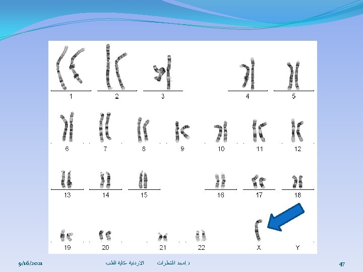
S E M O OS S OU M RO CH Notice that chromosomes number 23 are not homologous, what does this mean? OG HO L MO Notice that in this picture There are two chromosomes Numbered 1 and etc. These Chromosomes are called homologous chromosomes; one comes from the mother and the other comes from the father During fertilization 9/16/2021 Picture of the 46 chromosomes (23 pairs of chromosomes) ﻛﻠﻴﺔ ﺍﻟﻄﺐ - ﺍﻻﺭﺩﻧﻴﺔ ﺍﻣﺠﺪ ﺍﻟﺸﻄﺮﺍﺕ. ﺩ 17

Because the G phases are periods when there is no activity related to DNA duplication, they are thought of as gaps or interruptions in DNA duplication. The G 1 phase is the interval between the mitotic phase and the S phase. During G 1, the cell replicates most of its organelles and cytosolic components but not its DNA. ØReplication of centrosomes also begins in the G 1 phase. 9/16/2021 ﻛﻠﻴﺔ ﺍﻟﻄﺐ - ﺍﻻﺭﺩﻧﻴﺔ ﺍﻣﺠﺪ ﺍﻟﺸﻄﺮﺍﺕ. ﺩ 18

For a cell with a total cell cycle time of 24 hours, G 1 lasts 8 to 10 hours. However, the duration of this phase is quite variable. It is very short in many embryonic cells or cancer cells. Cells that remain in G 1 for a very long time, perhaps destined never to divide again, are said to be in the G 0 phase. Most nerve cells Th e. G 2 are in the G 0 phase. Once a cell enters the S phase, however, it is committed to go through the rest of the cell cycle. phase is the interval between the S phase and the mitotic phase. It lasts 4 to 6 hours. During G 2, cell growth continues, enzymes and other proteins are synthesized in preparation for cell division, and replication of centrosomes is completed. 9/16/2021 ﻛﻠﻴﺔ ﺍﻟﻄﺐ - ﺍﻻﺭﺩﻧﻴﺔ ﺍﻣﺠﺪ ﺍﻟﺸﻄﺮﺍﺕ. ﺩ 19

Cell Division Somatic cells Reproductive cells by by Mitosis Cell Cycle Meiosis To be discussed later Mitotic phase (Cell is dividing) Interphase (Cell is not dividing) Consists of four stages: Consist of three phases: 1 - The G 1 phase 2 - The S phase 3 - The G 2 phase 9/16/2021 ﻛﻠﻴﺔ ﺍﻟﻄﺐ - ﺍﻻﺭﺩﻧﻴﺔ ﺍﻣﺠﺪ ﺍﻟﺸﻄﺮﺍﺕ. ﺩ 1 -Prophase 2 -Metophase 3 -Anaphase 4 -Telophase 20
 One chromosome is")
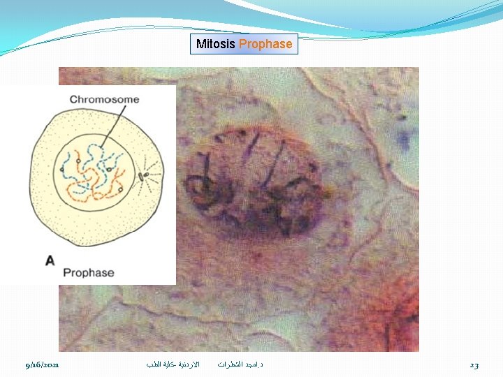
2 n Prophase it has two sets of Chromosomes (two copies) One chromosome is paternal and the other maternal Kinetochore Mitotic spindle microtubules Centromere (a constricted region holds the chromatid pair together) Outside of each centromere is a protein complex called Kinetochore Later in prophase tubulins in the pericentriolar material of the centrosomes start to form the mitotic spindle attaches to the Kinetochore As the mitotic spindle (microtubules) lengthen they push the centrosomes to the poles 9/16/2021 ﻛﻠﻴﺔ ﺍﻟﻄﺐ - ﺍﻻﺭﺩﻧﻴﺔ ﺍﻣﺠﺪ ﺍﻟﺸﻄﺮﺍﺕ. ﺩ 21
 2 n One chromosome is paternal")
it has two sets of Chromosomes (two copies) 2 n One chromosome is paternal and the other maternal Prophase Kinetochore Mitotic spindle microtubules Centromere (a constricted region holds the chromatid pair together) Outside of each centromere is a protein complex called Kinetochore Later in prophase tubulins in the pericentriolar material of the centrosomes start to form the mitotic spindle attaches to the Kinetochore As the mitotic spindle (microtubules) lengthen they push the centrosomes to the poles 9/16/2021 ﻛﻠﻴﺔ ﺍﻟﻄﺐ - ﺍﻻﺭﺩﻧﻴﺔ ﺍﻣﺠﺪ ﺍﻟﺸﻄﺮﺍﺕ. ﺩ 22

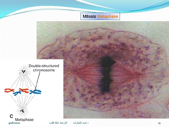
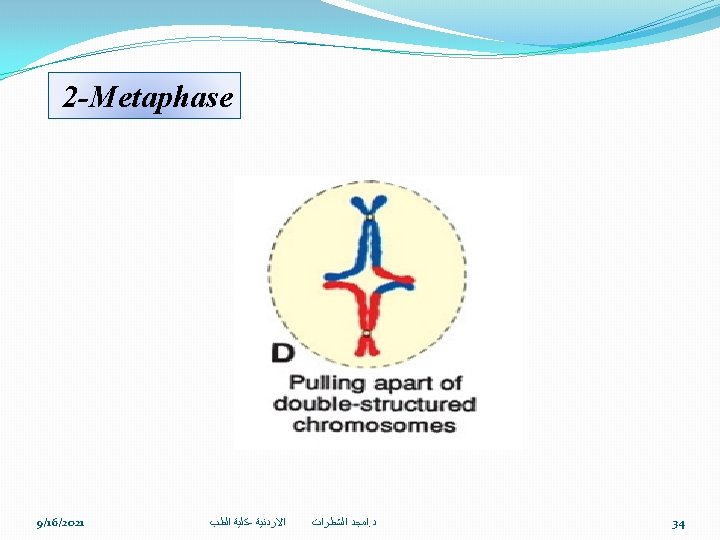
METAPHASE The Kinetochore microtubules align the centromeres at the exact center of the mitotic spindle This midpoint region called metaphase 9/16/2021 ﻛﻠﻴﺔ ﺍﻟﻄﺐ - ﺍﻻﺭﺩﻧﻴﺔ ﺍﻣﺠﺪ ﺍﻟﺸﻄﺮﺍﺕ. ﺩ plate 24

ANAPHASE 2 n 2 n The centromeres split leading to the separation of the two members of the chromatid pair once separated the chromatids are termed chromosomes 9/16/2021 ﻛﻠﻴﺔ ﺍﻟﻄﺐ - ﺍﻻﺭﺩﻧﻴﺔ ﺍﻣﺠﺪ ﺍﻟﺸﻄﺮﺍﺕ. ﺩ 26

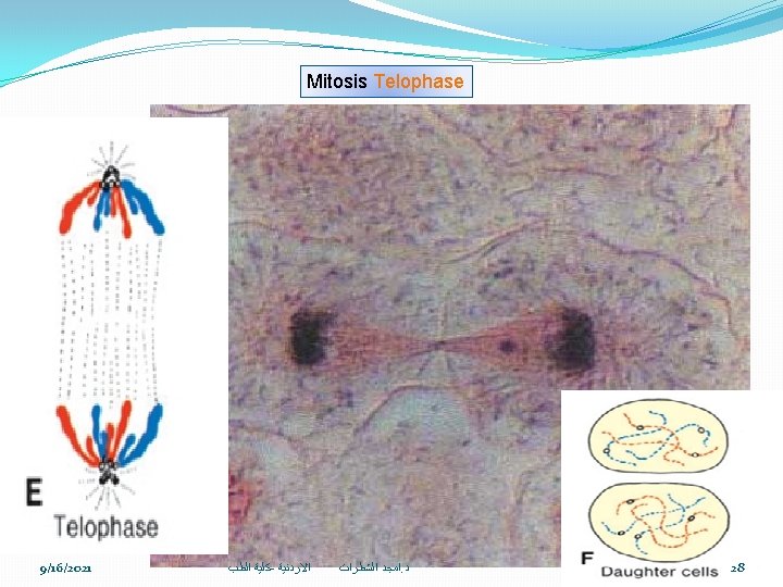
TELOPHASE The identical sets of chromosomes now at apposite poles of the cell A nuclear envelope forms around each chromatin mass The mitotic spindle disappears 9/16/2021 ﻛﻠﻴﺔ ﺍﻟﻄﺐ - ﺍﻻﺭﺩﻧﻴﺔ ﺍﻣﺠﺪ ﺍﻟﺸﻄﺮﺍﺕ. ﺩ 27
")
Meiosis occurs in two successive stages : Meiosis Ι (also known as reductional meiosis) which deals with the number of chromosomes it halves the number of chromosomes Meiosis Ι Ι (also known as equational meiosis) which deals with the conditions of chromosomes 2 n 46 Reductional Meiosis Ι Reproductive cell 9/16/2021 1 n 23 23 1 n Equational Meiosis Ι Ι 23 1 n ﻛﻠﻴﺔ ﺍﻟﻄﺐ - ﺍﻻﺭﺩﻧﻴﺔ ﺍﻣﺠﺪ ﺍﻟﺸﻄﺮﺍﺕ. ﺩ 29

Meiosis I is generally divided into four stages: 1 -Propahse 2 -metaphase 3 -Anophase 4 -Telophase 1 -Prophase is running into stages A- LEPTOTEN stage, (lepto means long) In this stage chromosomes are elongated and extended and become gradually visible 9/16/2021 ﻛﻠﻴﺔ ﺍﻟﻄﺐ - ﺍﻻﺭﺩﻧﻴﺔ ﺍﻣﺠﺪ ﺍﻟﺸﻄﺮﺍﺕ. ﺩ 30
 In this stage identical chromosomes pair up together")
B- ZYGOTEN stage, (zygo means joined) In this stage identical chromosomes pair up together (synapsis) C- PACHYTENE stage, (pachy means short) In this stage chromosomes become shorter and more condensed 9/16/2021 ﻛﻠﻴﺔ ﺍﻟﻄﺐ - ﺍﻻﺭﺩﻧﻴﺔ ﺍﻣﺠﺪ ﺍﻟﺸﻄﺮﺍﺕ. ﺩ 31

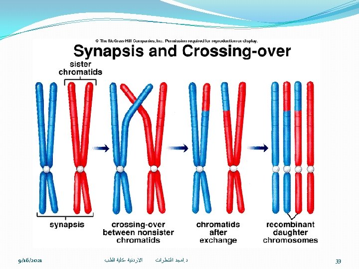
D- DIPLOTENE stage, Chromosomes come together and cross each other by certain segments of their bodies forming what we called CHIASMATA: X- shaped structure Formed by the junction of two chromatids of the for chromatids (tetrad) In Prophase I Crossing over of non-sister chromatids During prophase I, non-sister chromatids can undergo synapsis, in which the chromatids line up side-byside & exchange genetic information between them This allows new combination of genetic material which will become part of a new offspring 9/16/2021 ﻛﻠﻴﺔ ﺍﻟﻄﺐ - ﺍﻻﺭﺩﻧﻴﺔ ﺍﻣﺠﺪ ﺍﻟﺸﻄﺮﺍﺕ. ﺩ 32

3 -Anophase 1 n 1 n 4 -Telophase Notice that daughter cells after meiosis 1 are different from the original cell, the 9/16/2021 ﻛﻠﻴﺔ ﺍﻟﻄﺐ - ﺍﻻﺭﺩﻧﻴﺔ ﺍﻟﺸﻄﺮﺍﺕ ﺍﻣﺠﺪ a. ﺩ new combination chromosomes are of 35

Meiosis II runs into 4 stages: 1 -Prophase 2 -Metophase 3 -Anaphase 4 -Telophase 9/16/2021 ﻛﻠﻴﺔ ﺍﻟﻄﺐ - ﺍﻻﺭﺩﻧﻴﺔ ﺍﻣﺠﺪ ﺍﻟﺸﻄﺮﺍﺕ. ﺩ 36

iri n M eio go sis mo fc I log hro ou Da mo sc ug so hro hte me rc mo s ell s sepom sa a e re ha rates plo id Da ug ht Si ste Ho r c No M p h ito er ro air sis ce m in ati g lls ds ar ed se pa ip ra lo te, id Pa 9/16/2021 ﻛﻠﻴﺔ ﺍﻟﻄﺐ - ﺍﻻﺭﺩﻧﻴﺔ ﺍﻣﺠﺪ ﺍﻟﺸﻄﺮﺍﺕ. ﺩ 37

Chromosomal abnormalities may be numerical or structural Abnormalities in chromosome number may originate during meiotic or mitotic divisions. 9/16/2021 ﻛﻠﻴﺔ ﺍﻟﻄﺐ - ﺍﻻﺭﺩﻧﻴﺔ ﺍﻣﺠﺪ ﺍﻟﺸﻄﺮﺍﺕ. ﺩ 38

l a m r o N In meiosis, two members of a pair of homologous chromosomes normally separate during the first meiotic division so that each daughter cell receives one member of each pair 9/16/2021 ﻛﻠﻴﺔ ﺍﻟﻄﺐ - ﺍﻻﺭﺩﻧﻴﺔ ﺍﻣﺠﺪ ﺍﻟﺸﻄﺮﺍﺕ. ﺩ 39

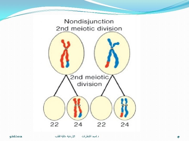
r ot n s cu c o e o d n io rat t c n a o tim e m So h es, ve e w ( ep , r s ) n io u j s di n o n Both members of a pair move into one cell. As a result of nondisjunction of the chromosomes, one cell receives 24 chromosomes, and the other receives 22 instead of the normal 23. 9/16/2021 ﻛﻠﻴﺔ ﺍﻟﻄﺐ - ﺍﻻﺭﺩﻧﻴﺔ ﺍﻣﺠﺪ ﺍﻟﺸﻄﺮﺍﺕ. ﺩ 40

Translocations Sometimes chromosomes break, and pieces of one chromosome attach to another. may be 1 - Balanced, in which case breakage and reunion occur between two chromosomes but no critical genetic material is lost and individuals are normal 2 -Unbalanced, in which case part of one chromosome is lost and an altered phenotype is produced. An example, unbalanced translocations between the long arms of chromosomes 14 and 21 during meiosis I or II produce gametes with an extra copy of chromosome 21, one of the causes of Down syndrome 9/16/2021 ﻛﻠﻴﺔ ﺍﻟﻄﺐ - ﺍﻻﺭﺩﻧﻴﺔ ﺍﻣﺠﺪ ﺍﻟﺸﻄﺮﺍﺕ. ﺩ 42

at fertilization, a gamete having 23 chromosomes fuses with a gamete having 24 or 22 chromosomes, the result is an individual with either 47 chromosomes Trisomy or 45 chromosomes Monosomy 9/16/2021 ﻛﻠﻴﺔ ﺍﻟﻄﺐ - ﺍﻻﺭﺩﻧﻴﺔ ﺍﻣﺠﺪ ﺍﻟﺸﻄﺮﺍﺕ. ﺩ 43

Is us ua co lly c py au of se ch d b ro y a m n os ex om tra Maternal e 2 Age Frequency 1 9/16/2021 e m dro w o D yn s n Trisomy 21 at birth 15 -191/1250 20 -241/1400 25 -291/1100 1/900 31 1/750 31 1/625 32 1/500 33 1/386 34 1/300 35 1/225 36 1/175 37 1/140 38 1/100 39 1/80 40 1/65 41 1/50 42 90%: Meiotic nondisjunction during meiosis II of oogenesis 1/40 43 10%: Meiotic nondisjunction during meiosis I of spermatogenesis 1/25 ﺍﻟﻄﺐ 44 ﻛﻠﻴﺔ - ﺍﻻﺭﺩﻧﻴﺔ 44 ﺍﻣﺠﺪ ﺍﻟﺸﻄﺮﺍﺕ. ﺩ

’ r e t l e f e n i l K e m o dr n y s. S XXY – Phenotypically male with an extra X chromosome 9/16/2021 ﻛﻠﻴﺔ ﺍﻟﻄﺐ - ﺍﻻﺭﺩﻧﻴﺔ ﺍﻣﺠﺪ ﺍﻟﺸﻄﺮﺍﺕ. ﺩ 45

Turner’s Syndrome XO – Phenotypically female missing an X chromosome is the only monosomy compatible with life. Even then, 98% of all fetuses with the syndrome are spontaneously aborted. The few that survive are unmistakably female in appearance and are characterized by the absence of ovaries (gonadal dysgenesis) 9/16/2021 ﻛﻠﻴﺔ ﺍﻟﻄﺐ - ﺍﻻﺭﺩﻧﻴﺔ ﺍﻣﺠﺪ ﺍﻟﺸﻄﺮﺍﺕ. ﺩ 46
- Slides: 48