General arthrology and myology Joints of the shoulder

: no gap, more stable but less mobile")
 articulating surfaces → determine available movements 2) articular")

 ginglymus")
 trochoid joint (pivot joint): axis of rotation is parallel to")
 trochoginglymus (hinge-and-pivot joint): combination of the former two types e.")
 ellipsoidal joint : two perpendicular axes e. g. : wrist")
 saddle joint: carpometacarpal joint of the thumb isthe only one")
 spheroid joint (ball-and-socket, free joint)")
 plane joint: joint limited gliding movement e. g. : joints")


 interclavicular lig. ant. and post. sternoclavicular lig. articular disc costoclavicular")
 joint in which movements are described around three axes: (1) vertical")
 composed of the conoid and trapezoid ligaments Acromial facet of")


 The socket: ial m cro a o ac t r")


 mainly acts upon: glenohumeral joint (minor effect on")

 They only act upon the")
 insert either on the greater or on the lesser tubercle of")


- Slides: 34
General arthrology and myology. Joints of the shoulder girdle and muscles acting upon them Mark Kozsurek, M. D. , Ph. D. assistant professor ED I. , 17 th Sept. ,
Joints between bones 1. Continuous joints (synarthroses): no gap, more stable but less mobile or completly immobile connections. a) Fibrous joint (syndesmosis) b) Cartilaginous joint (synchondrosis) c) Bony union (synostosis) 2. Discontinuous joints (synovial joints, diarthroses): hyalin cartilage-covered articulating surfaces isolated by a synovial gap filled by synovial fluid and enclosed by a capsule.
Obligatory components of synovial joints 1) articulating surfaces → determine available movements 2) articular capsule 3) synovial gap and fluid
Additional components of synovial joints • articular ligaments • articular lips (glenoid and acetabular labrum) • articular discs, menisci • articular muscles (knee joint only)
Classification of joints according to the number of axes 1. Uniaxial joints: a) ginglymus (hinge joint): axis of movement is perpendicular to the axis of articulating bones (e. g. interphalangeal joints ).
1. Uniaxial joints: b) trochoid joint (pivot joint): axis of rotation is parallel to the articulating bones e. g. : atlantooccipitalis joint
2. Biaxial joints: a) trochoginglymus (hinge-and-pivot joint): combination of the former two types e. g. : elbow joint
2. Biaxial joints: b) ellipsoidal joint : two perpendicular axes e. g. : wrist (radiocarpal) joint
2. Biaxial joint: c) saddle joint: carpometacarpal joint of the thumb isthe only one in the human body
3. Multiaxial joint: a) spheroid joint (ball-and-socket, free joint)
3. Multiaxial joint: b) plane joint: joint limited gliding movement e. g. : joints of carpal and tarsal bones
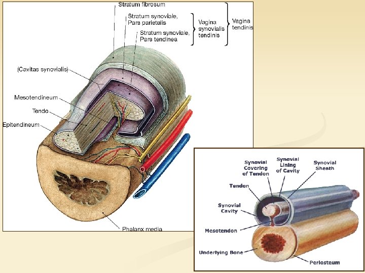
Bursae and tendon sheats Both of them are found at points of friction between moving structures (a tendon and a bone, typically). Both of them consists of an inner synovial layer and an outer fibrous layer. In fact, tendon sheats might be considered as special bursae into which the tendon completly invaginates. Supplying small vessels as well as nerve fibres reach the tendon through the
The shoulder girdle AC GH SC Three joints will attach the free upper extremity to the trunk and they will assure extended movements of it: the sternoclavicular (SC), acromioclavicular (AC) and glenohumeral (GH or simply shoulder)
The sternoclavicular joint (SC) interclavicular lig. ant. and post. sternoclavicular lig. articular disc costoclavicular lig. Sternal facet of clavicle articulates with the clavicular notch of the sternum. Capsule is reinforced by anterior and posterior sternoclavicular ligaments and the capsules of the two sides are also connected by the interclavicular ligament. Typically the synovial gap is devided into two non-communicating compartments by a complete articular disc. Movements of the joint are
Restricted multiaxial (ball-and-socket) joint in which movements are described around three axes: (1) vertical axis, (2) sagittal axis, (3) longitudinal axis
The acromioclavicular joint (AC) composed of the conoid and trapezoid ligaments Acromial facet of clavicle articulates with the acromion of scapula. Outside the capsule the acromioclavicular ligament supports the joint. Inside the capsule a frequently incomplete disc is found. Movements of the joint are restricted by the coracoclavicular ligament
vertical axis sagittal axis longitudinal axi
Stermoclavicular and acromioclavicular joints determine the position of the shoulder together! Movements of the sternoclavicular joint are followed by compensatory movements of the acromioclavicular joint, that will keep the scapula on the dorsal surface of the thoracic cage.
Instead of describing isolated movements of the sternoclavicular and acromioclavicular joints, we rather give how they affect the position of scapula together!
The glenohumeral joint (GH) The socket: ial m cro a o ac t r o c en m liga coracoid process glenoidal labrum (lip) acromion • pear-shaped, small and shallow • glenoid labrum made of fibrocartilage extends the socket a little bit • coracoacromial lig. provides an arch over the head of humerus making upward dislocation almost impossible The „ball”: • large and spherical, the head of humerus
Capsule is thick and strong but also loose and displays three major recesses: • synovial sheath of Biceps tendon: follows the tendon downward in the intertubercular sulcus • axillar recess: is considered as a reserve fold as without this, capsule would restrict abduction of arm • subscapular recess (or bursa): fits bellow the coracoid process and isolates that from the tendon of Subscapularis muscle
n Sagittal axis n abduction-adduction n Transvers axis n anteversion-retroversion (terms flexion and extension are also frequently used but are not pecise) n Longitudinal axis n inward-outward rotation
Basic concept of muscles acting upon the shoulder 1. Dorsal thoracoappendicular muscles Act predominantly on the SC+AC joints and determine the position of the shoulder. 2. Ventral thoracoappendicular muscles Exclusively act upon the glenohumeral joint. 3. Intrinsic shoulder muscles -
On the right side the Trapezius and Latissimus dorsi have been removed Dorsal thoracoappendicular muscles (or superficial back or spinohumeral Trapezius, muscles) Levator scapulae and Rhomboid muscles exclusively act upon sternoclavicular and acrommioclavicular joints, and determine the position of shoulder! Latissimus dorsi rather acts upon the glenohumeral joint. acts upon: glenohumeral joint ( minor effect on SC+AC joints) superficial layer act upon: SC+AC deep As it is indicated by the arrows Levator scapulae and Rhomboid muscles will elevate and retract the shoulder. Trapezius in general retracts the scapula, superior fibres elevate, inferior ones depress the shoulder. Superior fibres of Trapezius also rotate scapula turning the glenoid cavity
Ventral thoracoappendicular muscles (or thoracohumeral muscles) mainly acts upon: glenohumeral joint (minor effect on SC+AC) act exclusively upon: SC+AC Pectoralis minor, Subclavius and Serratus anterior exclusively act upon the sternoclavicular and acromioclavicular joints and mainly depress and protract the scapula. Serratus anterior will also rotate scapula and turns the glenoid cavity upward (as protraction affects the inferior angle of scapula much more). Pectoralis major acts rather upon the glenohumeral joint.
Latissimus dorsi ADDUCTION + RETROVERSION They are considered as the strongest ADDUCTORS as they pull the arm to the trunk. Pectoralis major ADDUCTION + ANTEVERSION
Intrinsic shoulder muscles (true shoulder muscles or scapulohumeral muscles) They only act upon the glenohumeral joint! Supraspinatus, Infraspinatus and Teres minor muscles arise from the dorsal surface of scapula and all of them insert on the greater tubercle of the humerus. ROTATE OUTWARD Subscapularis muscle covers the entire costal surface of the scapula and terminates on the lesser tubercle.
Muscles that (1) insert either on the greater or on the lesser tubercle of the humerus (2) are responsible for holding the head of the humerus in the glenoidal labrum and (3) rotate inward/outward are summarized as the rotator cuff or the SITS muscles (Supraspinatus, Infraspinatus, Teres minor, Subscapularis).
There are two further intrinsic muscles that do not contribute to the rotator cuff: Deltoid: arises from an arch constituded by the spine of scapula, acromion and clavicle and inserts on the deltoid tuberosity. Is considered as the strongest ABDUCTOR, but also contributes to inward and outward ROTATION of humerus with its anterior and posterior portions, respectively. Teres major: emerges from the inferior angle of scapula and inserts with the tendon of Latissimus dorsi along the crest of lesser tubercle.
Topographical anatomy of the shoulder region
Axillary fossa Metastasis of breast cancer - axillary lymph nodes n n anterior wall: Pectoralis major and minor medial wall: Serratus anterior attached to the external surface of ribs posterior wall: Subscapularis muscle filling the subscapular fossa, Teres major and Latissimus dorsi muscles lateral wall: intertubercular groove, surgical neck of humerus, flexors of
Triangular and quadrangular spaces Triangular space (Teres minor and major, long head of Triceps brachii): circumflex scapular artery Quadrangular space (Teres minor and major, long head of Triceps brachii, surgical neck of humerus): post. circumflex humeral artery and axillary nerve
Thank you for your attention!