General Approach in Investigation of Haemostasis Lecture 10


")


 Near Patient Testing Devices The INRatio single use test strip")

- Slides: 24
General Approach in Investigation of Haemostasis Lecture 10: Coagulation Instruments
Evaluation of Coagulation tests: q Manual Method: q Coagulometers: q Semiautomated Method. q Automated Method.
q Manual methods were used at the begining, then semi automatic equipments appeared based on photometric or mechanical principles to detect fibrin. More recently, fully automated instruments have become common in modern laboratories. q Today, new equipment connected to specific data processing systems can undertake clotting, Chromogenic, and immunological tests. q Automation has contributed to improvements in standardization and facilitating tests – improve lab efficiency and repertoire
Manual Method q All reagents and samples are added manually by the operator. q Temperature is maintained by a waterbath or heat block; q May require external measurement by operator, most often using a stopwatch.
Semi Automated Method. q All reagents and samples are added manually by the operator. q Usually contains a device for maintaining' constant 37°C temperature, Analyzer may or may not internally monitor temperature. q Has mechanism to automatically initiate timing device upon addition of final reagent and internal mechanism for detecting Clot formation.
Automated Method. q All reagents are automatically pipetted by the instrument. Samples may or may not be automatically pipetted. q Contains monitoring devices and internal mechanism to maintain and monitor constant 37οC temperature throughout testing sequence. q Timers are initiated and clot formation detected automatically
q Manual Method Sem iautomated Method Automated Method.
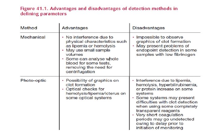
Methods of Endpoint Detection q Mechanical q Optical § Photo optical § Nephelometric § Chromogenic § Immunologic q Electrochemical
Mechanical Two primary methodologies are utilized for mechanical detection of clot formation. Ø Electromechanical (Impedance) Method. Ø Magnetic, Steel Ball Method.
Electromechanical Method. q When coagulation process takes place, the concentration of clotting factors (charges) and inorganic ions will change along the time and the measured impedance or conductance will be also changed correspondingly at the same time. q During the reaction, one probe moves in and out of the solution at constant intervals. The electrical circuit between the two probes is not maintained as the moving probe rises in and out of the solution. q When a clot (fibrin) is formed in the solution, the fibrin strands maintain electrical contact between the two probes when the moving probe leaves the solution, which stops the timer.
Magnetic, Steel Ball Method q Mechanical clot detection involves monitoring the movement of a steel ball within the test solution using a magnetic sensor. As clot formation occurs, the movement of the ball changes, which is detected by the sensor.
There are two variations of this principle used in current instrumentation. *A change in the movement of the steel ball may be detected when there is increased viscosity of the test solution, changing its range of motion, 2. *Or by a break in contact with the magnetic sensors when the steel ball becomes incorporated into a fibrin clot as the cuvette rotates. 1.
Photo-optical Method q Detection of clot formation measured by a change in OD of a test sample is the basis of photo optical instrumentation, which is also known as turbidometric methodology: q When a light source of a specified wavelength is passed through a test solution (plasma), a certain amount of light is detected by a photodetector or photocell located on the other side of the solution. q The amount of light detected is dependent on the color and clarity of the plasma sample and is considered to be the baseline light transmission value.
Photo-optical Method When soluble fibrino genbegins to polymerize into a fibrin clot, formation of fibrin strands causes light to scatter, allowing less light to fall on the photodetector (i. e. , the plasma becomes more opaque, decreasing the amount of light detected. q When the amount of light reaching the photodetector decreases to an exact point from the baseline value as predetermined by the instrument, this change in OD triggers the timer to stop, indicating clot formation. q
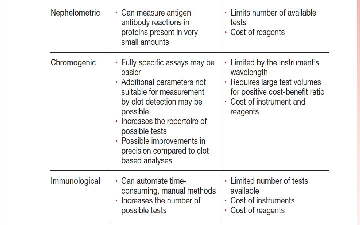
Nephelometric Quantifying plasma proteins based on the specific reaction of the protein being measured with highly specific antisera. q Precipitants are antigen antibody complexes, which show up in solutions as turbidity and scatter incident light. q The nephelometer uses a light emitting diode at a high wavelength (usually >600 nm) to detect variations in light scatter as antigen antibody complexes are formed. When the light rays encounter insoluble complexes such as fibrin strands, they are scattered in both forward (l 80 degree) and side (90 degree) angles. q
Instruments employing this type of measuring system detect the amount of agglutination of particles by reading the increasing amount of light scattered at a 70 degree angle as agglutinates are formed. The timer is triggered to stop when the amount of light scatter reaches a specific predetermined level. q This method of endpoint detection is in contrast to the photo -optical systems, which sense decreased light transmission at 180 degrees due to the opaqueness of the sample in a cuvette when fibrin is formed. Nephelometric q
Chromogenic, or amidolytic, methodology is based on the use of a specific color producing substance known as a chromophore. The chromophore normally used in the coagulation laboratory is para nitroaniline (p nitroaniline or p. NA), which has an optical absorbance peak at 405 nm on a spectrophotometer.
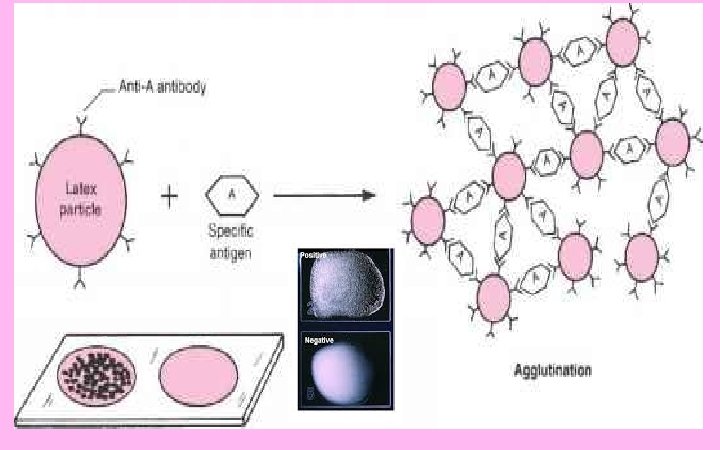
Immunologic Method Immunologic assays are based on antigen antibody reactions. q Latex Microparticles are coated with a specific antibody directed against the analyte (antigen) to be measured. q A beam of monochromatic light is then passed through the suspension of microlatex particles. When the wavelength of light is greater than the diameter of the particles in suspension, only a small amount of light will be absorbed by the particles. q
Immunologic Method When the latex microparticles coated with specific antibody come in contact with the antigen present in the solution, the antigen attaches to the antibody and forms bridges between the particles, causing them to agglutinate. q As the diameter of the agglutinates becomes larger and closer to the wavelength of the monochromatic light beam, the greater the amount of light that is absorbed. q The increase in light absorbance is proportional to the size of the agglutinates, which, in turn, is proportional to the antigen level present in the sample, which is read from a standard curve. q
Electrochemical INRatio Meter (Hemosense) Near Patient Testing Devices The INRatio single use test strip is made of laminated layers of transparent plastic. Each test strip has a sample well where blood is applied, three channels through which the blood sample flows to reach the testing areas, reagents to start the coagulation process, and electrodes that interface with the INRatio meter. The device detects a change in electrical resistance when blood clots.
http: //site. iugaza. edu. ps/wael/