Gene Expression CHAPTER 19 Gene expression Gene regulation


 of transcription Easiest place to control")
 proposed the operon model to")


- Slides: 49
Gene Expression CHAPTER 19
Gene expression § Gene regulation is the turning on and off of genes. § Gene expression is the overall process of information flow from genes to proteins. § The control of gene expression allows cells to produce specific kinds of proteins when and where they are needed
Gene Expression �Essential in both prokaryotes and eukaryotes Flow of information from DNA to protein Conversion of genotype to phenotype �Prokaryotes use it to take advantage of changing environments �In Eukaryotes, it is critical for directing development and maintaining homeostasis
Gene expression and regulation �Coiling of DNA into chromatin around histones to create nucleosome
Gene Expression �Usually occurs at the beginning (initiation) of transcription Easiest place to control the gene expression process �Regulatory proteins bind to either block transcription or stimulate transcription RNA polymerase need these proteins to bind to the promoter region on the DNA template (antisense) strand
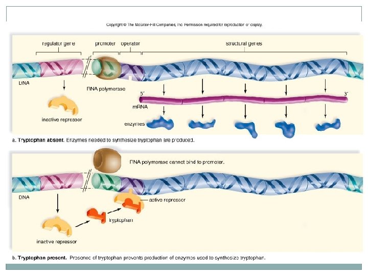
Prokaryotic Gene Expression �François Jacob and Jacques Monod (1961) proposed the operon model to explain regulation of gene expression in prokaryotes An operon is a group of structural and regulatory genes that function as a single unit Found ONLY in prokaryotes
Operons � An operon consists of three components Promoter � DNA sequence where RNA � Short segment of DNA polymerase first attaches Operator � DNA sequence where active repressor binds � Short segment of DNA Structural Genes � One to several genes coding for enzymes of a metabolic pathway � Transcribed simultaneously as a block � Long segment of DNA �A regulatory gene that codes for a repressor protein � The regulatory gene is normally located outside the operon repressor protein controls whether the operon is active or not
Prokaryotic Gene Expression �Have similar features as the eukaryotes have but also may differences �Can be either positive or negative gene expression Positive-increases the frequency of initiation Negative- decreases the frequency of initiation �Controlled by regulatory proteins Negative- proteins are called repressors and bind to a place called the operators Positive- proteins called activators
trp operon � The trp Operon The regulator codes for a repressor If tryptophan (an amino acid) is absent: � Repressor � RNA polymerase binds to the promoter � Enzymes is unable to attach to the operator (expression is normally “on”) for synthesis of tryptophan are produced If tryptophan is present: � It combines with the repressor protein as its corepressor � Repressor becomes functional when bound to tryptophan � Repressor blocks synthesis of enzymes in the pathway for tryptophan synthesis
lac operon �The lac Operon The regulator codes for a repressor If lactose (a sugar that can be used for food) is absent: � The repressor attaches to the operator � Expression is normally “off” If lactose is present: � It combines with the repressor and renders it unable to bind to operator � RNA � The polymerase binds to the promoter three enzymes necessary for lactose catabolism are produced
When lactose is present
When lactose is absence
Figure 11. 1 C trp operon lac operon Promoter Operator Gene DNA Active repressor Inactive repressor Lactose Inactive repressor Tryptophan
Gene Expression �Epigenetic inheritance �X chromosome inactivation � In female mammals, one of the two X chromosomes is highly compacted and transcriptionally inactive. � Either the maternal or paternal chromosome is randomly inactivated. � Inactivation occurs early in embryonic development, and all cellular descendants have the same inactivated chromosome. � An inactivated X chromosome is called a Barr body.
Figure 11. 2 B Early Embryo Adult Two cell populations X chromosomes Allele for orange fur Cell division and random X chromosome Active X inactivation Inactive X Allele for black fur Inactive X Active X Orange fur Black fur
Eukaryotic Gene Regulation �Regulate gene expression at every step of the central dogma � 5 primary levels of control: Nuclear levels � Chromatin Structure � Transcriptional Control � Posttranscriptional Control Cytoplasmic levels � Translational Control � Posttranslational Control
1. Chromatin Structure 2. Transcription 3. Posttranscription 4. Translation 5. Posttranslation
1. Chromatin structure regulation �Eukaryotic DNA is associated with histone proteins � Together make up chromatin �The levels of chromatin packing are determined by degree of nucleosome coiling �Adding acetyl groups to the ends of histones helps uncoil DNA and allow binding Euchromatin � Loosely coiled DNA � Transcriptionally active Heterochromatin � Tightly packed DNA � Transcriptionally inactive
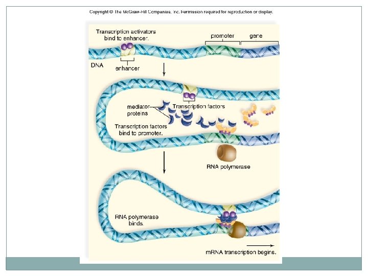
2. Transcription regulation �Transcriptional control is the most critical level of genetic control. �Transcription is controlled by DNA-binding proteins called transcription factors. A group of transcription factors binds to a promoter adjacent to a gene; the complex attracts and binds RNA polymerase, but transcription may still not begin. Transcription activators are often involved in controlling transcription in eukaryotes. � Bind to enhancer region
3. Posttranscriptional regulation �Posttranscriptional control includes m. RNA processing and the speed at which m. RNA leaves the nucleus. �Differential excision of introns and splicing of m. RNA can vary the type of m. RNA that leaves nucleus. Alternative splicing The hypothalamus and thyroid glands produce calcitonin but the m. RNA that leaves the nucleus is not the same in both types of cells.
4. Translational regulation The longer an active m. RNA molecule remains in the cytoplasm, the more product is produced. Ribonucleases are enzymes associated with ribosomes that degrade m. RNA. � Mature m. RNA has non-coding segments at 3' cap and 5' poly-A tail ends; differences in these segments influence how long the m. RNA avoids being degraded. Micro. RNAs are small, processed pieces of intron; after micro. RNAs are degraded, they combine with protein and the complex binds to m. RNAs, destroying them. � RNA interference (RNAi) � http: //www. youtube. com/watch? v=H 5 ud. Fj. WDM 3 E
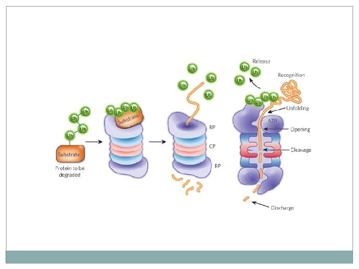
5. Posttranslational regulation �Affects the activity of a protein product �Posttranslational control is accomplished by regulating Activation � INSULIN Degradation rate
Used as an Evolutionary Link �Absolutely essential for organisms �There are features among cell communication processes that are similar between organisms Cells must communicate to coordinate their activities. �Biologists have discovered some universal mechanisms of cellular regulation, involving the same small set of cell-signaling mechanisms.
Used as an Evolutionary Link �signal-transduction pathway = the process by which a signal on a cell’s surface is converted into a specific cellular response consists of a series of steps Single-celled organisms-send pheromones to simulate reproduction (yeast) Multi-cellular organisms-Epinephrine stimulation of glycogen breakdown in mammals during Flight or Fight response
Used as an Evolutionary Link �The molecular details in both yeast and animal cells are strikingly similar, even though their last common ancestor was over a billion years ago. �Signaling molecules evolved first in ancient prokaryotes and were then adopted for new uses by single-celled eukaryotes and multicellular descendents.
Cell-Environment Interactions �Cells interact directly or indirectly by responding to extracellular chemicals �Always involves glycocalyx Cell adhesion molecules (CAMs) Plasma membrane receptors Voltage-gated channel proteins © 2013 Pearson Education, Inc.
Cells communicate with each other through direct contact with other cells or from a distance �Cell-to-cell communication Gap junctions in animals cells Plasmodesmata in plant cells �Short-Distance Neurotransmitters �Long-Distance signaling Occurs via chemical messengers (hormones) Endocrine signals are produced by endocrine cells that release signaling molecules (Insulin, TSH, Testosterone)
Roles of Cell Adhesion Molecules �Thousands on approximately every cell in body �Anchor to extracellular matrix or each other �Assist in movement of cells past one another �Attract WBCs to injured or infected areas �Stimulate synthesis or degradation of adhesive membrane junctions �Transmit intracellular signals to direct cell migration, proliferation, and specialization © 2013 Pearson Education, Inc.
Chemical Signaling �The origins of our understanding of cell signaling were pioneered by E. W. Sutherland his colleagues. �Reception Transduction Response © 2013 Pearson Education, Inc.
Overview of Signaling � Ligand binding receptor structural change protein alteration
Signal Transduction Pathway �Reception: Signaling begins with the recognition of a chemical messenger, a ligand, by a receptor protein. Different receptors recognize different chemical messengers, which can be peptides, small chemicals or proteins, in a specific one-to-one relationship.
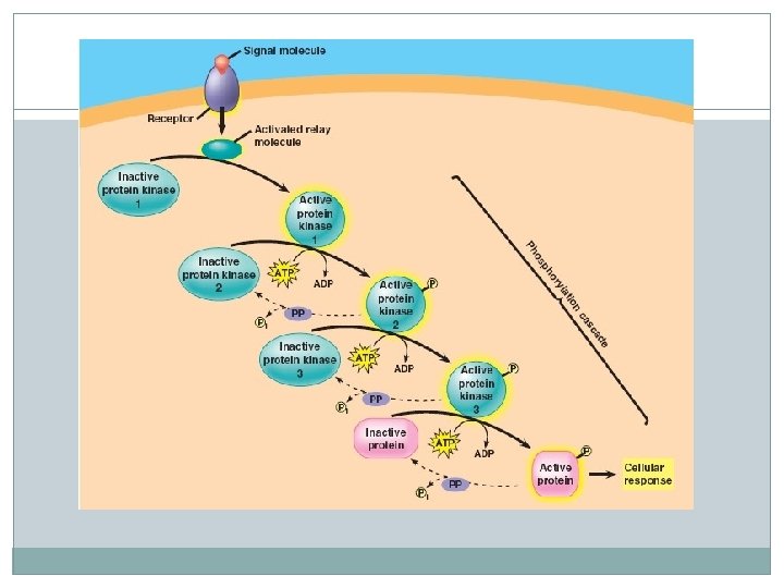
Signal Transduction Pathway �A receptor protein recognizes signal molecules, causing the receptor protein’s shape to change, which initiates transduction of the signal. � 3 main types of receptors: G-protein-linked Tyrosine kinases Ion channel
Signal Transduction Pathway �Transduction: a change in the receptor triggers changes along a signal-transduction pathway. �Signaling cascades relay signals from receptors to cell targets often amplifying the incoming signals �Leads to: A conformational change in a protein Phosphorylation cascades in which a series of protein kinases add a phosphate group to the next protein in the cascade sequence
Signal Transduction Pathway �Second messengers are often essential to the function of the cascade. Calcium is an important second messenger because cells are able to regulate it (most widely used) � Muscle contractions � Immune response with T cells and B cells � Insulin Cyclic AMP (c. AMP) is made from ATP � Glucagon � Adrenaline
Signal Transduction Pathway �Response: the transduced signal triggers a specific cellular activity. Can turn on or off genes (transcription) Regulate cellular activity
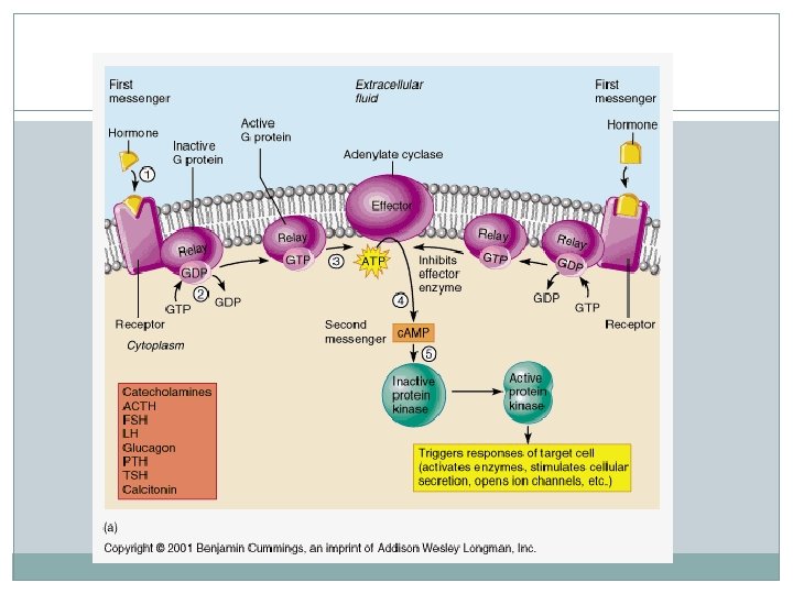
Figure 3. 16 G proteins act as middlemen or relays between extracellular first messengers and intracellular second messengers that cause responses within the cell. Slide 1 The sequence described here is like a molecular relay race. Instead of a baton passed from runner to runner, the message (a shape change) is passed from molecule to molecule as it makes its way across the cell membrane from outside to inside the cell. 1 Ligand* (1 st messenger) binds to the receptor. The receptor changes shape and activates. Ligand (1 st Receptor G protein Enzyme messenger) 2 The activated receptor binds to a G protein and activates it. The G protein changes shape (turns “on”), causing it to release GDP and bind GTP (an energy source). 2 nd messenger 3 Activated G protein activates (or inactivates) an effector protein by causing its shape to change. Extracellular fluid Effector protein (e. g. , an enzyme) Ligand Receptor G protein GDP Inactive 2 nd messenger Activated kinase enzymes * Ligands include hormones and neurotransmitters. © 2013 Pearson Education, Inc. 4 Activated effector enzymes catalyze reactions that produce 2 nd messengers in the cell. (Common 2 nd messengers include cyclic AMP and Ca 2+. ) 5 Second messengers activate other enzymes or ion channels. Cyclic AMP typically activates protein kinase enzymes. 6 Kinase enzymes activate other enzymes. Kinase enzymes transfer phosphate groups from ATP to specific proteins and activate a Cascade of cellular responses series of other enzymes that trigger (The amplification effect is various metabolic and structural tremendous. Each enzyme changes in the cell. catalyzes hundreds of reactions. ) Intracellular fluid
Figure 3. 16 G proteins act as middlemen or relays between extracellular first messengers and intracellular second messengers that cause responses within the cell. Slide 2 Ligand (1 st Receptor G protein Enzyme messenger) 2 nd messenger 1 Ligand* (1 st messenger) binds to the receptor. The receptor changes shape and activates. Extracellular fluid Ligand Receptor * Ligands include hormones and neurotransmitters. © 2013 Pearson Education, Inc. Intracellular fluid
Figure 3. 16 G proteins act as middlemen or relays between extracellular first messengers and intracellular second messengers that cause responses within the cell. Slide 3 Ligand (1 st Receptor G protein Enzyme messenger) 1 Ligand* (1 st messenger) binds to the receptor. The receptor changes shape and activates. Ligand 2 The activated receptor binds to a G protein and activates it. The G protein changes shape (turns “on”), causing it to release GDP and bind GTP (an energy source). 2 nd messenger Extracellular fluid Receptor G protein GDP * Ligands include hormones and neurotransmitters. © 2013 Pearson Education, Inc. Intracellular fluid
Figure 3. 16 G proteins act as middlemen or relays between extracellular first messengers and intracellular second messengers that cause responses within the cell. Slide 5 Ligand (1 st Receptor G protein Enzyme messenger) 1 Ligand* (1 st messenger) binds to the receptor. The receptor changes shape and activates. 2 The activated receptor binds to a G protein and activates it. The G protein changes shape (turns “on”), causing it to release GDP and bind GTP (an energy source). 2 nd messenger 3 Activated G protein activates (or inactivates) an effector protein by causing its shape to change. Extracellular fluid Effector protein (e. g. , an enzyme) Ligand Receptor G protein GDP * Ligands include hormones and neurotransmitters. © 2013 Pearson Education, Inc. Inactive 2 nd messenger Active 2 nd messenger 4 Activated effector enzymes catalyze reactions that produce 2 nd messengers in the cell. (Common 2 nd messengers include cyclic AMP and Ca 2+. ) Intracellular fluid
Figure 3. 16 G proteins act as middlemen or relays between extracellular first messengers and intracellular second messengers that cause responses within the cell. Slide 6 Ligand (1 st Receptor G protein Enzyme messenger) 1 Ligand* (1 st messenger) binds to the receptor. The receptor changes shape and activates. 2 The activated receptor binds to a G protein and activates it. The G protein changes shape (turns “on”), causing it to release GDP and bind GTP (an energy source). 2 nd messenger 3 Activated G protein activates (or inactivates) an effector protein by causing its shape to change. Extracellular fluid Effector protein (e. g. , an enzyme) Ligand Receptor G protein GDP Inactive 2 nd messenger Activated kinase enzymes * Ligands include hormones and neurotransmitters. © 2013 Pearson Education, Inc. 4 Activated effector enzymes catalyze reactions that produce 2 nd messengers in the cell. (Common 2 nd messengers include cyclic AMP and Ca 2+. ) 5 Second messengers activate other enzymes or ion channels. Cyclic AMP typically activates protein kinase enzymes. Intracellular fluid
Figure 3. 16 G proteins act as middlemen or relays between extracellular first messengers and intracellular second messengers that cause responses within the cell. Slide 7 Ligand (1 st Receptor G protein Enzyme messenger) 1 Ligand* (1 st messenger) binds to the receptor. The receptor changes shape and activates. 2 The activated receptor binds to a G protein and activates it. The G protein changes shape (turns “on”), causing it to release GDP and bind GTP (an energy source). 2 nd messenger 3 Activated G protein activates (or inactivates) an effector protein by causing its shape to change. Extracellular fluid Effector protein (e. g. , an enzyme) Ligand Receptor G protein GDP Inactive 2 nd messenger Activated kinase enzymes * Ligands include hormones and neurotransmitters. © 2013 Pearson Education, Inc. 4 Activated effector enzymes catalyze reactions that produce 2 nd messengers in the cell. (Common 2 nd messengers include cyclic AMP and Ca 2+. ) 5 Second messengers activate other enzymes or ion channels. Cyclic AMP typically activates protein kinase enzymes. 6 Kinase enzymes activate other enzymes. Kinase enzymes transfer phosphate groups from ATP to specific proteins and activate a Cascade of cellular responses series of other enzymes that trigger (The amplification effect is various metabolic and structural tremendous. Each enzyme changes in the cell. catalyzes hundreds of reactions. ) Intracellular fluid
Problems �Changes in signal transduction pathways can alter cellular response. Conditions where signal transduction is blocked or defective can be deleterious, preventative or protective. Diabetes Drugs (Birth Control) Effects of poisons and Antihistamines
Mutations § Mutations in two types of genes can cause cancer. 1. 2. Oncogenes Proto-oncogenes are normal genes that promote cell division. Mutations to proto-oncogenes create cancer-causing oncogenes that often stimulate cell division. Tumor-suppressor genes normally inhibit cell division or function in the repair of DNA damage. Mutations inactivate the genes and allow uncontrolled division to occur.
Mutations and Cancer p 53 � The normal product of the p 53 gene is a transcription factor that normally activates genes for factors that inhibit cell division. � In the absence of functional p 53, cell division continues because the inhibitory protein is not produced. ras � In a healthy cell, the product of the ras proto-oncogene relays a signal when growth factor binds to a receptor. � But in a cancerous condition, the product of the ras protooncogene relays the signal in the absence of a growth factor, leading to uncontrolled growth.