
Gel electrophoresis Dr Divya Menon K Assistant Professor
- Slides: 13
Gel electrophoresis Dr Divya Menon K Assistant Professor Department of Botany St. Mary’s College Thrissur
Gel electrophoresis v Gel electrophoresis is a technique used to separate DNA fragments according to their size. v. DNA samples are loaded into wells (indentations) at one end of a gel, and an electric current is applied to pull them through the gel. v. DNA fragments are negatively charged, so they move towards the positive electrode. Because all DNA fragments have the same amount of charge per mass, small fragments move through the gel faster than large ones. v. When a gel is stained with a DNA-binding dye, the DNA fragments can be seen as bands, each representing a group of same-sized DNA fragment Gel electrophoresis, Divya Menon k, St. Mary’s College
What is a gel? v. As the name suggests, gel electrophoresis involves a gel: a slab of Jello-like material. Gels for DNA separation are often made out of a polysaccharide called agarose, which comes as dry, powdered flakes. v. At one end, the gel has pocket-like indentations called wells, which are where the DNA samples will be placed: v. One end of the box is hooked to a positive electrode, while the other end is hooked to a negative electrode. The main body of the box, where the gel is placed, is filled with a salt-containing buffer solution that can conduct current. v. The end of the gel with the wells is positioned towards the negative electrode. The end without wells (towards which the DNA fragments will migrate) is positioned towards the positive electrode. Gel electrophoresis, Divya Menon k, St. Mary’s College
How do DNA fragments move through the gel? v. One well is reserved for a DNA ladder, a standard reference that contains DNA fragments of known lengths. v. Next, the power to the gel box is turned on, and current begins to flow through the gel. The DNA molecules have a negative charge because of the phosphate groups in their sugar-phosphate backbone, so they start moving through the matrix of the gel towards the positive pole. Gel electrophoresis, Divya Menon k, St. Mary’s College
v. When the power is turned on and current is passing through the gel, the gel is said to be running. v. A typical voltage for running an agarose DNA gel would be in the range of 8080 - 120 V 120 text{ V}120 V 120, space, V. A higher voltage will make the gel run faster, but may also melt it if it runs for a long period of time. Gel electrophoresis, Divya Menon k, St. Mary’s College
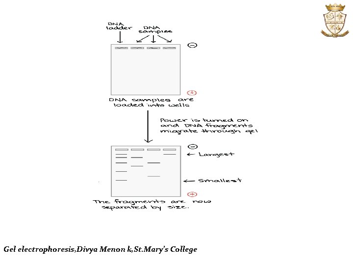
DNA samples are loaded into wells at negative electrode end of gel. Power is turned on and DNA fragments migrate through gel (towards the positive electrode). After the gel has run, the fragments are separated by size. The largest fragments are near the top of the gel (negative electrode, where they began), and the smallest fragments are near the bottom (positive electrode). As the gel runs, shorter pieces of DNA will travel through the pores of the gel matrix faster than longer ones. After the gel has run for awhile, the shortest pieces of DNA will be close to the positive end of the gel, while the longest pieces of DNA will remain near the wells. Very short pieces of DNA may have run right off the end of the gel if we left it on for too long. Gel electrophoresis, Divya Menon k, St. Mary’s College
Gel electrophoresis, Divya Menon k, St. Mary’s College
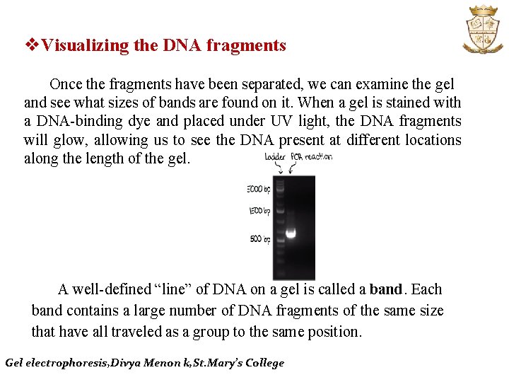
v. Visualizing the DNA fragments Once the fragments have been separated, we can examine the gel and see what sizes of bands are found on it. When a gel is stained with a DNA-binding dye and placed under UV light, the DNA fragments will glow, allowing us to see the DNA present at different locations along the length of the gel. A well-defined “line” of DNA on a gel is called a band. Each band contains a large number of DNA fragments of the same size that have all traveled as a group to the same position. Gel electrophoresis, Divya Menon k, St. Mary’s College
DNA as well as RNA are normally visualized by staining with Ethidium bromide which intercalates into the major grooves of the DNA and fluoresces under UV light. The ethidium bromide may be added to the agarose solution before it gels, or the DNA gel may be stained later after electrophoresis. SYBR Green requires the use of a blue-light transilluminator. DNA stained with crystal violet can be viewed under natural light without the use of a UV transilluminator which is an advantage, however it may not produce a strong band. Gel electrophoresis, Divya Menon k, St. Mary’s College
When stained with ethidium bromide, the gel is viewed with an UV transilluminator. The UV light excites the electrons within the aromatic ring of ethidium bromide, and once they return to the ground state, light is released, making the DNA and ethidium bromide complex fluoresce. ngths lluminators ndard use B), however exposure of DNA to UV radiation for as little as 45 seconds can produce damage to DNA and affect subsequent procedures. Gel electrophoresis, Divya Menon k, St. Mary’s College
Fragment patterns and molecular genotypes One application of gel electrophoresis in DNA analysis is that it can reveal an individual’s genotype at a specific genetic locus. Molecular genotypes can be inferred from the fragment banding pattern observed if one keeps in mind what is happening at the molecular level. Gel electrophoresis, Divya Menon k, St. Mary’s College
Homozygous individuals will have the same DNA sequence at this locus in both chromosomes. Heterozygous individuals will have two different versions of this DNA. If the DNA sequence differences that occur result in a longer DNA segment from one chromosome and a shorter segment from the other then we can visualize this difference in the electrophoresis banding pattern observed in the gel. Gel electrophoresis, Divya Menon k, St. Mary’s College
REFERENCE v. Brown TA. Gene Cloning and DNA Analysis. Blackwell Science Pub: Gel electrophoresis, Divya Menon k, St. Mary’s College