Gastrointestinal Tumors EsophagusGastric Tumors Symptomatology and Diagnosis Koray


 • Weight loss without")


 2. Barium esophagraphy 3.")


 Unlike CT, EUS allows visualization of the distinct layers")
 Resolution of EUS permits the distinction of T 1,")


 Ø weight")

 is a lymph node in the left")


 scan,")

 Physical Examination Anemia (Occult bleeding) Abdominal Pain Abdominal distention (obstruction) Unexplained, persistent")


")


- Slides: 51
Gastrointestinal Tumors Esophagus-Gastric Tumors Symptomatology and Diagnosis Koray Topgül, MD, Prof Department of General Surgey
Esophageal Cancer
Esophageal Cancer Frequency: In the US: • Cancer of the esophagus accounts for 7% of gastrointestinal tract cancers in the United States. • In 2000, approximately 12, 300 new cases of esophageal carcinoma occurred in the United States, and 12, 100 deaths resulted from the disease. • By the early 1990 s, adenocarcinoma had become the most common cancerous cell type among white Americans, accounting for approximately one half of esophageal malignancies in the United States and Europe. • Squamous cell cancers still predominate among African American patients. Internationally: • Esophageal cancer is the seventh leading cause of cancer death worldwide. • Unlike in the United States, squamous cell carcinoma is responsible for 95% of all esophageal cancer worldwide. Esophageal Squamous Cell Carcinoma Esophageal Adenocarcinoma
Diagnosis-Esophageal Cancer of the esophagus remains a devastating disease because it is usually not detected until it has progressed to an advanced incurable stage. Only surgical resection at a very early stage has been shown to improve survival rates in patients with this disease.
Signs and Symptoms of Esophageal Cancer • Difficulty swallowing *(dysphagia) • Weight loss without trying • Chest pain, pressure or burning • Worsening indigestion or heartburn • Coughing or hoarseness (due reflux)
Signs and Symptoms • Weight loss is the second most common symptom and occurs in more than 50% of people with esophageal carcinoma. • Pain can be felt in the epigastric or retrosternal area. It can also be felt over bony structures, representing a sign of metastatic disease. • Hoarseness caused by invasion of the recurrent laryngeal nerve is a sign of unresectability. • Respiratory symptoms can be caused by aspiration of undigested food or by direct invasion of the tracheobronchial tree by the tumor. The latter also is a sign of unresectability. • Stridor, cough and aspiration pneumonia as the result of erosion into the tracheobronchial tree
Dysphagia is the most common presenting symptom. Dysphagia is initially experienced for solids, but eventually it progresses to include liquids. Dysphagia usually occurs late in the course of the disease when the esophageal lumen has been narrowed by 50 -75%.
Modern diagnostic techniques, including: 1. Endoscopy and bx (pathologic diagnosis) 2. Barium esophagraphy 3. Contrast-enhanced computed tomography (CT) 4. Magnetic resonance imaging (MRI) 4. Endoscopic ultrasonography (EUS) 5. Positron-emission tomography (PET) are powerful tools in the detection, diagnosis, and staging of this malignancy.
Esophageal Cancer X-ray findings: Barium esophagraphy is unique among esophageal studies for assessing both morphology and motility. Barium esophagraphy remains the study of choice for characterization of esophageal strictures. Esophageal carcinoma may demonstrate a variety of appearances on barium esophagrams: • Lesions may be annular and constricting; intraluminal, polypoid, or masslike; infiltrative; ulcerating; or varicoid. A mixed pattern is most common. • Early esophageal carcinoma may present as a small polypoid lesion or as coalescent plaques or nodules. TNM staging of esophageal carcinoma. M=metastasis; N=lymph node; T 1 -T 4=depth of esophageal wall invasion.
Esophageal Cancer Barium esophagram demonstrates an abrupt change in the caliber and contour of the esophagus caused by an irregular circumferential stricture containing focal ulcerations An ulcerative tumour growth measuring about 5 cm longitudinally is present involving most of the circumference of oesophagus. The tumour has fairly sharply demarcated margins.
Esophageal Cancer Anteroposterior barium esophagram demonstrates an abrupt change in the caliber of the esophagus, with a long, irregular, annular stricture of the lower esophagus. The masslike shouldering at the proximal extent of the lesion at which filling defects are present within the dilated esophageal lumen. Findings are most consistent with esophageal carcinoma.
Esophageal Cancer Coil-type stent placed in a 75 year-old man with midesophageal squamous cell carcinoma. Coil-type stent placed in a 65 year-old woman with squamous cell carcinoma. Upper portion of stent on the left; view through the stent on the right.
Esophageal Cancer CAT SCAN Findings: Contrast-enhanced CT plays an important role in the staging of esophageal carcinoma. Attention is directed to determining the extent of the local tumor; invasion of mediastinal structures; involvement of supraclavicular, mediastinal, or upper abdominal lymph nodes; and distant metastases. These observations are useful in distinguishing between T 3 and T 4 lesions and in determining the N and M status. Key findings include the following: 1. Eccentric or circumferential wall thickening is greater than 5 mm. 2. Peri-esophageal soft tissue and fat stranding may be demonstrated. 3. A dilated fluid- and debris-filled esophageal lumen is proximal to an obstructing lesion.
Esophageal Cancer CT Findings Enhanced axial CT image demonstrates wall thickening of the esophagus. No significant loss of the fat plane is noted between the esophageal mass and the descending thoracic aorta, indicating the absence of aortic invasion.
Esophageal Cancer CT Findings Enhanced axial CT image demonstrates irregular wall thickening of the esophagus. A heterogeneously enhancing mass to the right of the esophagus represents a markedly enlarged metastatic lymph node. Lack of the fat plane between the esophageal mass and the descending thoracic aorta, indicates the aortic invasion.
Esophageal Cancer CT Findings Enhanced axial CT image demonstrates wall thickening of the esophagus on the level of esophageal hiatus. Carcinoma infiltrate the fundus of the stomach
Esophageal Cancer CT Findings Nonenhanced axial CT image demonstrates dilated esophagus with a large amount of retained ingested food and a slight trace of barium. Nonenhanced axial CT image demonstrates wall thickening of the esophagus and complete obstruction of the esophageal lumen.
Esophageal Cancer MRI presents the advantage of direct multiplanar imaging capabilities, which may be of particular use in assessing tracheobronchial, aortic, and pericardial invasion. Preliminary studies have shown that the sensitivity and specificity of MRI for the determination of tumor invasion are equivalent to those of CT. r - thymus s - ascending aorta e - superior vena cava u - pulmonary trunk v - left pulmonary vein b - lung q - left main bronchus p - right main bronchus x - azygos vein g - esophagus c - descending aorta
Esophageal Cancer Endoscopic ultrasonography (EUS) Unlike CT, EUS allows visualization of the distinct layers within the esophageal wall. Alternating circumferential layers define: the mucosal interface (hyperechoic), the mucosa (hypoechoic), the submucosa (hyperechoic), the muscularis propria (hypoechoic), and the adventitial interface (hyperechoic).
Esophageal Cancer Endoscopic ultrasonography (EUS) Resolution of EUS permits the distinction of T 1, T 2, T 3, and T 4 tumors. Esophageal carcinoma appears as a hypoechoic lesion disrupting the normal circumferential layers. At this time, only EUS is useful in distinguishing T 1 and T 2 lesions.
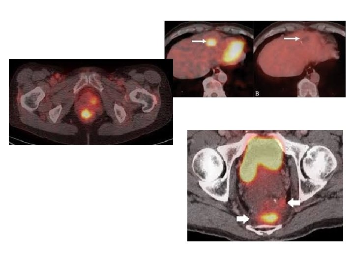
Esophageal Cancer Nuclear medicine PET - Positron Emission Tomography PET is quickly becoming a standard oncologic imaging modality. The technique is useful not only for the primary detection of tumor and metastases but also for the further characterization of abnormalities discovered by using other imaging modalities. 2 -[Fluorine 18]-fluoro-2 -deoxy-D-glucose (FDG) is the most commonly used radiopharmaceutical. FDG PET scanning remains the technique of choice for the detection of metastatic abdominal lymph nodes. The sensitivity of FDG PET in assessing nodal metastasis is reportedly 33 -83%, but studies have shown the superiority of FDG PET to CT and EUS for determining the N status FDG PET is more sensitive than CT for the detection of distant metastases.
Esophageal Cancer Nuclear medicine PET - Positron Emission Tomography From: http: //www. petscaninfo. com/
Gastric Cancer
Gastric Cancer Ø Stomach cancer is the second cause of death among cancers in the world. Ø The incidence of cancer of the stomach continues to decrease in the United States; however, it still accounts for 12, 400 deaths occur in people older than 40 years of age, but they Sometimes occur in younger people. Ø Men have a higher incidence of gastric cancers than women do. Ø The incidence of gastric cancer is much greater in Japan, which has instituted mass screening programs for earlier diagnosis
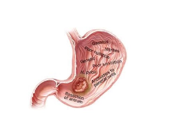
Ø Diet appears to be a significant factor. Ø A diet high in smoked foods and low in fruits and vegetables may increase the risk of gastric cancer. Ø Other factors related to the incidence of gastric cancer include chronic inflammation of the stomach, anemia, gastric ulcers, H. pylori infection, genetics, Smoking, a diet poor in fiber, and Drink alcohol
Applied anatomy of the stomach
Pathophysiology Ø Most gastric cancers are adenocarcinomas and can occur in any portion of the stomach. Ø The tumor infiltrates the surrounding mucosa, penetrating the wall of the stomach and adjacent organs and structures. Ø The liver, pancreas, esophagus, and duodenum are often affected at the time of diagnosis. Ø Metastasis through lymph to the peritoneal cavity occurs later in the disease.
Clinical Manifestations Ø In the early stages of gastric cancer, symptoms may be absent Ø Early symptoms are seldom definitive because most gastric tumors begin on the lesser curvature, where they cause little disturbance of gastric functions. Ø Some studies show that early symptoms, such as pain relieved with antacids, resemble those of benign ulcers.
Ø Symptoms of progressive disease may include: Ø anorexia Ø dyspepsia (indigestion) Ø weight loss Ø abdominal pain Ø anemia Ø nausea and vomiting Ø constipation Ø These can be symptoms of other problems such as a gastric ulcer. Diagnosis should be done by a gastroenterologist or an oncologist
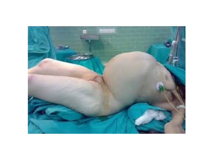
• Cachexia • Abdominal/epigastric mass • Ascites
• Virchow's node (or signal node) is a lymph node in the left supraclavicular fossa (the area above the left clavicle). • It takes its supply from lymph vessels in the abdominal cavity. • It is a finding/symptom of distant metastasis of gastric/GIS cancers.
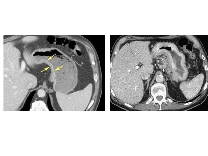
Assessment and Diagnostic Findings Ø The first diagnostic tests performed for patients who complain of the symptoms above is radiography of the stomach. Ø Different radiographic appearance of tumors of the stomach are wide variations. May appear in the form of a block in the bright spot, ulcerated or not ulcerated, or be in the form of thickening of a nonstretch in the wall of the stomach.
Ø Is a gastroscopy with multiple biopsies taken from the area suspected the method adopted for the diagnosis of carcinoma of the stomach. Ø Must be supported by taking swabs brushing during endoscopy and sent for testing cell, since the cytological may be positive (about 15% of cases) at the time the biopsies do not reveal the presence of carcinoma
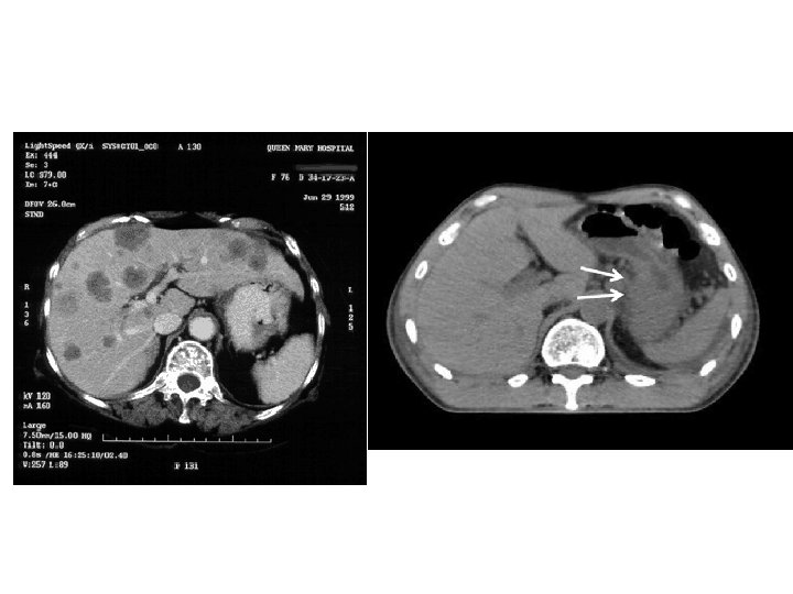
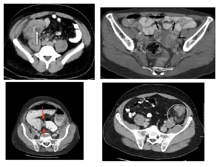
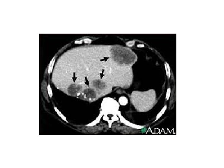
Ø Because metastasis often occurs before warning signs develop, a computed tomography (CT) scan, bone scan, and liver scan are valuable in determining the extent of metastasis. Ø A complete x-ray examination of the GI tract should be performed when any person older than 40 years of age has had indigestion (dyspepsia) of more than 4 weeks’ duration
Gastrointestinal Tumors Colon-Rectum Symptomatology and Diagnosis Koray Topgül, MD, Prof Department of General Surgey
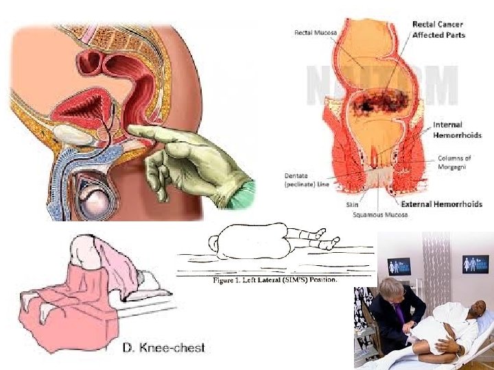
Bleeding (Rectal) Physical Examination Anemia (Occult bleeding) Abdominal Pain Abdominal distention (obstruction) Unexplained, persistent nausea or vomiting Unexplained weight loss Rectal pain (Rectal ceancer) Abdominal mass
• People commonly attribute all rectal bleeding to hemorrhoids, thus preventing early diagnosis owing to lack of concern over bleeding hemorrhoids. • Rectal bleeding may be hidden and chronic and may show up as an iron deficiency anemia. • It may be associated with fatigue and pale skin due to the anemia. • It usually, but not always, can be detected through a fecal occult (hidden) blood test, in which samples of stool are submitted to a lab for detection of blood. • If the tumor gets large enough, it may completely or partially block your colon. You may notice the following symptoms of bowel obstruction (abdominal distention, nause, pain. . )
Perforation---acute abdomen and sepsis
Colonoscopy Rectosigmoidoscpy Rectoscopy Bx
Colonography barium contrast CT MRI PET/CT Endo Rectal US (ERUS)
Apple core sign - describes a short segment irregular circumferential stricture of the large bowel that has abrupt “shouldered” margins resembling an apple core.