Gastrointestinal Physiology Secretion Fig 24 26 Functions Provided


.")


 help in lubrication")


 and mucos cells. - Optimal activity at")


. Essential for B")


: vagal activation during cephalic and gastric phases ( via long")
: vagal activation during cephalic and gastric phases ( via long")

 H 2 receptors on")


 - Cells of mucosal epithelium secrete mucus, water and")


: activated by enterokinase from the duodenum acts as (endopeptidase.")


 blood ductal cells increase water and HCO 3")


- Slides: 74
Gastrointestinal Physiology Secretion
Fig. 24. 26
Functions Provided by secretory glands which serve 2 functions: - Digestive enzymes. - Lubrication and protection of the mucosa.
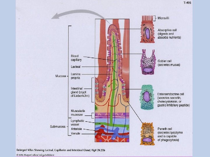
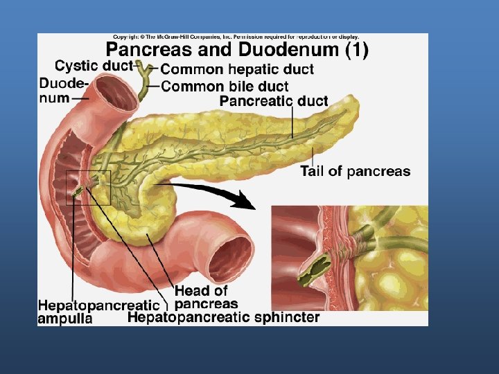
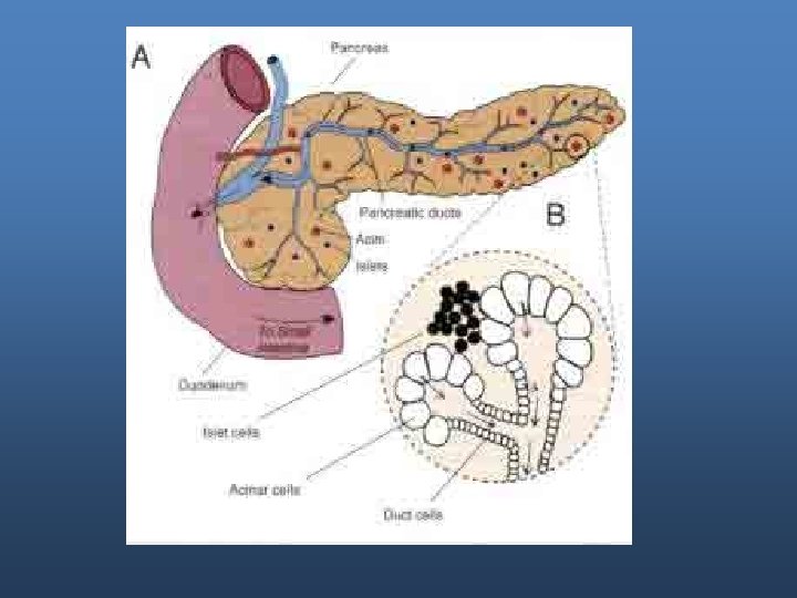
Types of secretory structures The types of secretory glands: - Single-cell secretory glands (goblet cells). - Pits that represent invaginations of the epithelium in the submucosa in small intestine are known as crypts of Lieberkühn. - Complex glands : in stomach and duodenum. - Organs: salivary, pancreas and liver. Located outside the tubular structure of the GI.
Control of secretion Neural Control ENS: ANS: Parasympathetic: Sympathetic: - moderate increase - it reduces secretion by reducing blood flow.
Hormonal regulation Some hormones are secreted by the presence of food or other local changes in the digestive organs.
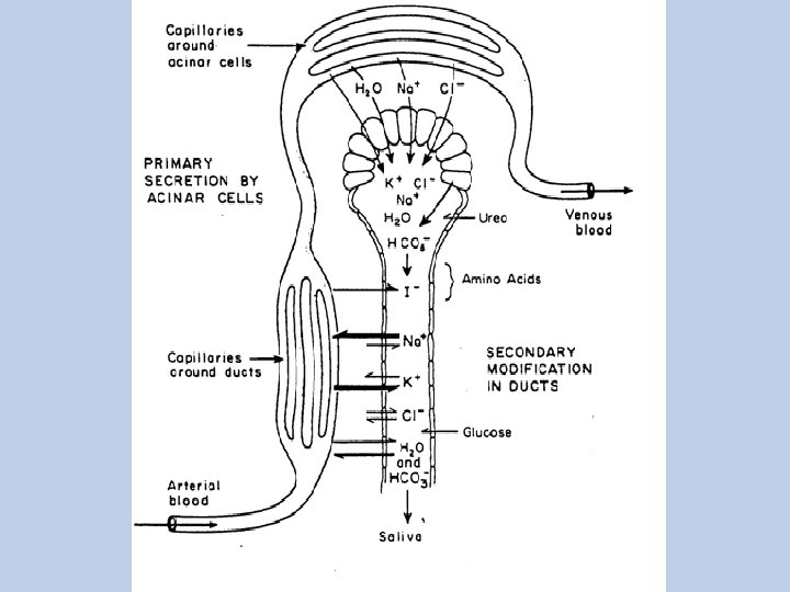
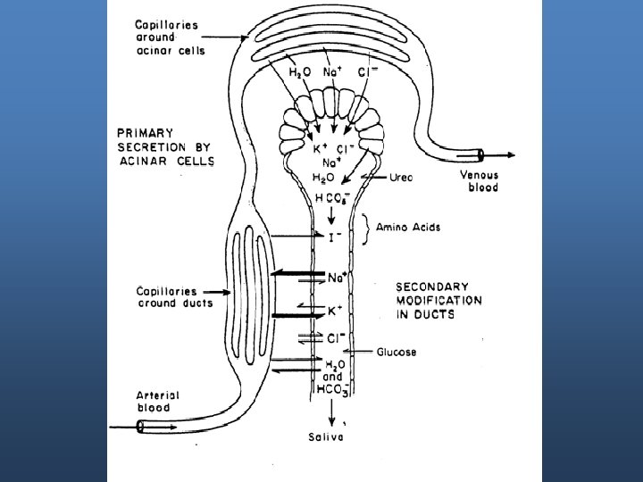
Salivary Secretions
Mechanism of Secretion • • Active transport of Cl- at the basal portion of the membrane. Increase in negativity of membrane potential which attract the positive ion (Na+). Increase osmotic pressure inside the cell >> pull water inside >> increase hydrostatic pressure. This increase results in minute ruptures at the lumenal part of the membrane which causes flushing of water,
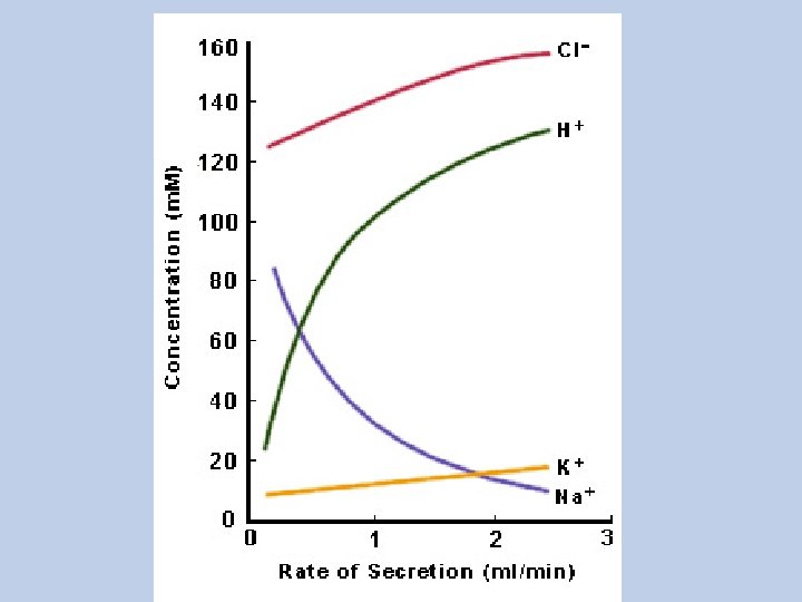
Changes in Composition in Final Saliva ↓ the Na+ and Cl- concentration to the 1/10 of their plasma concentration ↑ 7 folds increase in K+ concentration. ↑ HCO 3 - concentration also increases 2 -3 times.
Rate of Secretion The amount of salivary secretion is about 1500 ml/day. Resting secretion rate 0. 025 -0. 5 ml/min (during basal conditions). The p. H = 7. 0
DURING MAXIMAL STIMULATION The primary saliva increasing 20 folds. - Flow rate of saliva is increased PH=8
Control of salivary Secretion Autonomic nervous system. - Both sympathetic and parasympathetic increase salivation but by different mechanisms - parasympathetic increase water and electrolyte secretion. - Sympathetic increase mucin synthesis. An increase in the sympathetic activity reduces salivation
Control of salivary Secretion Salivation is increased by: - Unconditioned salivary reflex (dental procedures). - Conditioned salivary reflex (learned – response).
Functions of Saliva - Saliva begins digestion of carbohydrates in the mouth: Amylase that breaks polysaccharide into maltose (disaccharide consists of 2 glucose). - Facilitate swallowing by: Moistening the food particles. Lubrication
Functions of Saliva - Antibacterial actions: Lysozyme: an enzyme that lyses or destroys certain bacteria. - oral hygiene keeping mouth and teeth clean by the constant flow and secretion of Ig. A which helps in the destruction of bacteria
Functions of Saliva - Solvent for molecules that stimulate taste buds. - Aids speech. - Bicharbonate neutralizes acids preventing cari
Esophageal secretion - Simple mucus glands and solitary cells (mucoid character) help in lubrication and protection. - Compound mucus glands near the esophago-gasrtic junction and protect the esophagus from reflux.
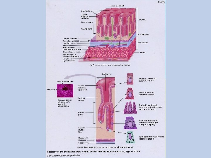
Gastric Secretions
Mechanism of HCl Secretion
Functions of HCl - Conversion of pepsinogen to pepsin - Helps in the decomposition of connective tissue. - Defense (killing most microorganisms ingested with food).
Secretion of pepsinogen Secreted by peptic (chief) and mucos cells. - Optimal activity at p. H (1. 8 -3. 5). Function: - Pepsin cleaves the peptide linkage protein into smaller peptide fragments.
Mucus secreting cells
Mucus secreting cells Function: - Lubricating functions. - Protect the mucosa from the chemical injury by: - Preventing the activity of the proteolytic enzymes to act on the mucosa - Neutralizing HCl by its alkaline character.
Gastrin Secretion Secreted by G cells stimulated by: - gastric distention. - presence of proteins in chyme. - vagal stimulation. Functions: - Increases HCl and pepsinogen secretion. - trophic effect on gastric mucosa to maintain growth of mucosal cells.
Secretion of Intrinsic factor Is secreted by parietal cells (oxyntic cells). Essential for B 12 absorption
Control of Gastric Secretion
Neural Control ENS: Ach neurons parietal and peptic cells.
Neural Control ANS (Parasympathetic): vagal activation during cephalic and gastric phases ( via long arc reflex)
Neural Control ANS (Parasympathetic): vagal activation during cephalic and gastric phases ( via long arc reflex) - enteric excitatory neurons to release Ach. - enteric neurons enterochromaffin-like cells Histamine. - enteric neurons that release GRP Gastrin Releasing Peptide G Cells Gastrin.
Control of Gastric Secretion Hormonal control Gastrin parietal cells increase HCl secretion. Gastrin stimulate CCK-B receptor on oxyntic cells to secrete HCl. This receptor can also be activated by CCK (cholecystokinin).
Control of Gastric Secretion Paracrine Histamine (secreted by enterochromaffin-like cells) H 2 receptors on parietal cells increased c. AMP increased HCl secretion. Somatostatin (SS) SS receptors on parietal cells decrease c. AMP decrease HCl secretion.
Role of HCl in controlling secretion - HCl acts indirectly by initiating enteric reflexes that causes an increase in pepsinogen secretion by peptic cell. - Excess of acids - causes feed back inhibition of gastric secretions by 2 ways: * Reduction of gastrin release * Initiation of inhibitory reflexes. This maintains the p. H from falling below 3.
Summary of Control • Cephalic phase • Gastric phase • Intestinal phase
3 phases of control of gastric secretions : - Cephalic phase: stimuli before food reaching the stomach via parasympathetic NS - Gastric phase: Food in stomach - Distension and the presence of proteins local and long reflexes increased gastric secretion. - Caffeine and alcohol also stimulate acid secretions via ENS, ANS and Hormones - Intestinal phase: - Excitatory - Inhibitory •
Intestinal Secretions
Small Intestinal Secretions (1500 ml/day) - Cells of mucosal epithelium secrete mucus, water and electrolytes. Tubular glands (crypts of Leiberkuhn) secrete serous secretion.
Small Intestinal Secretions Regulation Neural mechanisms (mediated by Ach and VIP. Hormonal: Secretin: increases duodenal secretion.
Colonic secretions - Mostly mucus secretion - Small amount of serous secretions which is high in K+ and HCO 3 -.
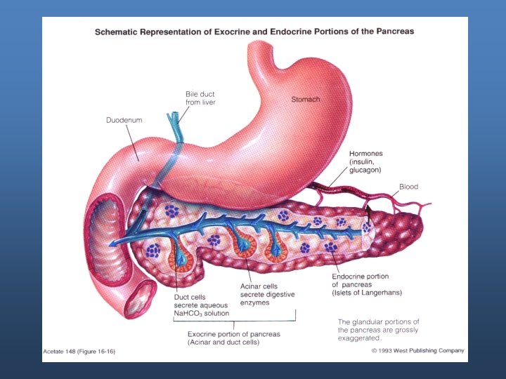
Pancreatic Secretions
Exocrine portion - Enzymes: secreted by acinar cells. - Water and bicarbonate are secreted by duct cells.
Enzyme Secretion by acinar cells
Protelytic enzymes: - Trypsin (ogen): activated by enterokinase from the duodenum acts as (endopeptidase. As long as this enzyme is in pancreas remains inactive by trypsin inhibitor. - Chemotrypsin(ogen): activated by trypsin and acts as endopeptodase. - (Pro) carboxypeptidase: activated by trypsin and acts as exopeptidase.
Enzyme for Digestion of Carbohydrates Pancreatic amylase: secreted as active enzyme to convert Starch (polysaccharide) disaccharides.
Lipolytic enzymes - Lipase that split Triglycerides monglyceride + free fatty acids. Their activity requires an oil/water interface, bile salts (secreted by liver) and other co-lipase secreted by the pancreas. - Phospholipase. - Cholesterol ester hydroxylase.
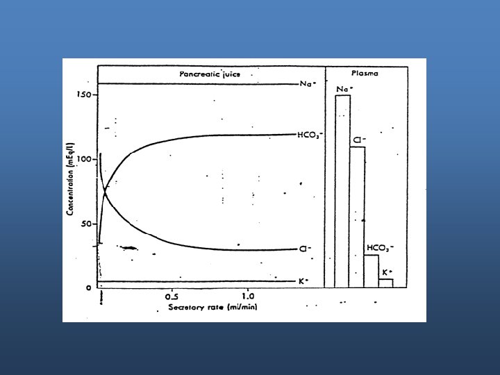
Water and bicarbonate secretion by duct cells.
Control of pancreatic secretion: - Neural - Hormonal
Neural Control - Parasympathetic: Vagal stimulation enteric nervous system release of Ach, VIP, and GRP (Gastrin releasing peptide). - Sympathetic: indirect inhibition via vasoconstriction
Hormonal Control - Secretin (duodenal mucosa) blood ductal cells increase water and HCO 3 secretion. -CCK (Cholecystokinin): * CCK-A receptors (acinar cells) enzyme secretion. * vago-vagal reflex to stimulate enzyme secretions.
Hormonal Control - Pancreatic polypeptide: inhibits the release of enzymes by its inhibitory effect *- Inhibits Ach release from enteric nervous system. *- Inhibits vagal output of the CNS.
Control of pancreatic secretion: - Cephalic phase - Gastric phase - Intestinal phase
3 phases of control of pancreatic secretions Cephalic phase: sight, smell, taste or hearing. Mediated by vagus. Gastric phase: Distension. Mediated by vagus. Intestinal phase: Aminoacids (aa), Fatty acids, H+, Distension. Mediated by CCK, secretin, enteropancreatic reflexes, other hormones.
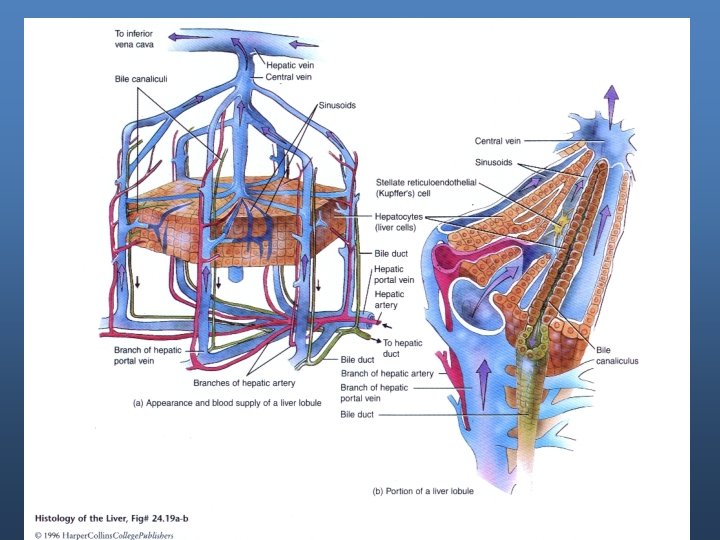
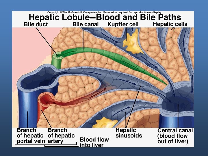
Liver Secretions
Liver functions - Metabolic processing: Process all nutrients after their absorption. - Detoxification of body wastes, hormones, drugs, and other foreign bodies. - Synthesis of plasma proteins, including clotting factors (their synthesis requires vit. K), hormone transporters. - Storage organ of glycogen, iron (ferritin), copper, and vitamines. - Removal of bacteria and foreign materials by reticuloendothelial cells (Kupffer cells). - Excretion of cholesterol and bilirubin.
Bile secretion - Bile acts as detergent to emulsify lipids and make them soluble. Bile is composed of bile salts, water & electrolytes, cholesterol, phosphlipids and wastes intended for excretion, (bilirubin).
Liver functions - Metabolic processing: Process all nutrients after their absorption. - Detoxification of body wastes, hormones, drugs, and other foreign bodies. - Synthesis of plasma proteins, including clotting factors (their synthesis requires vit. K), hormone transporters. - Storage organ of glycogen, iron (ferritin), copper, and vitamines. - Removal of bacteria and foreign materials by reticuloendothelial cells (Kupffer cells). - Excretion of cholesterol and bilirubin.
Excretion of bilirubin in the bile Bilirubin results from the catabolism of hemoglobin Heme + Globin Heme ring iron + biliverdin Biliverdin bilirubin secreted with bile as conjugated (glucoronide, sulfate, other substances).
Bile formation - Bile salts are synthesized by the liver, concentrated in the gallbladder and modified in the lumen. -Synthesized as primary bile acids from cholesterol (cholic and chenodeoxycholic acid)
Bile - Between meals, bile gallbladder where it is stored. The epithelium of the gallbladder removes water and electrolytes 5 -20 fold concentration of bile.
Bile salts
Fig. 24. 21
Enterohepatic circulation