Gastrointestinal Functions q Introduction q Mouth digestion q


 tract Accessory glandular organs")


,")


 Mucous cells Mucus,")
 1. 1 Secretion - It is secreted")


 - help to")


 AT P cartoon")


 - The proximal half of")
- Slides: 90
Gastrointestinal Functions
q Introduction q Mouth digestion q Stomach digestion q Small intestinal digestion q Large intestinal digestion q Absorption
Section 1 Introduction n Physiological characteristics of digestive smooth muscle n Secretion of digestive gland n Neural control of digestive tract § Gastrointestinal hormones
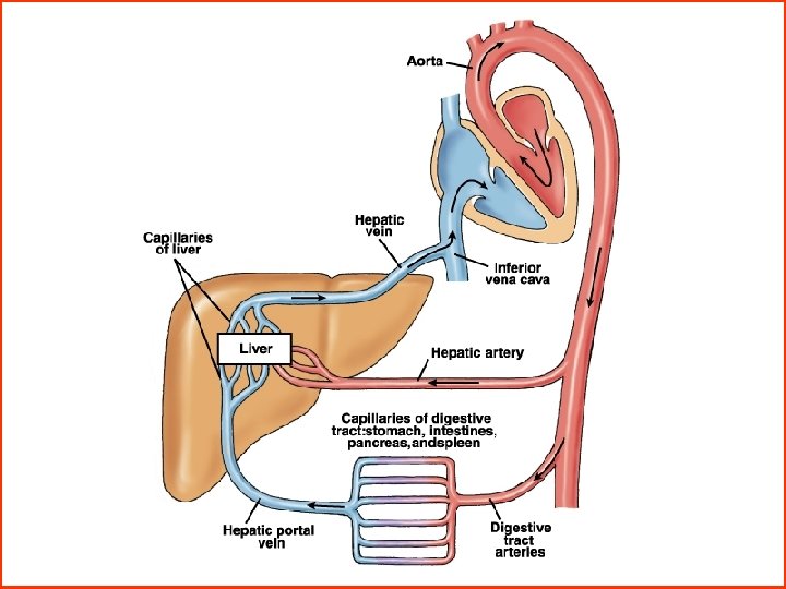
Gastrointestinal system consists of Gastrointestinal (GI) tract Accessory glandular organs
Anatomy and functions of the GI tract mouth, pharynx, esophagus, stomach, small intestine, large intestine, anus Accessory Glandular Organs salivary glands, liver, gallbladder, pancreas
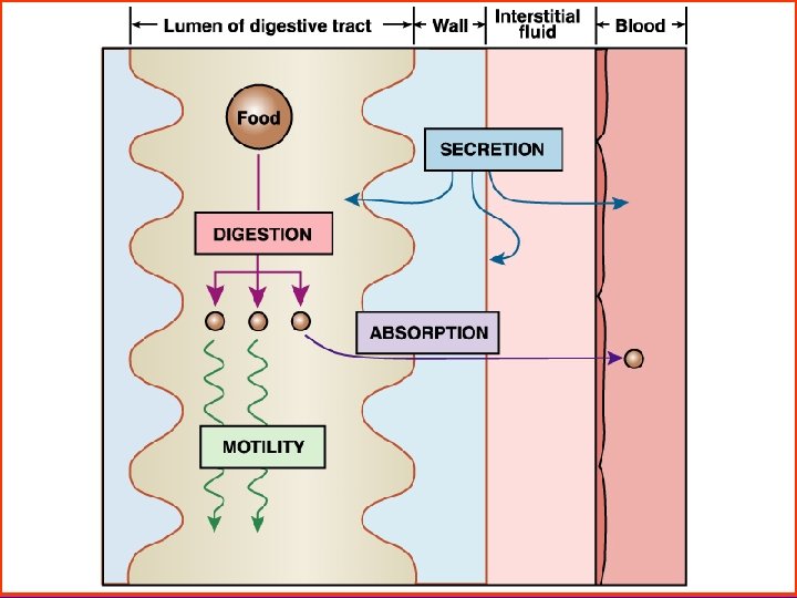
1. Definition - Digestion It shows the large molecular substances were changed into the small molecular substances. - Absorption It indicates the small molecular substances were absorbed to blood & lymph. 2. Digestive modes - Mechanical digestion: by smooth muscles of digestive tracts - Chemical digestion: by digestive enzyme
Physiological Characteristics of Digestive Smooth Muscle 1. General characteristics - The intestinal wall is composed of 5 layers. - Excitation is lower, contraction is slow. - Have autorhythmicity, but is not stable. - Have large dilation. - It is not sensitive to electric stimulus and is sensitive to stretch, T and chemical stimulus
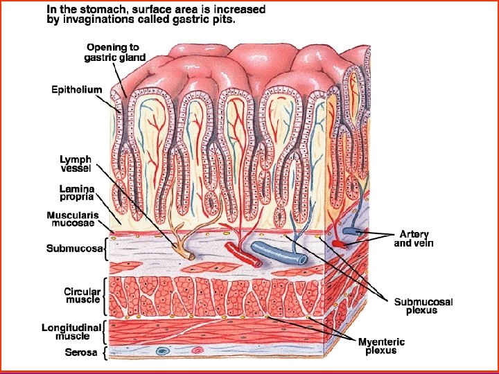
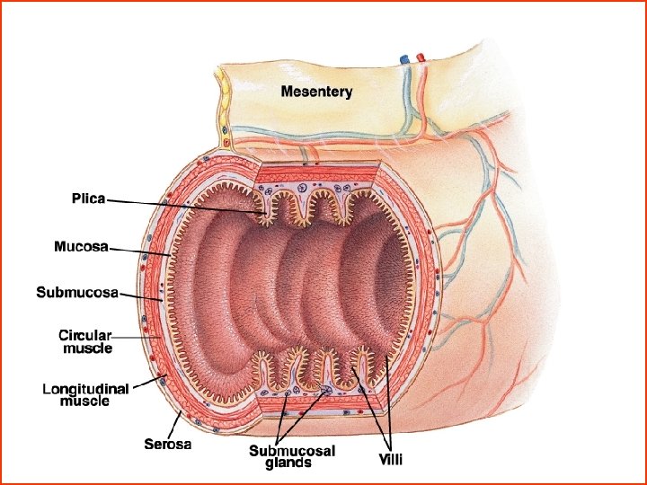
Layers of Alimentary Canal Submucosa Mucosa Circular muscle layer Longitudinal muscle layer Serosa
Layers of Alimentary Canal Serosa Myenteric plexus Mucosa Submucosal plexus Longitudinal muscle Submucosa Circular muscle
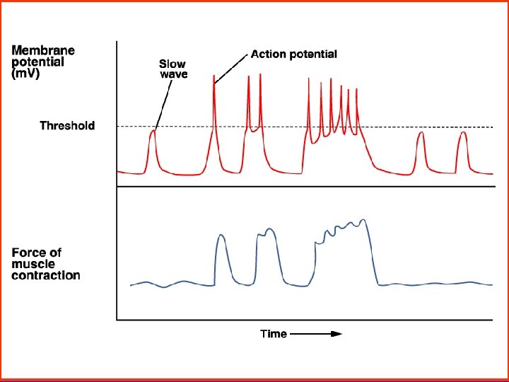
2. Characteristic of electrical activity muscle - Resting potential: - 50 ~ - 60 m. V - Slow wave potential: it is not AP, it is undulating changes in the RP, about 10 ~ 15 m. V. - Action or spike potential: They occur when the RP becomes more positive than about – 40 m. V, the channels responsible for the AP allow Ca ions an Na ions enter.
Cells and Electrical Events in the Muscularis Insert fig. 18. 16
Secretion of Digestive Gland 1. Component: Water, digestive enzyme, mucus and antibody ets. 2. Function: - digest food - provide p. H, - dilute food - protect mucosa of digestive tract.
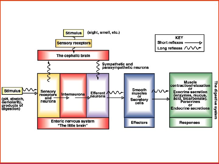
Neural Control of Gastrointestinal Function 1. Enteric nervous system - The Myenteric plexus, or Auerbach’s plexus Stimulation causes increased “tone” of the gut wall, increased intensity and rate of rhythmical contractions - The submucosal plexus, or Meissner’s plexus It is mainly concerned with controlling function within the inner wall of each minute segment of the intestine.
CNS to gut connections BRAIN STEM nodose ganglion Vagal afferent Vagal efferent dorsal root ganglion Spinal afferent SPINAL CORD
2. Autonomic nerve The parasympathetic nerves - It increases the activity of the enteric nervous system. This in turn enhances the activity of most gastrointestinal functions. The sympathetic nervous - to a slight extent by a direct action that inhibits the smooth muscles of gastrointestinal wall. - to a major extent by an inhibitory effect on the neurons of the enteric nervous system. 3. Gastrointestinal reflexes
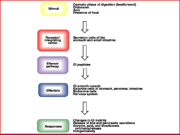
The Reflex Pathway of the Digestive System Central nervous system Sympathetic, parasympathetic nerve Enteric nervous system Stretch, chemical, T receptor smooth muscle (GI) secreting cells endocrine cells
Gastrointestinal Hormones 1. Gastrointestinal tract produces many hormones All known GI hormones are peptides with molecular weights in most cases ranging between 2, 000 to 5, 000. 2. Effects: - Regulate secretion of digestive glands and motility of digestive tract. - Regulate the other hormone release. - Trophic action 3. The three major hormones are in the below table:
Three Major GI Hormones Hormone Effects Gastrin Stimulates acid secretion and promote digestive motility. CCK Stimulates gallbladder contraction and pancreatic enzyme secrete Secretin Stimulates HCO 3 - and water secrete of pancretic liquid. Site G cells I cells S cells
Section 2 Mouth and Esophagus § Saliva & function § Digestion of food in mouth
Saliva & Function 1. Saliva - Secretion Saliva is secreted by three pairs of large glands, the parotid, submaxillary, and sublingual gland. - Components: Water 99%, ptyalin, bacteriolysis enzyme, mucus, Ig A, Na+, K+, HCO 3 - , p. H 6. 6 ~ 7. 1. Control of secretion - It includes conditional and unconditional reflex. - It is controlled mainly by vagus and sympathetic signals.
Salivary gland
2. Functions of saliva - The serous secretion contains ptyalin ( an αamylase ), which is an enzyme for digesting starch. - Mucous secretion contains mucus for surface protection and lubrication. - Solubilizes dry food - Keep oral hygiene: bacteriolysis enzyme, Ig A.
Esophagus peristaltic contractions During swallowing, boluses of food are propelled through the esophagus by strong peristaltic contractions.
Section 3 Gastric Digestion § Functional structure of the stomach § Gastric secretion § Regulation of gastric secretion § Motor functions of the stomach
Functional Anatomy of Stomach Fundus Body Antrum • Storage • • • Oesophagus Lower Oesophageal Sphincter Storage Duodenum Pylorus Mucus HCl Pepsinogen Intrinsic factor • Mixing/Grinding • Gastrin Fundus Body Antrum
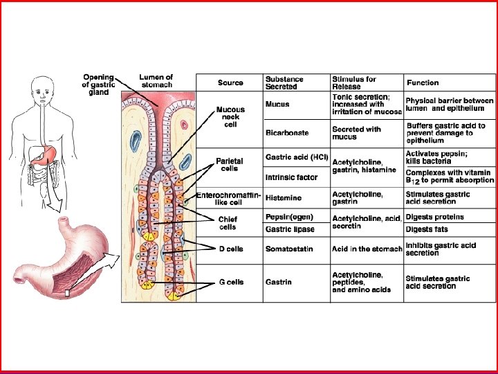
Functional Structure of Stomach Region Cell type Secretion Cardiac gland (fundus) Mucous cells Mucus, HCO 3 - Oxyntic gland (body) Parietal cells, Chief cells HCl, intrinsic factor Pepsinogen, Pyloric gland (antrum) G cells Mucus cells D cells Gastrin Mucus Somatostatin
Gastric Secretion 1. Gastric acid ( HCl) 1. 1 Secretion - It is secreted by parietal cells, p. H 0. 9 ~ 1. 5. - Excreting volume: u Basal acid secretion : 0 ~ 5 mmol / h fasting overnight u The maximal secretion: 20 ~ 25 mmol / h - Determine acid content Normal value: total HCl 10 ~ 50 u fasting overnight
Proton pump Alkaline tide
1. 2 Effects - turning pepsinogen into activated pepsin, - changing the character of the protein, - killing many swallowed virulent organisms, - promoting absorption of ferrum and Ca 2+, - promoting secretion of pancreas and bile. 2. Pepsinogen - It is secreted by chief cell, and is activated to the active form pepsin by HCl. - It hydrolyzes protein molecules to form proteoses, peptones.
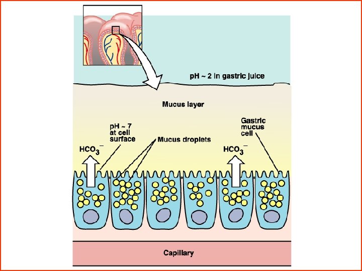
3. Intrinsic factor - It is a glucoprotein secreted by the parietal cell. - It is essential to absorption of vitamin B 12 in the ileum. 4. Mucus - The surface epithelial cells of the stomach secrete a viscous mucus, they also secrete HCO 3 -. - It is widely believed that mucus and bicarbonate form a mucus bicarbonate barrier. - The mucus bicabonate barrier serves an important role in protecting the epithelium from acid - pepsin and other chemical insults.
Regulation of Gastric Secretion 1. Stimulating factor of gastric secretion - Ach It excites secretion of gastric secretion. - Gastrin G cells secrete gastrin which stimulates pariatal cells strongly and peptic cells to a lesser extent. - Histamine( enterochromaffin like cell) It can enhances acid secretion by stimulating parietal cells.
2. There are three phases of gastric secretion Cephalic phase - It is 30% of all gastric juice, HCl and pepsinogen are higher. - Vagus directly stimulates parietal cells and G cell releasing gastrin. Gastric phase - It is 60 % of all gastic juice, HCl is high but pepsinogen is lower than cephalic’. Intestinal phase - It is 10%, HCl and pepsinogen are low.
Cephalic phase Gastric phase
enterochromaffin-like cell
3. Inhibitory factor of gastric secretion - Excess acid When the p. H of gastric acid falls, gastrin secretion decrease by inhibit G cells and nervous reflex. - Chyme ( acid in the upper intestine, protein break down products, or the irritation of the mucosa ) in the small intestine inhibits secretion. - Hormones Secretin, bulbogastrone, enterogastrone, gastric inhibitory peptide are especially important in the inhibition of gastric secretion.
Motor Functions of the Stomach 1. Motility in head region - The stomach relaxes when food enters it. This is called receptive relaxation. - Its function is storage of food. 2. Motility in tail region - Peristalsis: It occurs when food enters stomach, 3 times / min. “Retropulsion” is an important mixing mechanism of the stomach. - It can propels food into the duodenum. - Migrating motility complex
Gastric Motility Peristaltic waves: Body Antrum Body Thin muscle weak contraction No mixing Antrum Thick muscle powerful contraction A Mixing B Contraction of pyloric sphincter 1 Only small quantity of gastric content (chyme) entering duodenum 2 Further mixing as antral contents forced back towards body
The contraction of the gastric pump can be differentiated into three phases: A: phase of propulsion, B: phase of emptying, C: phase of retropulsion and grinding Phase of propulsion Phase of emptying Phase of retropulsion Antrum Bulge Rapid flow of liquids with suspended small particles and delayed flow of large particles towards pylorus Emptying of liquids with small particles whereas large particles are retained in the buldge of the terminal antrum Retropulsion of large particles and clearing of the terminal antrum
Thin wall, holds large volumes of food by receptive relaxation Thick wall with strong and contraction that mix and propel food into duodenu cartoon
3. Empting of food: - The food is emptied into small intestine at a rate suitable for proper digestion and absorption. - Promoting empting factor: Gastric factor promotes gastric emptying. - Inhibitting empting factor: Gastric emptying is inhibited by enterogastric reflexes from the duodenum and cholecystokinin which is released from the mucosa of the jejunum in response to fatty substances.
Section 4 Digestion of the Small Intestine § Pancreatic secretion § Bile secretion n Secretion of the small intestine § Movement of the small intestine
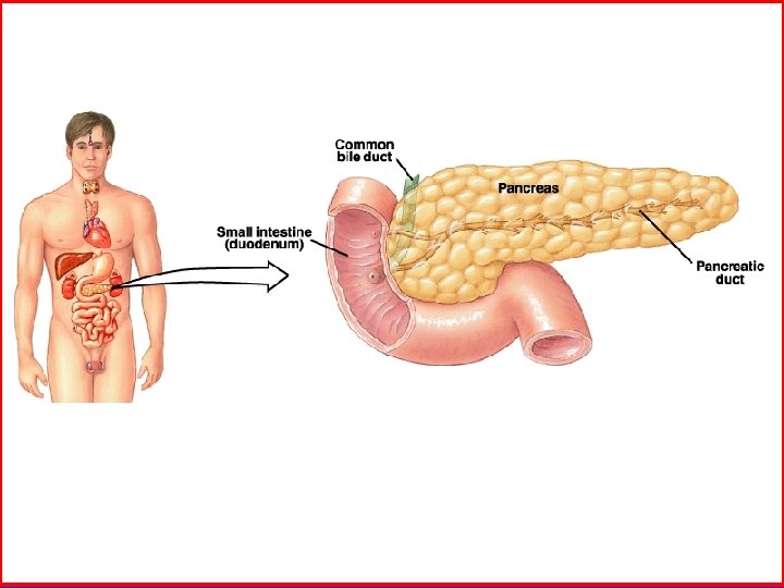
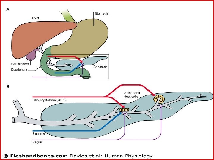
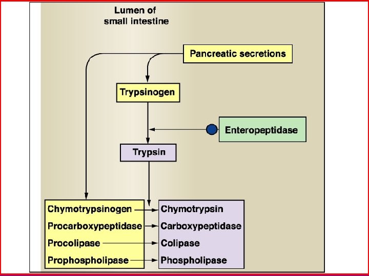
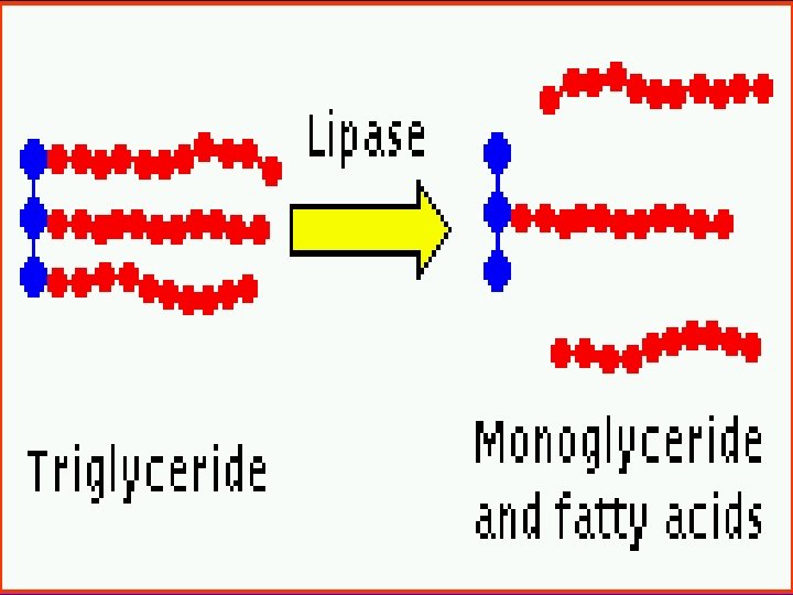
Pancreatic Secretion 1. Pancreatic fluid: p. H 7. 8 ~ 8. 4, 1 ~ 2 L/day. 1. 1 Digestive enzymes - For digestion of proteins are chymotrypsin, trypsin. - For carbohydrates is pancreatic amylase which hydrolyzes starches to form disaccharides. - For fat is pancreatic lipase which hydrolyzes neutral fat into fatty acids and monoglycerides. 1. 2 Bicarbonate ions and water They are secreted by epithelial cells of ductules.
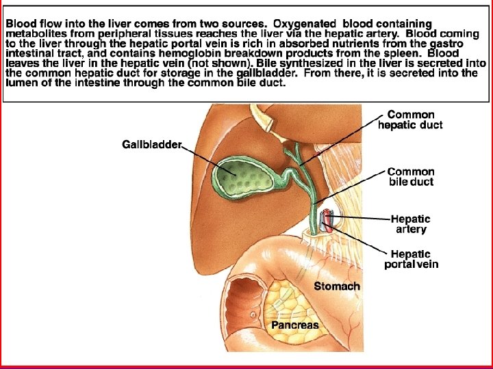
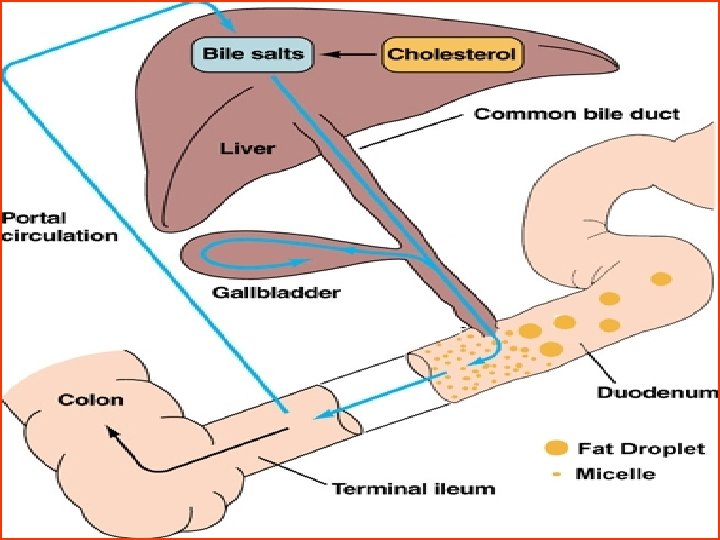
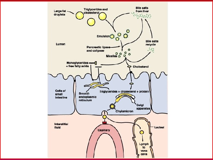
Bile Secretion 1. Components: Bile acids, cholesterol, bile salt, lecithin, bilirubin, electroytes. Bile contains no digestive enzyme. 2. Secretion: - Bile synthesized in the liver and is acidic as it flows through the bile ducts, is alkalescent in gallbladder. - Bile is concentrated about fivefold to tenfold in the gallbladder.
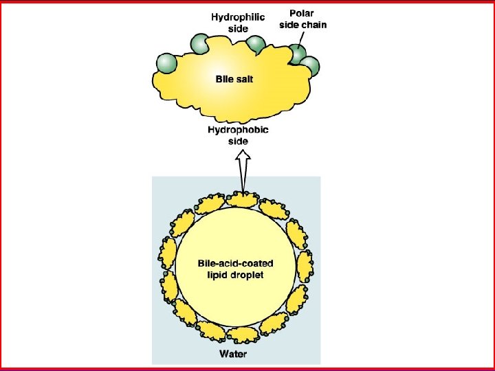
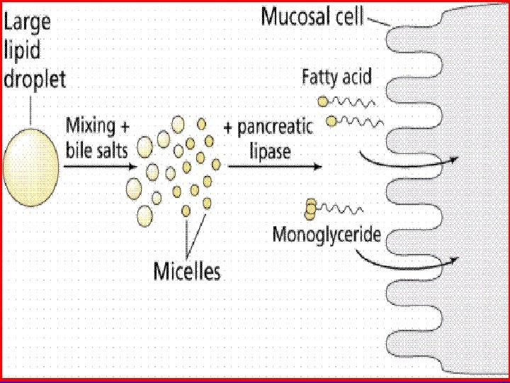
3. Action of bile salts: - promotes fat digestion (fat droplet) - help to emulsify the large fat particles into minute particles that can be attacked by the lipase enzyme. - promotes fat , fatsoluble vitamins absorption. 4. Regulation of bile: - Fatty foods that enter the duodenum cause CCK to be released from the local glands. - CCK cause rhythmical contractions of the gallbladder and simultaneous relaxation of the sphincter of Oddi.
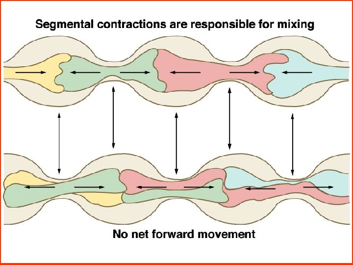
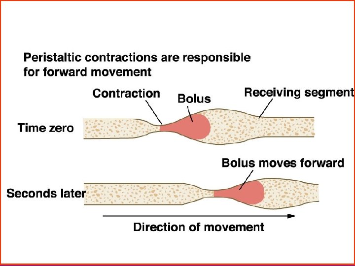
Movements of the Small Intestine 1. Motility patterns - Segment contraction It promotes mixing of the solid food particles with the secretions of the small intestine. - Peristalsis Chyme is propelled through the small intestine by peristaltic waves at a velocity of 0. 5 to 2. 0 cm/s. 2. Regulation - Nervous signals are caused by the chyme into the duodenum but also by a gastroenteric reflex. - Hormonal signals: Gastrin, CCK enhance; secretin inhibits.
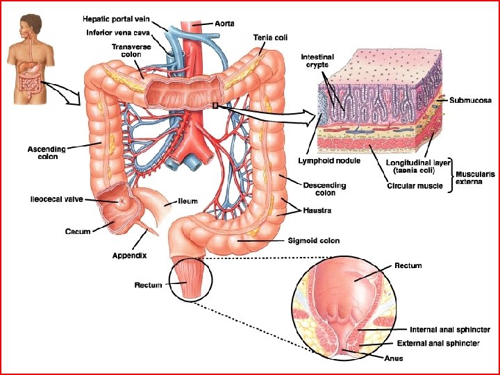
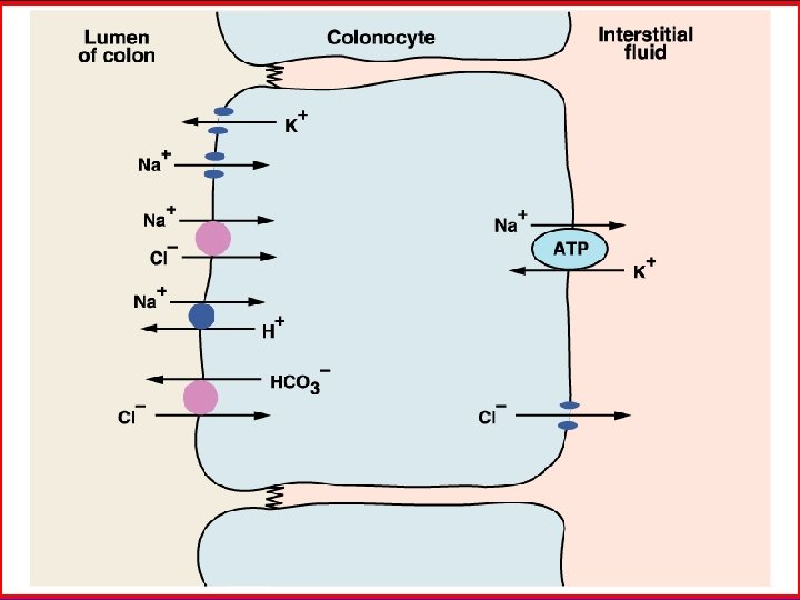
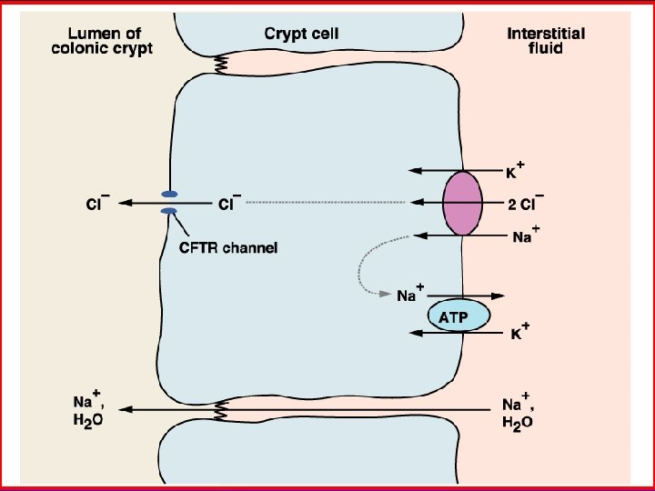
Secretion of the Large Intestine 1. Secretion: p. H 8. 3 -8. 4 - Most of the secretion in large intestine is mucus. - Mucus protects the intestinal wall against friction and bacterial activity, provides the adherent medium for fecal matter and barrier to keep acids from attacking the intestinal wall. 2. Bacterias action It can synthesize vitamine B ( B 1, B 2, B 12 ), K and folacin by ferment and decay of bacterias.
Section 6 Absorption in the Gastrointestinal Tract n The site of the absorption n Absorption of foods
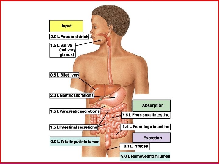
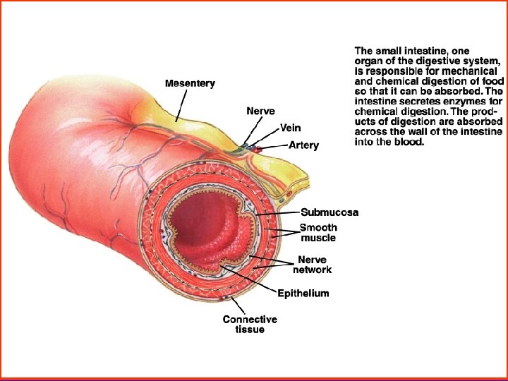
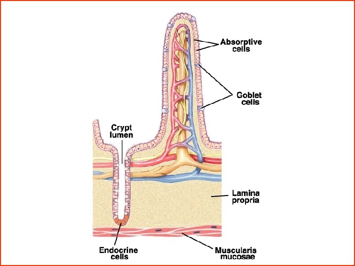
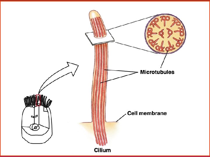
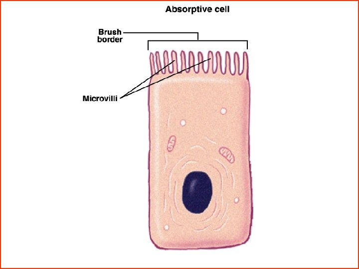
The site of Absorption - in the Small Intestine - The folds of plica, villi, and microvilli increase the mucosal absorptive area. The total area of the small intestinal mucosa is 250 m 2 about the surface area of a tennis court. - Foods have been digested to small molecules. - Foods stay in small intestine for long time, about 3 to 8 hours. - Every villi has a capillary and lymph duct, also has smooth muscle.
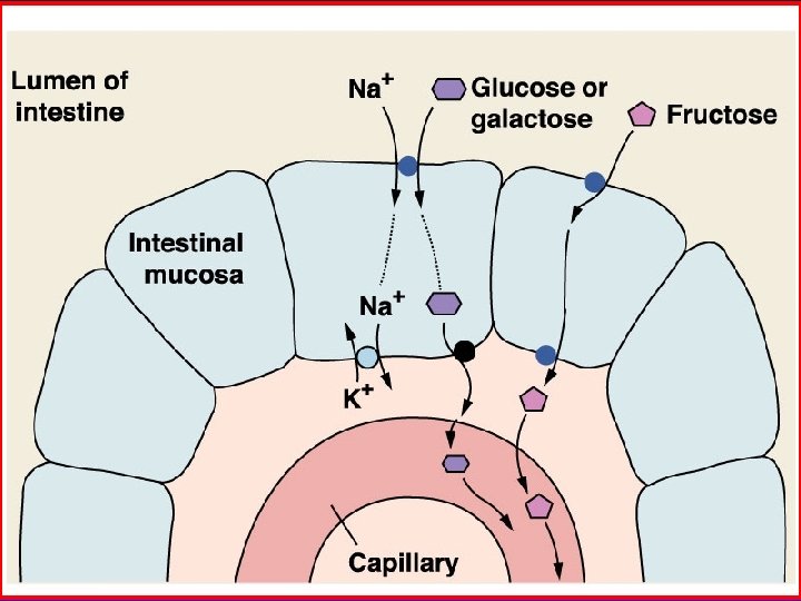
Absorption of Foods 1. Absorption of carbohydrates - Essentially all carbohydrates are absorbed in the form of monosaccharides. The most abundant of the absorbed monosaccharides is glucose which is absorbed to blood. - Glucose is transported by a sodium co-transport mechanism. - It is secondary active transport.
Carbohydrate and protein absorption (secondary active transport) AT P cartoon
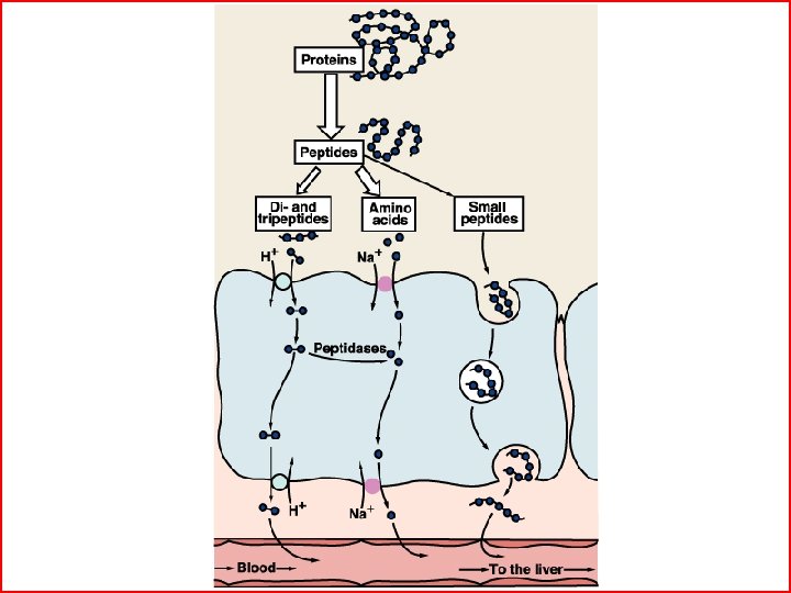
2. Absorption of proteins - Most proteins are absorbed to blood through the membranes of the intestinal epithelial cells in the form of dipeptide, tripeptide, and a few free amino acids. - The energy for most of this transport is supplied by a sodium co-transport mechanism in the same way that sodium co-transport of glucose occurs.
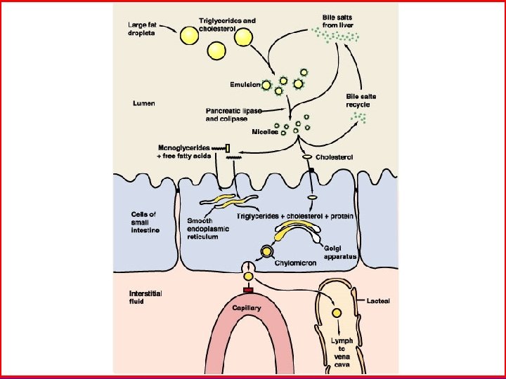
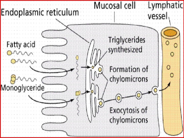
3. Absorption of fats - Monoglycerides and fatty acids diffuse passively to the interior of the enterorocyte. - After entering the enterocyte, the fatty acids and monoglycerides are mainly recombined to form new triglycerides. - The reconstituted triglycerides that contain cholesterol and phospholipids. These globules are called chylomicrons which enter the lymph, then upward through the thoracic duct to be emptied into the great veins of the neck.
Chylomicron
chylomicron
4. Electrolytes - Sodium: active transport by Na+ pump - Iron: Fe 3+ Vit C Fe 2+ Except the Vitamine C, HCl can dissolved iron, thereby helping absorption of iron. - Ca 2+ HCl can promote Ca 2+ dissociate, vitamine D make Ca 2+ dissolve.
5. Absorption of water ( in the large intestine) - The proximal half of the colon is important for absorption of electrolytes and water. - It can absorb a maximum of about 5 to 7 L of fluid and electrolytes each day. - It is passive and follows the absorption of osmotic particles.