Gastrointestinal bleeding Mohammad Jomaa CLASSIFICATION OF G I

Gastrointestinal bleeding Mohammad Jomaa

CLASSIFICATION OF G. I. BLEEDING GIB Appearance Acuity Site Apparent Acute Upper Obscure Chronic Lower

UPPER&LOWER G. I. BLEEDING

UPPER G. I. BLEEDING causes:

Acute upper gastrointestinal haemorrhage most common gastrointestinal emergency Accounting for 50– 170 admissions to hospital per 100 000 of the population each year in the UK mortality of patients admitted to hospital is about 10%

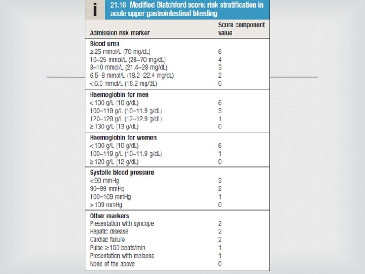
Modified Blatchford score Risk scoring systems have been developed to stratify the risk of needing endoscopic therapy or of having a poor outcome. The advantage of the Blatchford score is that it may be used before endoscopy to predict the need for intervention to treat Bleeding. Low scores (2 or less) are associated with a very low risk of adverse outcome

Clinical assessment Haematemesis is red with clots when bleeding is rapid and profuse, or black (‘coffee grounds’) when less severe. Syncope may occur and is caused by hypotension from intravascular volume depletion. Symptoms of anaemia suggest chronic bleeding. Melaena is the passage of black, tarry stools containing altered blood; it is usually caused by bleeding from the upper gastrointestinal tract, although haemorrhage from the right side of the colon is occasionally responsible. The characteristic colour and smell are the result of the action of digestive enzymes and of bacteria on haemoglobin. q Volumes as little as 50 to 100 m. L of blood in the stomach can result in melena. Severe acute upper gastrointestinal bleeding can sometimes cause maroon or bright red stool (Hematochezia)……. . hemodynamic instable q Usually in lower GI bleeding
 T • Treatment")
Management S E • Stabilization • Evaluation • (Endoscopy) T • Treatment

1. Intravenous access The first step is to gain intravenous access using at least one large-bore cannula. 2. Initial clinical assessment Define circulatory status. Severe bleeding causes tachycardia, hypotension and oliguria. The patient is cold and sweating, and may be agitated. Seek evidence of liver disease. Jaundice, cutaneous stigmata, hepatosplenomegaly and ascites may be present in decompensated cirrhosis. Identify comorbidity. The presence of cardiorespiratory, cerebrovascular or renal disease is important, because these may be worsened by acute bleeding and because they increase the hazards of endoscopy and surgical operations. These factors can be combined using the Blatchford score, which can be calculated at the bedside. A score of 2 or less is associated with a good prognosis, while progressively higher scores are associated with poorer outcomes.

3. Basic investigations q Full blood count. Chronic or subacute bleeding leads to anaemia but the haemoglobin concentration may be normal after sudden, major bleeding until haemodilution occurs. Thrombocytopenia may be a clue to the presence of hypersplenism in chronic liver disease. q Urea and electrolytes. This test may show evidence of renal failure. The blood urea rises as the absorbed products of luminal blood are metabolised by the liver; an elevated blood urea with normal creatinine concentration implies severe bleeding. q Liver function tests. These may show evidence of chronic liver disease. q Prothrombin time. Check when there is a clinical suggestion of liver disease or patients are anticoagulated. q Cross-matching. At least 2 units of blood should be cross-matched if a significant bleed is suspected.

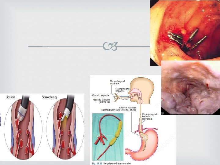
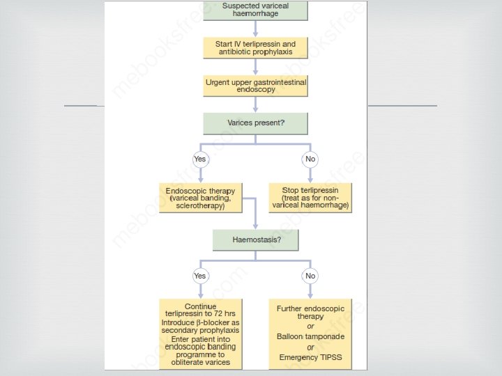
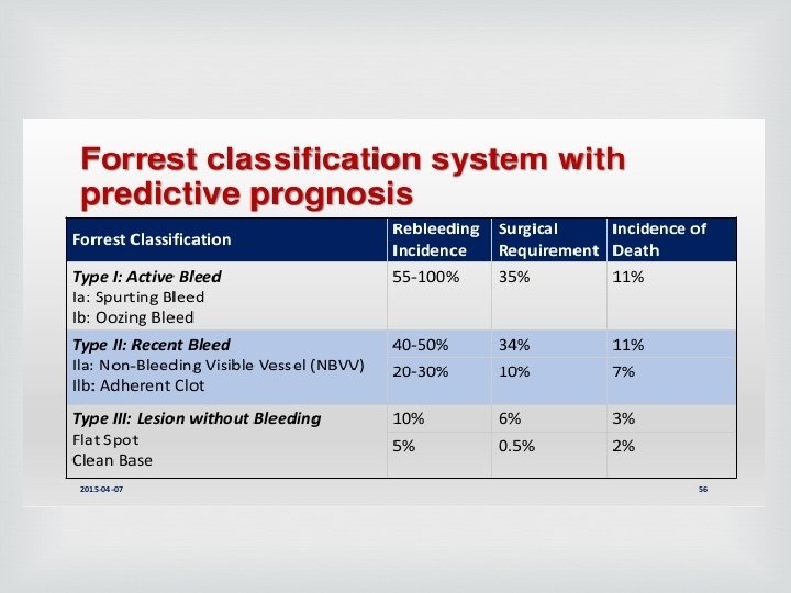
4. Resuscitation q Intravenous crystalloid fluids should be given to raise the blood pressure, and blood should be transfused when the patient is actively bleeding with low blood pressure and tachycardia. Comorbidities should be managed as appropriate. Patients with suspected chronic liver disease should receive broad-spectrum antibiotics. 5. Oxygen q This should be given to all patients in shock. 6. Endoscopy q This should be carried out after adequate resuscitation, ideally within 24 hours, and will yield a diagnosis in 80% of cases. q Patients who are found to have major endoscopic stigmata of recent haemorrhage can be treated endoscopically using a thermal or mechanical modality, such as a ‘heater probe’ or endoscopic clips, combined with injection of dilute adrenaline (epinephrine) into the bleeding point (‘dual therapy’). A biologically inert haemostatic mineral powder (‘haemospray’) can be used as rescue therapy when standard therapy fails. This may stop active bleeding and, combined with intravenous PPI therapy, may prevent rebleeding, thus avoiding the need for surgery. Patients found to have bled from varices should be treated by band ligation if this fails, balloon tamponade is another option, while arrangements are made for a transjugular intrahepatic portosystemic shunt (TIPSS)

7. Monitoring q Patients should be closely observed, with hourly measurements of pulse, blood pressure and urine output. 8. Surgery q Surgery is indicated when endoscopic haemostasis fails to stop active bleeding and if rebleeding occurs on one occasion in an elderly or frail patient, or twice in a younger, fitter patient. q If available, angiographic embolisation is an effective alternative to surgery in frail patients. q The choice of operation depends on the site and diagnosis of the bleeding lesion. Duodenal ulcers are treated by under-running, with or without pyloroplasty. Under-running for gastric ulcers can also be carried out (a biopsy must be taken to exclude carcinoma). Local excision may be performed, but when neither is possible, partial gastrectomy is required. 9. Eradication q Following treatment for ulcer bleeding, all patients should avoid non-steroidal anti -inflammatory drugs (NSAIDs) and those who test positive for H. pylori infection should receive eradication therapy. Successful eradication should be confirmedby urea breath or faecal antigen testing.

Treatment of UGIB-Summary MODALITY OF TREATMENT NON-VARICEAL I. V. PPI Medical Tx Mechanical(N/Sadrenalin injection, Haemoclip) Thermal (Heatprobe, Argon Endoscopic Tx VARICEAL Somatostatin or it's analogue Terlipressin Sclerotherapy Banding Rescue Tx. Hemospray Sengstaken tube Interventional Radiology Tx. Angiograghy. Embolization TIPSS Surgical Tx. Ligation of bleeding vessel Porto-systemic shunt

LOWER G. I. BLEEDING causes:

Severe acute lower gastrointestinal bleeding This presents with profuse red or maroon diarrhoea and with shock. Diverticular disease is the most common cause and is often due to erosion of an artery within the mouth of a diverticulum. Bleeding almost always stops spontaneously, but if it does not, the diseased segment of colon should be resected after confirmation of the site by angiography or colonoscopy. Angiodysplasia is a disease of the elderly, in which vascular malformations develop in the proximal colon. Bleeding can be acute and profuse; it usuallystops spontaneously but commonly recurs. Diagnosis is often difficult. Colonoscopy may reveal characteristic vascular spots and, in the acute phase, visceral angiography can show bleeding into the intestinal lumen and an abnormal large, draining vein. In some patients, diagnosis is achieved only by laparotomy with on-tablecolonoscopy. The treatment of choice is endoscopic thermal ablation but resection of the affected bowel may be required if bleeding continues.

Bowel ischaemia due to occlusion of the inferior mesenteric artery can present with abdominal colic and rectal bleeding. It should be considered in patients (particularly the elderly) who have evidence of generalised atherosclerosis. The diagnosis is made at colonoscopy. Resection is required only in the presence of peritonitis. Meckel’s diverticulum with ectopic gastric epithelium may ulcerate and erode into a major artery. The diagnosis should be considered in children or adolescents who present with profuse or recurrent lower gastrointestinal bleeding. • A Meckel’s 99 m. Tc-pertechnetate scan is sometimes positive but the diagnosis is commonly made only by laparotomy, at which time the diverticulum is excised.

Subacute or chronic lower gastrointestinal bleeding This can occur at all ages and is usually due to haemorrhoids or anal fissure. Ø Haemorrhoidal bleeding is bright red and occurs during or after defecation. q Proctoscopy can be used to make the diagnosis, but subjects who have altered bowel habit and those who present over the age of 40 years should undergo colonoscopy to exclude coexisting colorectal cancer. Ø Anal fissure should be suspected when fresh rectal bleeding and anal pain occur during defecation.

Major gastrointestinal bleeding of unknown cause In some patients who present with major gastrointestinal bleeding, upper endoscopy and colonoscopy fail to reveal a diagnosis. When severe life-threatening bleeding continues, urgent CT mesenteric angiography is indicated. This will usually identify the site if the bleeding rate exceeds 1 m. L/min and then formal angiographic embolisation can often stop the bleeding. Wireless capsule endoscopy is often used to define a source of bleeding prior to enteroscopy. If angiography is negative or bleeding is less severe, push or double balloon enteroscopy can visualise the small intestine and treat the bleeding source. When all else fails, laparotomy with on-table endoscopy is indicated.

Chronic occult gastrointestinal bleeding In this context, occult means that blood or its breakdown products are present in the stool but cannot be seen by the naked eye. Occult bleeding may reach 200 m. L per day and cause iron deficiency anaemia. Any cause of gastrointestinal bleeding may be responsible but the most important is colorectal cancer, particularly carcinoma of the caecum, which may produce no gastrointestinal symptoms. In clinical practice, investigation of the upper and lower gastrointestinal tract should be considered whenever a patient presents with unexplained iron deficiency anaemia. Testing the stool for the presence of blood is unnecessary and should not influence whether or not the gastrointestinal tract is imaged because bleeding from tumours is often intermittent and a negative faecal occult blood (FOB) test does not exclude the diagnosis. The only value of FOB testing is as a means of population screening for colonic neoplasia in asymptomatic individuals

- Slides: 24