GASTRIC CANCER Dr Saurabh Pathak Professor Department of

GASTRIC CANCER Dr. Saurabh Pathak Professor, Department of Surgery CIMS&H, Lucknow

ANATOMY & PHYSIOLOGY
, two curvatures")
ANATOMY The stomach J-shaped. It has two surfaces (the anterior & posterior), two curvatures (the greater & lesser), two orifices (the cardia & pylorus). It has fundus, body and pyloric antrum. Blood supply � Theleft gastricartery � Right gastro-epiploicartery � Left gastro-epiploicartery � Shortgastricarteries

GASTRIC CANCER �Stomach cancer begins when cancer cells form in the inner lining of your stomach. These cells can grow into a tumor. Also called gastric cancer, the disease usually grows slowly over many years. �It could be: �malignant or benign �primary or secondary

TYPES Tumor Type Percent Malignant tumors 93. 0 Carcinoma 87. 9 Lymphoma 3. 0 Leiomyosarcoma 1. 7 Carcinoid 0. 3 Others 0. 1 Benign tumors 7. 0 Polyp 3. 1 Leiomyoma 2. 0 Inflammatory lesions 0. 7 Heterotopic pancreas 0. 4 Others 0. 8
 : Cancer")
The four most common primary malignant gastric neoplasms are: � Adenocarcinoma (95%) : Cancer that begins in the glandular cells. � Lymphoma (4%) : Cancer that begins in immune system cells. � Carcinoid cancer(3%) : Cancer that begins in hormone-producing cell. � Gastrointestinal stromal tumor (GIST) (1%) : Cancer that begins in nervous system tissues of stomach.

EARLY GASTRIC CANCER �Defined as a tumor confined to the mucosal or sub-mucosal layer, with or without lymph node metastasis

ADVANCED GASTRIC CANCER � invasion depth beyond sub-mucosal layer

BORMANN CLASSIFICATIONS � Gross classification • • phymatoid type ulcerative type infiltrative ulcerative diffuse infiltrative type

TNM T: Primary tumor Tis Carcinoma in situ; intraepithelial tumor without invasion of lamina propria T 1 T 2 T 3 Tumor invades lamina propria or submucosa Tumor invades muscularis propria or subserosa Tumor penetrates serosa (visceral peritoneum) without invasion of adjacent structures T 4 Clinical Manifestations Tumor invades adjacent structures N: Regional lymph node N 0 N 1 N 2 N 3 M: Distant metastasis M 0 M 1 No regional lymph node metastasis Metastasis in 1 to 6 regional lymph nodes Metastasis in 7 to 15 lymph nodes Metastasis in more than 15 regional lymph nodes No distant metastasis Distant metastasis

SPREAD PATTERNS � Direct invasion � Lymph node dissemination � Blood spread � Intraperitoneal colonization

ETIOLOGY �Gastric cancer is more common Stomach surgery for an ulcer in patients with pernicious anemia. blood group A. Gastric ulcer. A family history of gastric cancer. Smoking Being overweight or obese Epstein-Barr virus infection Working in coal, metal, timber, or rubber industries Exposure to asbestos Infection with Helicobacter pylori Long-term stomach inflammation Had a polyp larger than 2 centimeters in your stomach A diet high in smoked, pickled, or salty foods

SIGNS AND SYMOTOMS Early Gastric Cancer �Asymptomatic or silent �Peptic ulcer symptoms �Nausea or vomiting � Anorexia �Early satiety �Abdominal pain �Gastrointestinal blood loss �Weight loss � Dysphagia 80% 10% 8% 8% 5% 2% <2% <1%

Advanced Gastric Cancer �Weight loss 60% �Abdominal pain 50% �Nausea or vomiting 30% � Anorexia 30% � Dysphagia 25% �Gastrointestinal blood loss 20% �Early satiety 20% �Peptic ulcer symptoms 20% �Abdominal mass or fullness 5% �Asymptomatic or silent <5%

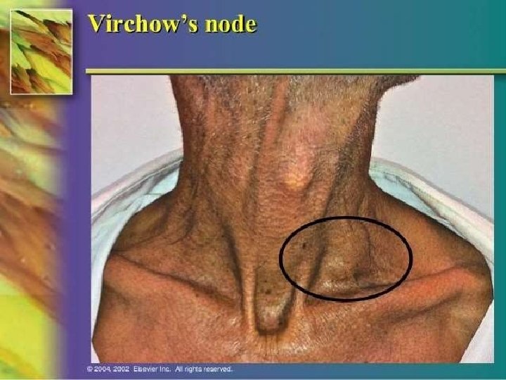
SPECIAL SIGNS �Linitis plastica: --- diffusely infiltrating with a rigid stomach �Virchow’s node: --- left supraclavicular lymph node �Sister Mary Joseph’s node: --- umbilical lymph node �prerectal pouch mass (Blumer shelf) --- seeding metastasis

SISTER MARY JOSEPH’S NODE

COMPLICATIONS Peritoneal and pleural effusion Obstruction of gastric outlet or small bowel Bleeding Intrahepatc jaundice by hepatomegaly

STAGES OF GASTRIC CANCER �Stage I. At this stage, the tumor is limited to the layer of tissue that lines the inside of the stomach. Cancer cells may also have spread to a limited number of nearby lymph nodes. �Stage II. The cancer at this stage has spread deeper, growing into the muscle layer of the stomach wall. Cancer may also have spread to more of the lymph nodes. �Stage III. At this stage, the cancer may have grown through all the layers of the stomach and spread to nearby structures. Or it may be a smaller cancer that has spread more extensively to the lymph nodes. �Stage IV. This stage indicates that the cancer has spread to distant areas of the body.
 �P-factor(Peritoneal dissemination) �H-factor(Thepresenceofhepaticmetastases) �N-factor(Lymphnodesinvolvement) �S-factor(Serosalinvasion)")
. PHNS Staging System (Japanese) �P-factor(Peritoneal dissemination) �H-factor(Thepresenceofhepaticmetastases) �N-factor(Lymphnodesinvolvement) �S-factor(Serosalinvasion)

Physical examination LABORATORY TESTS �blood test • check for anemia(may be caused by bleeding, liver dysfunction, or poor nutrition. • test for the presence of H. pylori bacteria. �Fecal occult blood test (FOBT) �Tumor markers • CEA: carcino-embryonic antigen • CA 19 -9: carbohydrate antigen • CA 724: carbohydrate antigen

�The best way to stage the tumor locally is via endoscopic ultrasound , it gives (80%) information about the depth of tumor penetration into the gastric wall, and can usually show enlarged (>5 mm) perigastric and celiac lymph nodes.
 is")
ENDOSCOPIC MUCOSAL RESECTION Gastric cancer lesion confined to mucosa layer Endoscopic ultrasound (EUS) is helpful in staging GC
 endoscopy has become the gold standard")
ENDOSCOPIC FEATURES OF GASTRIC CANCER Esophagogastro duodenoscopy (EGD) endoscopy has become the gold standard for the diagnosis of gastric malignancy.

�Biopsy. taking a small piece of tissue from the stomach to look at under a microscope for signs of cancer cells. It might done during an endoscopy.

RADIOLOGIC DIAGNOSIS Distal GC Proximal GC Linitis plastica

X-ray test. with barium. The fluid coats the stomach and makes it show up more clearly on X-rays.

� CT SCAN TEST

� MRI : benign regional lymph nodes

Positron Emission Tomography Scanning Whole-body PET scanning uses the principle that tumor cells preferentially accumulate positron-emitting 18 Ffluorodeoxyglucose. This modality is most useful in the evaluation of distant metastasis in gastric cancer but can also be useful in locoregional staging. PET scan is most useful when combined with spiral CT (PET-CT)95 and should be considered before major surgery in patients with particularly high-risk tumors or multiple medical comorbidities
.")
POSITRON EMISSION TOMOGRAPHY (PET).

PET CT SCAN

DETECTION OF EARLY GASTRIC CANCER Endoscopic screening general population or high risk persons • Careful observation • Japan is the only country that had conducted large nationwide mass population screening of asymptomatic individuals for gastric malignancy •

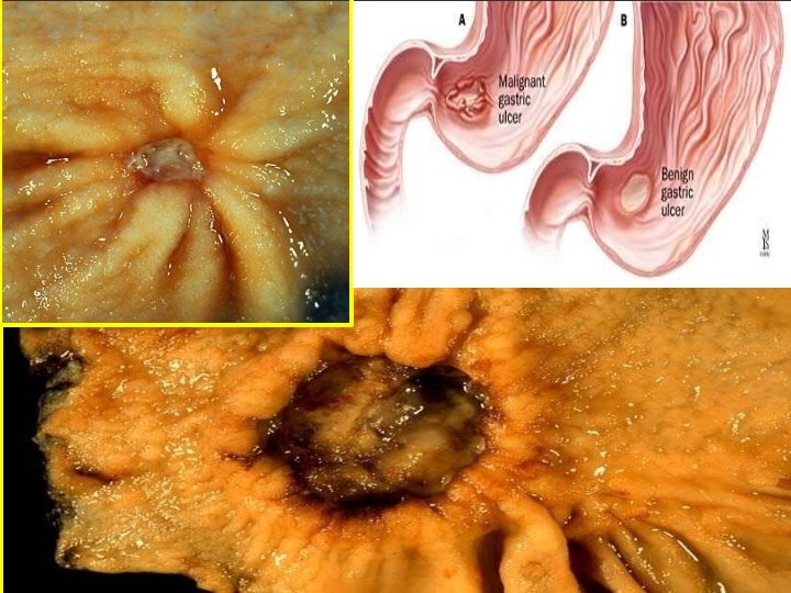
DIFFERENTIAL DIAGNOSIS Gastric Cancer Gastric Ulcer

X-ray showing Gastric ulcer With symmetrical radiating Mucosal folds. By histology, no evidence of Malignancies was observed. X-ray showing Extensive carcinoma involving the cardia & Fundus Pyloric stenosis

IF YOU SEE ULCER ASK UR SELF…BENIGN OR MALIGNANT? BENIGN MALIGNANT Round to oval punched out lesion with straight walls & flat smooth base Irregular outline with necrotic or hemorrhagic base Smooth margins with normal surrounding mucosa Irregular & raised margins Mostly on lesser curvature Anywhere Majority<2 cm Any size Normal adjoining rugal folds that extend to the margins of the base Prominent & edematous rugal folds that usually do not extend to the margins

Treatment Surgical resection is the only curative treatment for gastric cancer. Obvious exceptions include patients who cannot tolerate an abdominal operation, and patients with overwhelming metastatic disease. The goal of curative surgical treatment is resection of all tumor. Thus, all margins (proximal, distal, and radial) should be negative and an adequate lymphadenectomy performed.

Treatment Generally, the surgeon strives for a grossly negative margin of at least 5 cm. Some gastric tumors, particularly the diffuse variety, are quite infiltrative and tumor cells can extend well beyond the tumor mass; thus, gross margins beyond 5 cm may be desirable. More than 15 resected lymph nodes are required for adequate staging. The primary tumor may be resected en bloc with adjacent involved organs (e. g. , distal pancreas, transverse colon, or spleen) during the course of curative gastrectomy. Palliative gastrectomy may be indicated in some patients with obviously incurable disease, but most patients presenting with stage IV gastric cancer can be managed without major operation.

Extent of Gastrectomy The standard operation for gastric cancer is radical subtotal gastrectomy. Subtotal gastric resection typically entails ligation of the left and right gastric and gastroepiploic arteries at the origin, as well as the en bloc removal of the distal 75% of the stomach, including the pylorus and 2 cm of duodenum, the greater and lesser omentum, and all associated lymphatic tissue. Reconstruction is usually by Billroth II gastrojejunostomy, but if a small gastric remnant is left (<20%), a Roux-en-Y reconstruction is considered. The operative mortality is around 2 to 5%. Radical subtotal gastrectomy is generally deemed to be an adequate cancer operation in most Western countries, provided that the contingencies stated result in tumor-free margins, >15 lymph nodes, and the resection of all gross tumor. In the absence of involvement by direct extension, the spleen and pancreatic tail are not removed.

Extent of Lymphadenectomy The Japanese Research Society for Gastric Cancer has numbered the lymph node stations that potentially drain the stomach. Generally these are grouped into level D 1 (i. e. , stations 3 to 6), level D 2 (i. e. , stations 1, 2, 7, 8, and 11), and level D 3 (i. e. , stations 9, 10, and 12) nodes. Generally, D 1 nodes are perigastric, D 2 nodes are along the hepatic and splenic arteries, and D 3 nodes are the most distant. The operation described above (radical subtotal gastrectomy in the Extent of Gastrectomy section), which is by far the most commonly performed procedure in the United States for gastric cancer, is called a D 1 resection because it removes the tumor and the perigastric D 1 nodes.

Extent of Lymphadenectomy The standard operation for gastric cancer in Asia and specialized U. S. centers is the D 2 gastrectomy, which involves a more extensive lymphadenectomy (removal of the D 1 and D 2 nodes). In addition to the tissue removed in a D 1 resection, the standard D 2 gastrectomy removes the peritoneal layer over the anterior mesocolon and selectively over the pancreas, along with nodes along the hepatic and splenic arteries, and the crural nodes.

Extent of Lymphadenectomy Splenectomy and distal pancreatectomy are not routinely performed, because this clearly has been shown to increase the morbidity of the operation. The purported survival advantage of D 2 gastrectomy in gastric cancer is shown in Table 26 -20, which shows the 5 -year survival rates for gastric cancer stratified by pathologic stage for the United States and Japan. Unfortunately, the randomized prospective trials that have been performed have not confirmed this survival advantage, but the morbidity and mortality in the D 2 group was higher This was mostly attributable to the splenectomy and distal pancreatectomy, which are no longer routinely done as part of the D 2 gastrectomy.

Gastric Cancer 5 -Year Survival and Operative Mortality in the USA and Japan Maruyama (Japan), 1971– 1985 American College of Surgeons, 1982– 1987 No. patients 3176 18, 365 Stage I 91% 50% Stage II 72% 29% Stage III 44% 13% Stage IV 9% 3% Operative mortality 1% 7%

Randomized Trials Comparing D 1 and D 2 Gastrectomy for Gastric Cancer Authors Numbe Type of Postoperative 5 -Year r of Surgery Complications Mortality (%) Survival (%) Patients (%) Bonenkamp et al. 711 Cuschieri et al. 400 D 1 D 2 25 43 28 46 4 10 6. 5 13 45 47 35 33

Chemotherapy and Radiation for Gastric Cancer In general, the actuarial 5 -year survival for resected gastric adenocarcinoma stages 1, 2, and 3 is about 75%, 50%, and 25%, respectively. Because most surgical patients have stage 2 disease or greater, it is common to refer gastric cancer patients postoperatively to a medical and/or radiation oncologist. Adjuvant treatment with chemotherapy (5 -fluorouracil and leucovorin) and radiation (4500 c. Gy) has demonstrated a survival benefit in resected patients with stage II and III adenocarcinoma of the stomach.

Chemotherapy and Radiation for Gastric Cancer • Adequacy of lymphadenectomy has clearly been shown to impact survival, particularly in patients with stage III gastric cancer it has been suggested that the benefits of adjuvant chemoradiation shown in this study would be vitiated by an adequate operation. • A recently published study from the Japan Clinical Oncology Group showed a 69% overall 5 -year survival rate in patients with clinically curable T 2 b, T 3, and T 4 gastric cancer, treated with D 2 gastrectomy alone (no chemotherapy) • There was no incremental benefit from para-aortic lymph node dissection. There is no indication for the routine use of radiation alone in the adjuvant setting, but in certain patients, it can be effective palliation for bleeding or pain. In patients with gross unresectable, metastatic, or recurrent disease, palliative chemotherapy has not been demonstrated to conclusively prolong survival, but occasionally, a patient has a dramatic response. These patients should be considered for clinical trials. Agents that have shown activity against gastric cancer include 5 -fluorouracil, cisplatin, doxorubicin, and methotrexate. Neoadjuvant treatment of gastric adenocarcinoma is being evaluated, particularly in patients with clinical T 3 or N 1 disease.

Endoscopic Resection • It has been demonstrated initially at numerous East Asian centers that some patients with early gastric cancer can be adequately treated by an EMR. • Small tumors (<3 cm) confined to the mucosa have an extremely low chance of lymph node metastasis (3%), which approaches the operative mortality rate for gastrectomy. • If the resected specimen demonstrates no ulceration, no penetration of the muscularis mucosae, no lymphatic invasion, and size <3 cm, then the risk of lymph node metastases is less than 1%. • Thus, some patients with early gastric cancer might be better treated with the endoscopic technique. • Currently, this should be limited to patients with tumors <2 cm in size that are node negative and confined to the mucosa on EUS, in the absence of other gastric lesions.

- Slides: 49