GAS EXCHANGE in Animals Cells require O 2


 – Skin must be moist – organisms")


 extend")


 Air Lungs Muscles contract")


 and closed (right) stoma of a spider plant")


- Slides: 52
GAS EXCHANGE in “Animals” • Cells require O 2 for aerobic respiration and expel CO 2 as a waste product
Fick’s Law of Diffusion • Gas exchange involves the diffusion of gases across a membrane • Rate of diffusion (R) is governed by Fick’s Law: • R = DA p d D= diffusion constant (size of molecule, membrane permeability, etc) A= area over which diffusion occurs p = pressure difference between sides of the membrane d = distance across which diffusion must occur
Fick’s Law of Diffusion R = DA p d To maximize diffusion, R can be increased by: Increasing A (area over which diffusion occurs) Increasing p (pressure difference between sides of the membrane) Decreasing d (distance across which diffusion must occur) Evolutionary changes have occurred to maximize R
Figure 42. 18 The role of gas exchange in bioenergetics
GAS EXCHANGE in “Animals” • The part of the organism across which gases are exchanged with the environment is the respiratory surface
Respiratory Surfaces • Must be moist – plasma membranes must be surrounded by water to be stable • Must be sufficiently large – maximize A in Fick’s Law
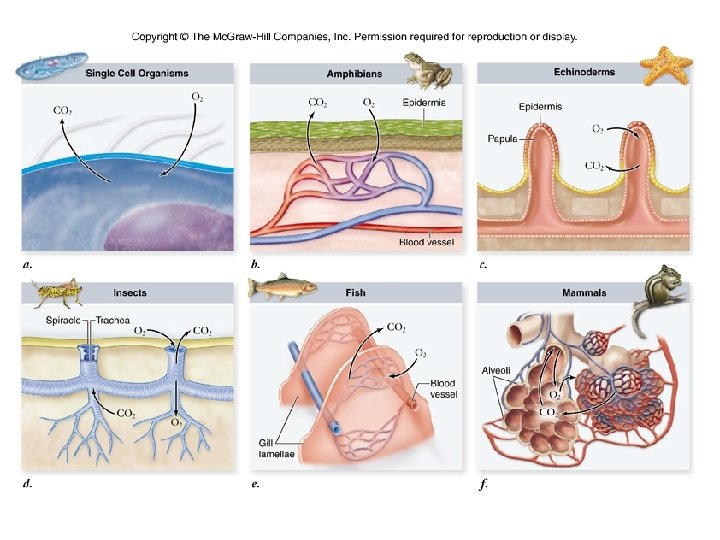
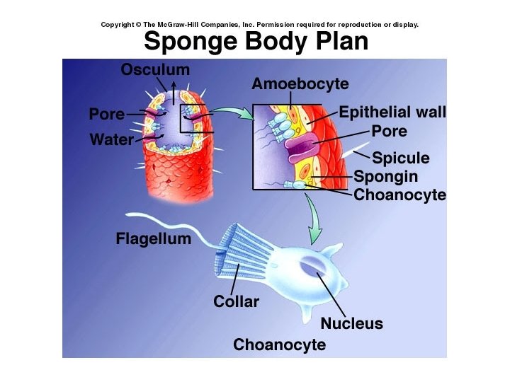
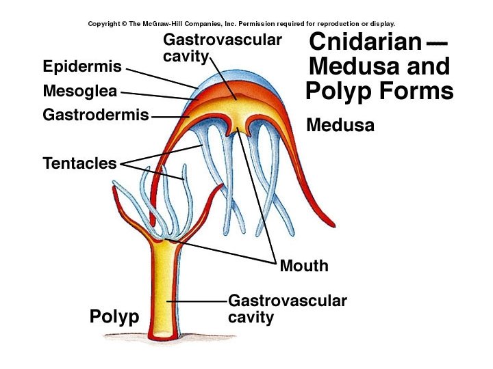
Comparative Respiratory Systems • Cell Membranes – in unicellular organisms – some simpler animals (sponges, cnidarians, flatworms)
Comparative Respiratory Systems • Respiratory surface = a single layer of epithelial cells – separates outer respiratory medium (air or water) from the organism’s transport system (blood)
Comparative Respiratory Systems • Skin (cutaneous respiration) – Skin must be moist – organisms with flat or wormlike bodies so skin in sufficient surface area – or in frogs and some turtles to supplement respiration using lungs
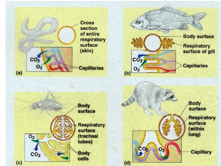
Comparative Respiratory Systems • Specialized region of body is folded and branched to provide large surface area • This maximizes A in Fick’s Law • Also decrease d by bringing the respiratory medium close to the internal fluid • Three such systems: – Gills (Aquatic organisms) – Trachea (insects) – Lungs (terrestrial vertebrates)
Figure 42. 19 Diversity in the structure of gills, external body surfaces functioning in gas exchange
Gills • most aquatic organisms • outfoldings of the body surface specialized for gas exchange • Water is the respiratory medium
Water as Respiratory Medium • Respiratory surface always moist • Oxygen content of water is much less than that of air • denser medium so harder to ventilate
Ventilation • Any method that increases the flow of the respiratory medium across the respiratory surface • This maximizes p in Fick’s Law – By constantly have new air or new water with more oxygen
Ventilation • requires a lot of energy to ventilate gills b/c water is denser than air • pumping operculum, ram ventilation
Gills Copyright © The Mc. Graw-Hill Companies, Inc. Permission required for reproduction or display. Buccal cavity Operculum Oral valve Water Mouth opened, jaw lowered Gills Opercular cavity Mouth closed, operculum opened
Copyright © The Mc. Graw-Hill Companies, Inc. Permission required for reproduction or display. Operculum Gills Water flow Gill arch Water flow Gill raker Oxygenrich blood Oxygen- Oxygenrich deficient blood Gill filaments Oxygendeficient blood Water flow Gill filament Lamellae with capillary networks Blood flow
Countercurrent Exchange • Enhances gas exchange in the gills of fish • blood is continually loaded with O 2 b/c it meets water with increasing O 2 concentration – Increases p in Fick’s Law
Copyright © The Mc. Graw-Hill Companies, Inc. Permission required for reproduction or display. Countercurrent Exchange Concurrent Exchange Water (100% Blood (85% O 2 saturation) Water (50% Blood (50% O 2 saturation) 85% 100% 80% 90% 70% 80% 60% 70% 50% 60% 50% 40% 60% 30% 40% 30% 70% 20% 30% 20% 80% 15% 10% 90% No further net diffusion Blood (0% O 2 saturation) Water (15% O 2 saturation) a. Blood (0% O 2 saturation) Water (100% O 2 saturation) b.
Air as respiratory medium • Higher oxygen concentration • ventilation is easier b/c air is less dense • respiratory surface loses water to air by evaporation
Air as respiratory medium • Solution… – fold respiratory surface inside the body
Trachea • Air tubes that branch throughout the body • finest tubes (tracheoles) extend to nearly every cell in the body • gas diffuses across moist epithelium that lines the terminal ends
Figure 42. 22 Tracheal systems
Trachea • Found in insects • Open Circulatory system of insects is NOT involved in transporting gases • Ventilation – diffusion – body movements
Lungs • Localized in one area of body – circulatory system must transport gases
Lungs • Ventilation – Positive pressure breathing - frogs
Copyright © The Mc. Graw-Hill Companies, Inc. Permission required for reproduction or display. Nostrils open External nostril Air Buccal cavity Esophagus Lungs a. Nostrils closed Air b.
Lungs • Ventilation – Negative pressure breathing- mammals
Negative pressure breathing
Negative pressure breathing Inspiration Sternocleidomastoid muscles contract (for forced inspiration) Air Lungs Muscles contract Diaphragm contracts a. Expiration Air Muscles relax b. Abdominal muscles contract Diaphragm (for forced expiration) relaxes
Lungs Blood flow Bronchiole Smooth muscle Nasal cavity Nostril Glottis Larynx Trachea Right lung Pharynx Pulmonary venule Left lung Pulmonary arteriole Left bronchus Alveolar sac Diaphragm Capillary network on surface of alveoli Alveoli
Lungs • Ventilation – air sacs act as bellows in birds • air flows in one direction during both inhalation & exhalation
Lungs of Birds Cycle 1 Inspiration Expiration Parabronchi of lung Anterior air sacs Posterior Trachea Anterior air sacs Lung Trachea Posterior air sacs Cycle 2 Inspiration a. b. Expiration
Transport of Gases • Occurs in the circulatory system when needed
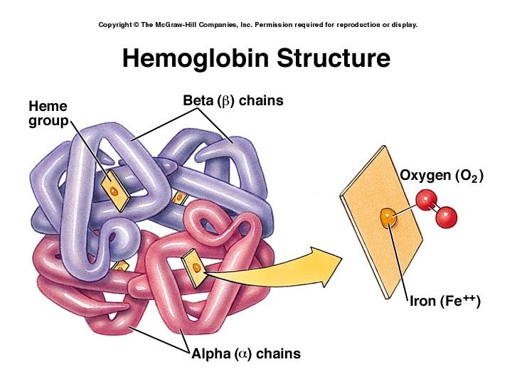
Transport of Gases • O 2 is transported by respiratory pigments – hemoglobin on red blood cells or hemocyanin in the plasma
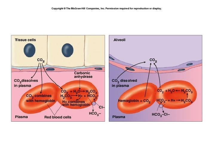
Transport of Gases • CO 2 is transported by respiratory pigments and dissolved in the plasma and in red blood cells as bicarbonate ion (HCO 3 -)
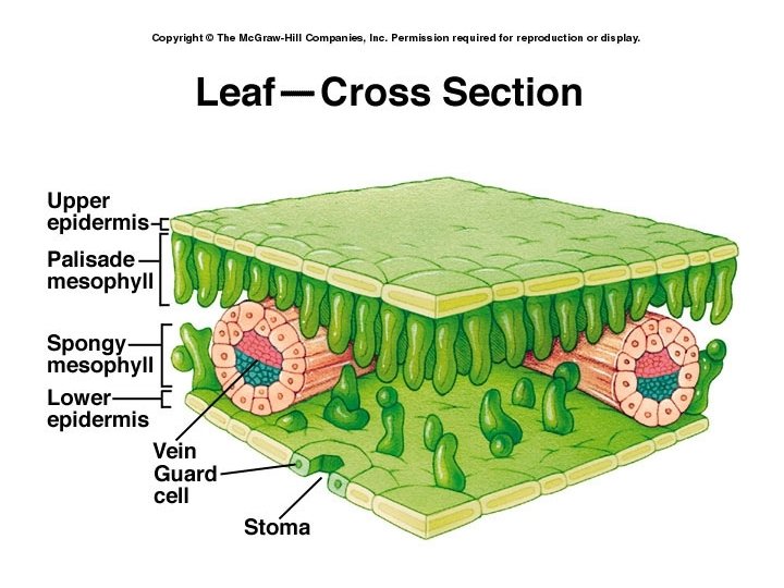
Gas Exchange in Plants • Stomata – tiny pores on the underside of leaves – lead to air spaces in the mesophyll
Gas Exchange in Plants • Guard cells – regulate the opening & closing of stomata – turgid - stomata open, flaccid - stomata close
Figure 36. 12 x Stomata on the underside of a leaf
Figure 36. 12 An open (left) and closed (right) stoma of a spider plant (Chlorophytum colosum) leaf
Figure 36. 13 a The mechanism of stomatal opening and closing
Figure 36. 13 b The mechanism of stomatal opening and closing
Guard cells’ turgor pressure • K+ into guard cells -- water follows due to osmosis, cells become turgid • K+ out of guard cells -- water moves out and cells become flaccid
Stomata • Generally open during the day & closed at night • Cues: – Light – depletion of CO 2 – Circadian rhythms – biological clock