GALLBLADDER By Dr Mahadevi A L Dept of

GALLBLADDER By Dr. Mahadevi A. L Dept of Physiology, SKHMC

• The gallbladder is a hollow organ that sits in a shallow depression below the right lobe of the liver, which is grey-blue in life. • In adults, the gallbladder measures approximately 7 to 10 centimetres in length and 4 centimetres (1. 6 in) in diameter when fully distended. • The gallbladder has a capacity of about 50 millilitres (1. 8 imperial fluid ounces). • The gallbladder is shaped like a pear, with its tip opening into the cystic duct. • The gallbladder is divided into three sections: the fundus, body, and neck. The fundus is the rounded base, angled so that it faces the abdominal wall. The body lies in a depression in the surface of the lower liver. The neck tapers and is continuous with the cystic duct, part of the biliary tree.

• The gallbladder fossa, against which the fundus and body of the gallbladder lie, is found beneath the junction of hepatic segments IVB and V. • The cystic duct unites with the common hepatic duct to become the common bile duct. At the junction of the neck of the gallbladder and the cystic duct, there is an out-pouching of the gallbladder wall forming a mucosal fold known as "Hartmann's pouch". • Lymphatic drainage of the gallbladder follows the cystic node which is located between cystic duct and common hepatic ducts. • Lymphatics from the lower part of the drain into lower hepatic lymph nodes. All the lymph finally drains into celiac lymph node.

Function: • The main function of the gallbladder is to store bile, also called gall, needed for the digestion of fats in food. • Produced by the liver, bile flows through small vessels into the larger hepatic ducts and ultimately through the cystic duct (parts of the biliary tree) into the gallbladder, where it is stored. • At any one time, 30 to 60 millilitres (1. 0 to 2. 0 US fl oz) of bile is stored within the gallbladder. • When food containing fat enters the digestive tract, it stimulates the secretion of cholecystokinin (CCK) from I cells of the duodenum and jejunum. In response to cholecystokinin, the gallbladder rhythmically contracts and releases its contents into the common bile duct, eventually draining into the duodenum.

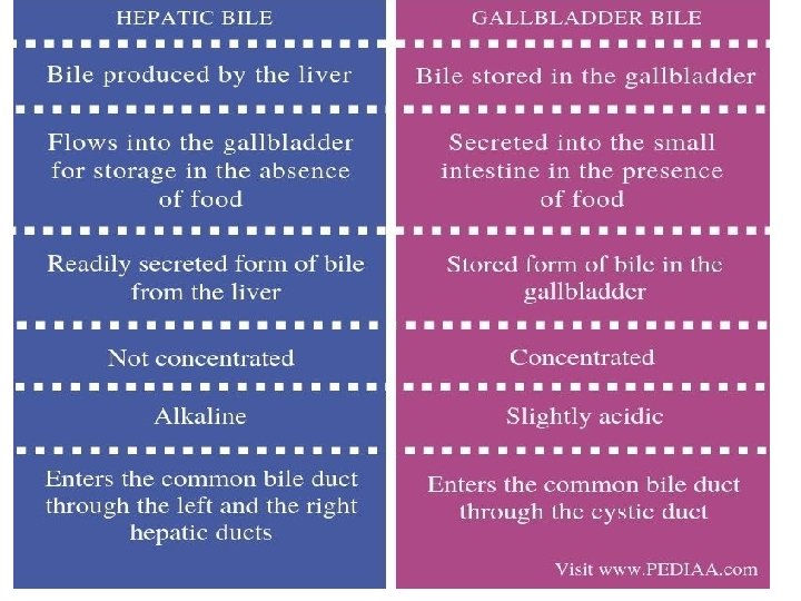
• The bile emulsifies fats in partly digested food, thereby assisting their absorption. • Bile consists primarily of water and bile salts, and also acts as a means of eliminating bilirubin, a product of hemoglobin metabolism, from the body. • The bile that is secreted by the liver and stored in the gallbladder is not the same as the bile that is secreted by the gallbladder. • During gallbladder storage of bile, it is concentrated 310 fold by removal of some water and electrolytes. • This is through the active transport of sodium and chloride ions across the epithelium of the gallbladder, which creates an osmotic pressure that also causes water and other electrolytes to be reabsorbed.

Gallstones form when the bile is saturated, usually with either cholesterol or bilirubin • Most gallstones do not cause symptoms, with stones either remaining in the gallbladder or passed along the biliary system. • When symptoms occur, severe "colicky" pain in the upper right part of the abdomen is often felt. • If the stone blocks the gallbladder, inflammation known as cholecystitis may result. • If the stone lodges in the biliary system, jaundice may occur; if the stone blocks the pancreatic duct, pancreatitis may occur. Gallstones are diagnosed using ultrasound. • When a symptomatic gallstone occurs, it is often managed by waiting for it to be passed naturally. Given the likelihood of recurrent gallstones, surgery to remove the gallbladder is often considered. • Some medication, such as ursodeoxycholic acid, may be used; lithotripsy, a procedure used to break down the stones, may also be used.

Inflammation • Known as cholecystitis, inflammation of the gallbladder is commonly caused by obstruction of the duct with gallstones, which is known as cholelithiasis. • Blocked bile accumulates, and pressure on the gallbladder wall may lead to the release of substances that cause inflammation, such as phospholipase. There is also the risk of bacterial infection. • An inflamed gallbladder is likely to cause pain, fever, and tenderness in the upper, right corner of the abdomen, and may have a positive Murphy's sign. • Cholecystitis is often managed with rest and antibiotics, particularly cephalosporins and, in severe cases, metronidazole.

Tests • Abdominal ultrasonography showing biliary sludge and gallstones • Tests used to investigate for gallbladder disease include blood tests and medical imaging. • A full blood count may reveal an increased white cell count suggestive of inflammation or infection. • Tests such as bilirubin and liver function tests may reveal if there is inflammation linked to the biliary tree or gallbladder, and whether this is associated with inflammation of the liver, and a lipase or amylase may be elevated if there is pancreatitis.

• Bilirubin may rise when there is obstruction of the flow of bile. A CA 19 -9 level may be taken to investigate for cholangiocarcinoma. • An ultrasound is often the first medical imaging test performed when gallbladder disease such as gallstones are suspected. An abdominal X-ray or CT scan is another form of imaging that may be used to examine the gallbladder and surrounding organs. • Other imaging options include MRCP (magnetic resonance cholangiopancreatography), • ERCP and percutaneous or intraoperative cholangiography. • A cholescintigraphy scan is a nuclear imaging procedure used to assess the condition of the gallbladder.

Thank u
- Slides: 11