
Fundamentals of Sensation and Perception THE VIS UAL



- Slides: 33
Fundamentals of Sensation and Perception THE VIS UAL B RAIN ERIK C HEVRIER NOVE MB ER 16, 2015
Video What are all those Floaty Things in Your Eye? The Evolution of the Human Eye Optical Illusions Show How We See
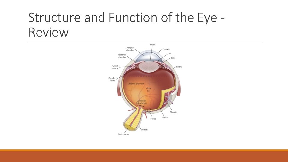
Structure and Function of the Eye Review
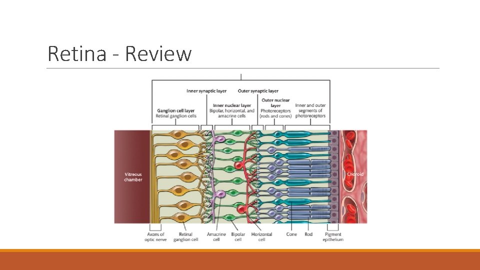
Retina - Review
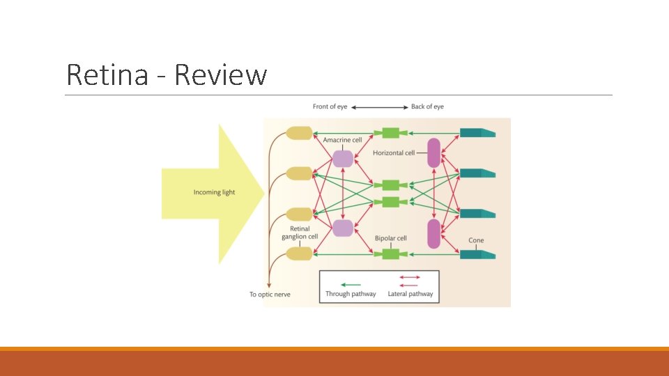
Retina - Review
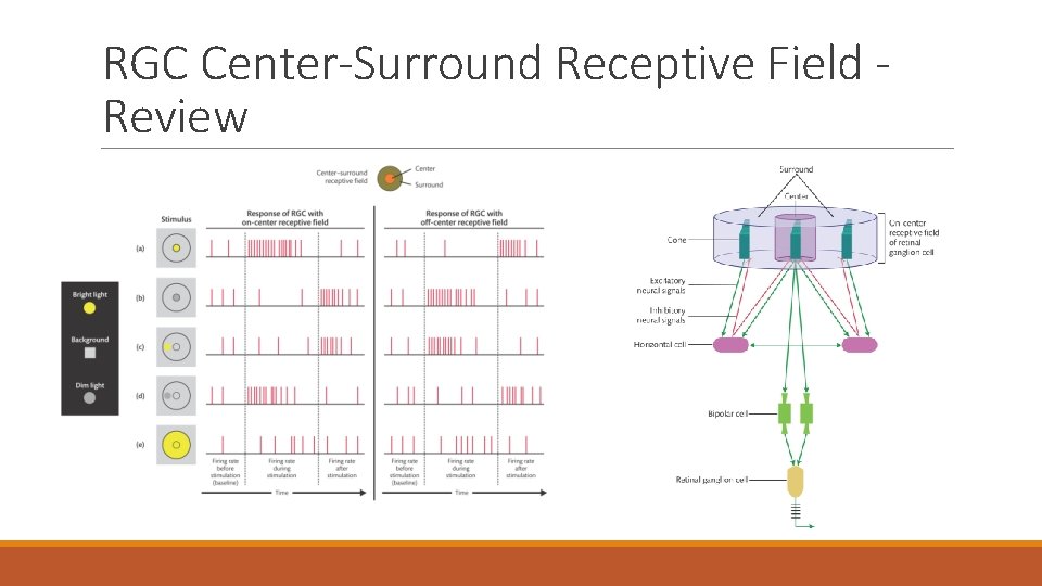
RGC Center-Surround Receptive Field Review
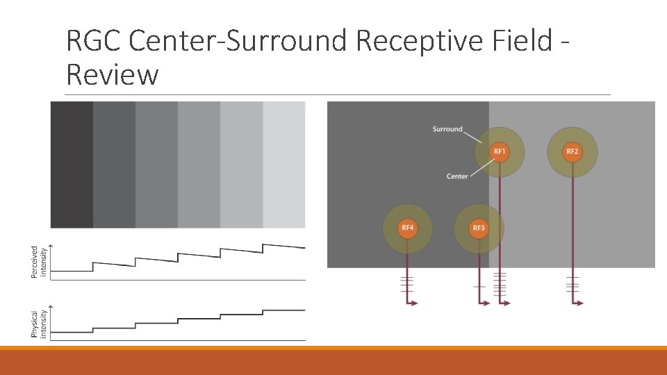
RGC Center-Surround Receptive Field Review
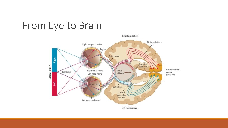
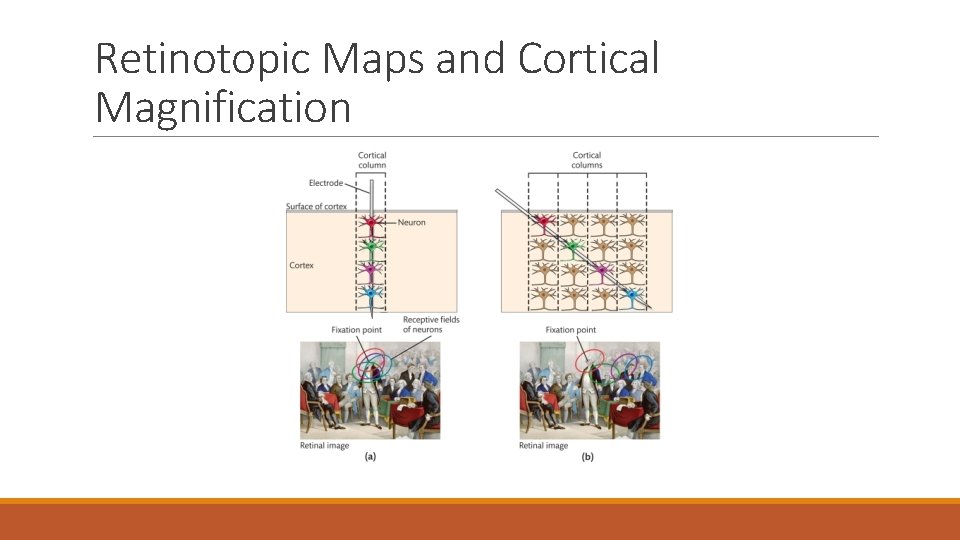
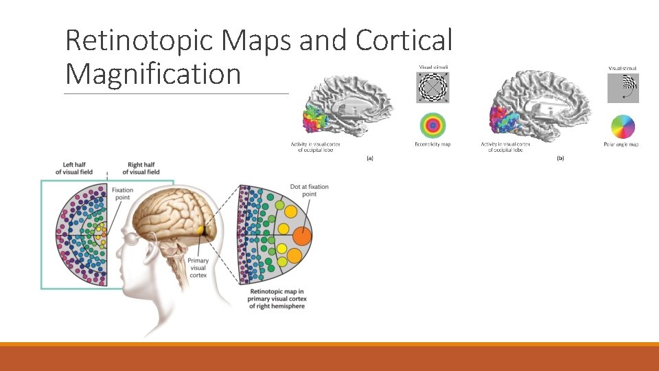
The Visual Brain Pathways and networks are organized by their: ◦ Functional specialization Shape, colour, motion, depth ◦ Retinotopic mapping Spatial arrangements of brightness and colour in the retinal image are echoed by the arrangement of the neurons throughout the visual system
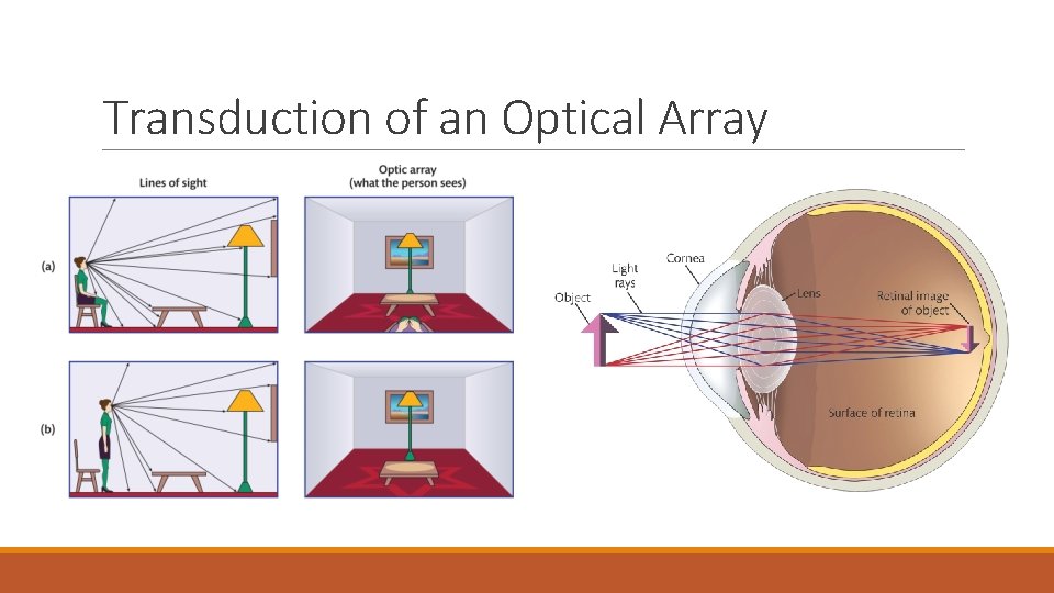
Transduction of an Optical Array
From Eye to Brain
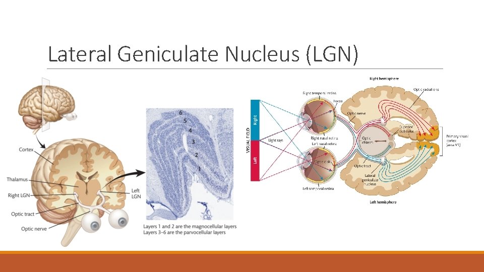
Lateral Geniculate Nucleus (LGN)
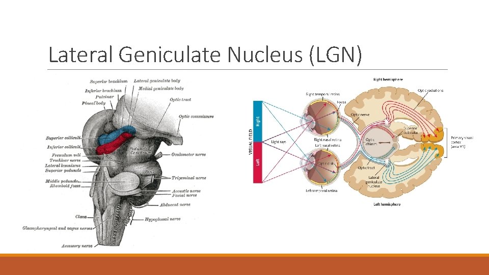
Lateral Geniculate Nucleus (LGN)
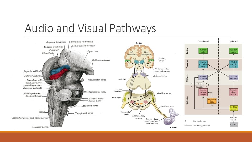
Audio and Visual Pathways
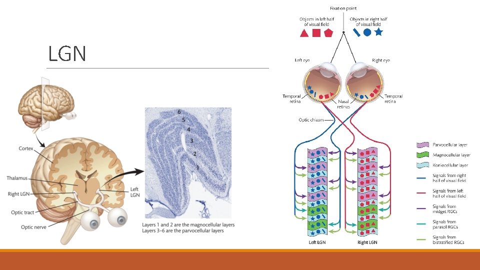
LGN
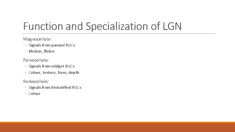
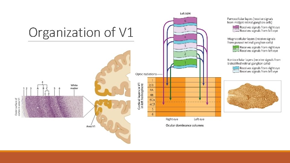
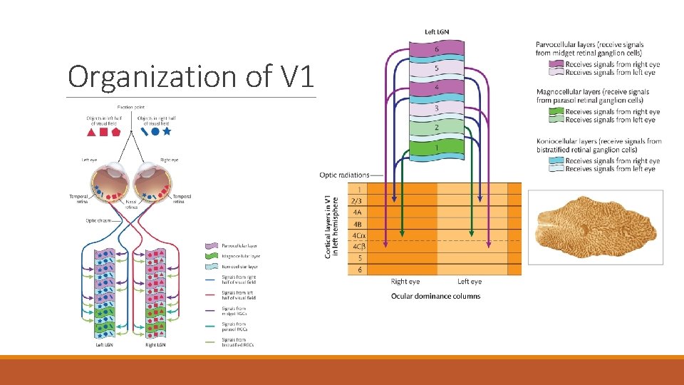
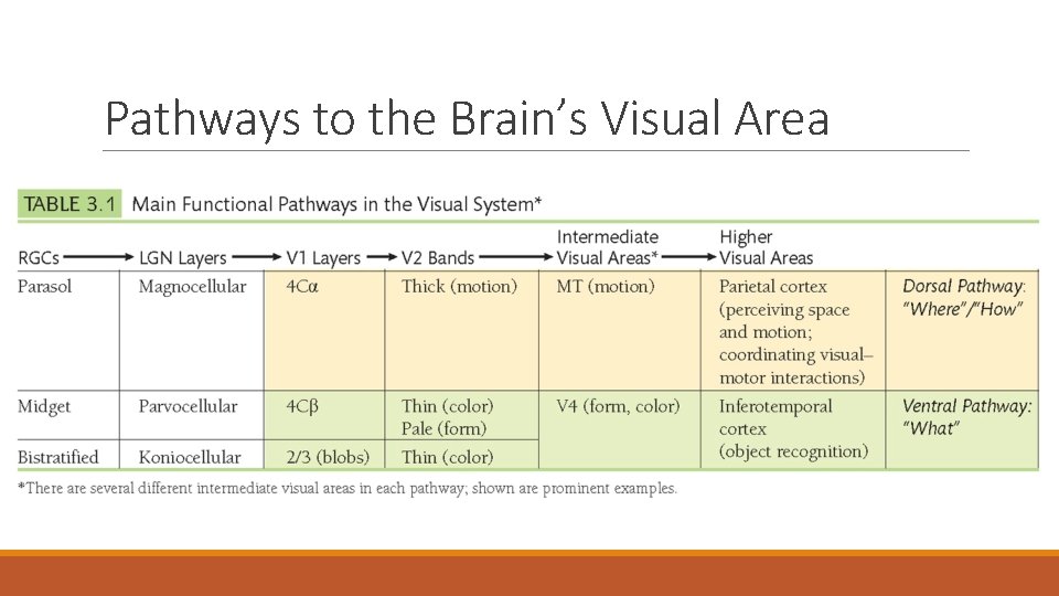
Function and Specialization of LGN Magnocellular ◦ Signals from parasol RGCs ◦ Motion, flicker Parvocellular ◦ Signals from midget RGCs ◦ Colour, texture, form, depth Koniocellular ◦ Signals from bistratified RGCs ◦ Colour
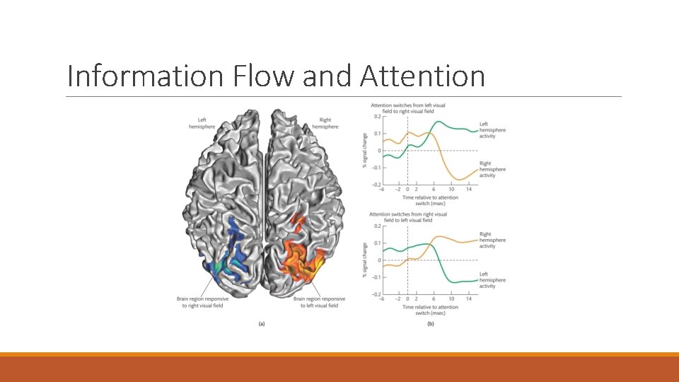
Information Flow and Attention
Information Flow and Attention
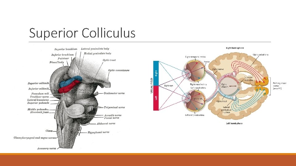
Superior Colliculus
Superior Colliculus Principal function is to control rapid eye movements to visual targets Cells are more concerned with where things are rather than what they are Arrive almost immediately after leaving the eye Travels from retina to areas beyond V 1 receptor (Blindsight) Receives signals from auditory and somatosensory systems (multisensory organ) ◦ Systems reinforce each other
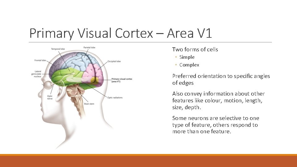
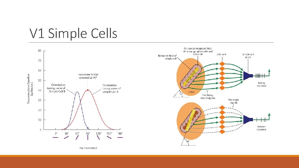
Primary Visual Cortex – Area V 1 Two forms of cells ◦ Simple ◦ Complex Preferred orientation to specific angles of edges Also convey information about other features like colour, motion, length, size, depth. Some neurons are selective to one type of feature, others respond to more than one feature.
V 1 Simple Cells
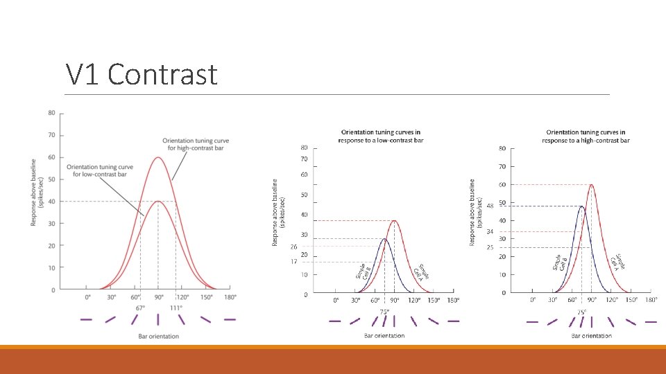
V 1 Contrast
Complex Cells Complex cells are like simple cells, they are tuned for orientation – they respond well to bars within a specific range of orientations flashed on their receptive field. They are unlike simple cells in two main ways: 1 – Complex cells respond as well to a light on a dark background as to a dark bar on a light background. Simple cells respond well to one or the other, but not both. 2 – Complex cells respond equally well to a bar at almost any location within their receptive field. Simple cells respond best to a bar at a very specific location within their receptive field.
Organization of V 1
Organization of V 1
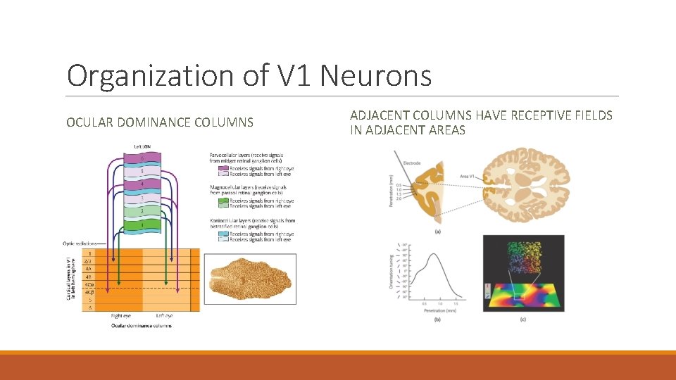
Organization of V 1 Neurons OCULAR DOMINANCE COLUMNS ADJACENT COLUMNS HAVE RECEPTIVE FIELDS IN ADJACENT AREAS
Retinotopic Maps and Cortical Magnification
Retinotopic Maps and Cortical Magnification
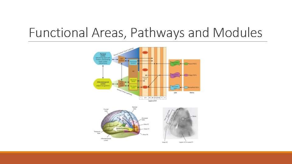
Functional Areas, Pathways and Modules
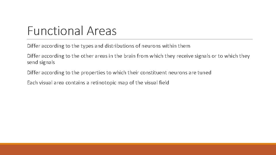
Functional Areas Differ according to the types and distributions of neurons within them Differ according to the other areas in the brain from which they receive signals or to which they send signals Differ according to the properties to which their constituent neurons are tuned Each visual area contains a retinotopic map of the visual field
Pathways to the Brain’s Visual Area
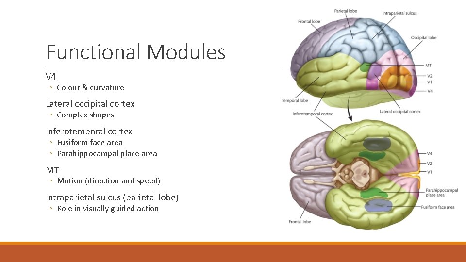
Functional Modules V 4 ◦ Colour & curvature Lateral occipital cortex ◦ Complex shapes Inferotemporal cortex ◦ Fusiform face area ◦ Parahippocampal place area MT ◦ Motion (direction and speed) Intraparietal sulcus (parietal lobe) ◦ Role in visually guided action
Questions? Have a great day!