FUNDAMENTALS OF ECG READINGS Learning objectives On completion

FUNDAMENTALS OF ECG READINGS.

Learning objectives On completion of this lecture the students will be able to : Ø Explain the CARDIAC CONDUCTION SYSTEM Ø Explain ELECTROCARDIOGRAPHIC LEADS. Ø Define THE ELECTRICITY OF THE HEART. Ø Describe THE ECG PAPER. Ø Explain ECG WAVES AND INTERVALS. Ø Incorporate CHARACTERISTICS OF ECG COMPONENTS, ELECTRICAL AND MYOCARDIAL ACTIVITY. Ø Interpret the ECG waves on ECG PAPER. 03/09/2021 2

OUTLINE: Ø CARDIAC CONDUCTION SYSTEM Ø ELECTROCARDIOGRAPHIC LEADS. Ø THE ELECTRICITY OF THE HEART. Ø THE ECG PAPER. Ø ECG WAVES AND INTERVALS. Ø CHARACTERISTICS OF ECG COMPONENTS, ELECTRICAL AND MYOCARDIAL ACTIVITY. Ø INTERPRETATIONS OF ECG.
, atrio-ventricular (AV), and intraventricular (IV) conduction.")
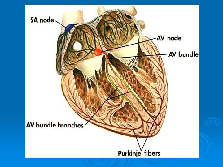
"Normal“ conduction implies normal sino-atrial (SA), atrio-ventricular (AV), and intraventricular (IV) conduction.

03/09/2021 6

ECG Intervals and Waves Ø The P wave represents atrial activation; the PR interval is the time from onset of atrial activation to onset of ventricular activation. Ø The QRS complex represents ventricular activation; the QRS duration is the duration of ventricular activation. Ø The ST-T wave represents ventricular repolarization. The QT interval is the duration of ventricular activation and recovery. The U wave probably represents "after depolarizations" in the ventricles. 03/09/2021 7
 PR interval")
ECG Intervals and Waves Heart rate (state atrial and ventricular, if different) PR interval (from beginning of P to beginning of QRS) QRS duration (width of most representative QRS) 03/09/2021 The P wave represents atrial activation; the PR interval is the time from onset of atrial activation to onset of ventricular activation. The QRS complex represents ventricular activation; the QRS duration is the duration of ventricular activation. The STT wave represents ventricular repolarization. The QT interval is the duration of ventricular activation and recovery. The U wave probably represents "afterdepolarizations" in the ventricles. 8

03/09/2021 9

1. Heart Rate Ø In normal sinus rhythm, a resting heart rate of below 60 bpm is called bradycardia and a rate of above 90 bpm is called tachycardia. 2. PR Interval Ø (measured from beginning of P to beginning of QRS in the frontal plane) Ø Normal: 0. 12 - 0. 20 s Ø Short PR: <0. 12 s Ø Prolonged PR: >0. 20 s (First degree AV block (PR interval usually constant) 03/09/2021 10
: Ø Normal: 0. 06")
3. QRS Duration (duration of QRS complex in frontal plane): Ø Normal: 0. 06 - 0. 10 s Ø Prolonged QRS Duration (>0. 10 s): QRS duration 0. 10 - 0. 12 s a. Incomplete right or left bundle branch block b. Nonspecific intraventricular conduction delay (IVCD) c. Some cases of left anterior or posterior fascicular block 03/09/2021 11

4. QT Interval Ø Ø Ø a. b. c. d. e. measured from beginning of QRS to end of T wave in the frontal plane Normal: heart rate dependent Long QT Syndrome Drugs (many antiarrhythmics, tricyclics, phenothiazines, and others) Electrolyte abnormalities ( K+, Ca++, Mg++) CNS disease (especially subarrachnoid hemorrhage, stroke, trauma) Hereditary LQTS (e. g. , Romano-Ward Syndrome) Coronary Heart Disease (some post-MI patients) 03/09/2021 12

A "Method" of ECG Interpretation NORMAL SINUS RHYTHM Ø Rate: 60 -100 beats per minute. Ø Rhythm: regular Ø P waves: present ; normal (smoothly rounded ; upright in leads I, II, a. VF; inverted in a. VR. ) Ø PR interval: 0. 12 -0. 20 sec. Ø QRS : normally less than o. 12 sec. Ø Conduction : each P wave is followed by one QRS. 03/09/2021 13

The Standard 12 Lead ECG Waves and Intervals: Ø P wave: the sequential activation (depolarization) of the right and left atria Ø QRS complex: right and left ventricular depolarization (normally the ventricles are Ø activated simultaneously) Ø ST-T wave: ventricular repolarization 03/09/2021 14

Orientation of the 12 Lead ECG Ø It is important to remember that the 12 lead ECG provides special information Ø about the heart's electrical activity in 3 approximately orthogonal directions: Ø Right Left Ø Superior Inferior Ø Anterior Posterior 03/09/2021 15

Each of the 12 leads represents a particular orientation in space, (RA = right arm; LA = left arm, LF = left foot) Bipolar limb leads (frontal plane): Ø Lead I: RA (-) to LA (+) (Right Left, or lateral) Ø Lead II: RA (-) to LF (+) (Superior Inferior) Ø Lead III: LA (-) to LF (+) (Superior Inferior) 03/09/2021 16
: Ø Lead a. VR: RA (+) to [LA &")
unipolar limb leads (frontal plane): Ø Lead a. VR: RA (+) to [LA & LF] (-) (Rightward) Ø Lead a. VL: LA (+) to [RA & LF] (-) (Leftward) Ø Lead a. VF: LF (+) to [RA & LA] (-) (Inferior) 03/09/2021 17

• The short PR interval is due to a bypass track, also known as the Kent pathway. • By bypassing the AV node the PR shortens. • The delta wave represents early activation of the ventricles from the bypass tract. • The fusion QRS is the result of two activation sequences, one from the bypass tract and one from the AV node. • The ST-T changes are secondary to changes in the ventricular activation sequence. 03/09/2021 18
- Slides: 18