Fundamentals of Blood Biochemistry BCH 220 DR MANSOUR
 DR. MANSOUR GATASHEH Biochemistry Department, Science College King")
Fundamentals of Blood Biochemistry (BCH 220) DR. MANSOUR GATASHEH Biochemistry Department, Science College King Saud University

Class 7: Anemia T

Objectives for this lecture l Discuss the definition and types anemias. l understand the action of G-6 -PD deficiency; polycythemia.
 l means deficiency of hemoglobin in the blood. l caused by: -")
Anemia (definition) l means deficiency of hemoglobin in the blood. l caused by: - too few RBCs - too little hemoglobin in the cells

Type of Anemia Ø Ø Ø Ø Blood Loss Anemia Aplastic Anemia Due to Bone Marrow Dysfunction Megaloblastic Anemia Hemolytic Anemia G-6 -PD deficiency Iron deficiency anemia Polycythemia


Blood Loss Anemia l l l After rapid hemorrhage. Body replaces the fluid portion of the plasma in 1 to 3 days, but this response results in a low concentration of RBCs. RBC concentration usually returns to normal within 3 to 6 weeks.

Blood Loss Anemia l In case of chronic blood loss: 1. Absorption of iron from the intestines is not enough. 2. Formation of hemoglobin is not rapidly as it is lost. 3. RBCs produced are smaller than normal and have too little hemoglobin inside them. 4. Rise to microcytic, hypochromic anemia.

Aplastic Anemia Due to Bone Marrow l l Bone marrow aplasia means lack of functioning bone marrow. Caused by damage stem cells due to: Ø exposure to high-dose radiation or chemotherapy for cancer treatment. Ø high doses of toxic chemicals, such as insecticides or benzene in gasoline. Ø autoimmune disorders. Ø half of cases the cause is unknown.

Aplastic Anemia Due to Bone Marrow l l People with severe aplastic anemia usually die. Treated by: - Blood transfusions. - Bone marrow transplantation.

Megaloblastic Anemia l l l Slow reproduction of erythroblasts. Result in forming too large RBCs, with odd shapes, and have fragile membranes. Caused by: Ø Loss vit B 12, folic acid, and intrinsic factor (glycoprotein) from stomach mucosa (gastrectomy). Ø Poor absorption of vit B compounds and folic acid by intestine.

Hemolytic Anemia l l l Inhered abnormal RBCs, make the cells fragile. number of RBCs normal, life span is so short. Type of it: ü Hereditary spherocytosis: small RBC, spherical, easily ruptured by compression. ü Erythroblastosis fetalis: Rh(+) RBCs in the fetus are attacked by antibodies from an Rh(-) mother, lead to rupture and causing the child to be born with a anemia.
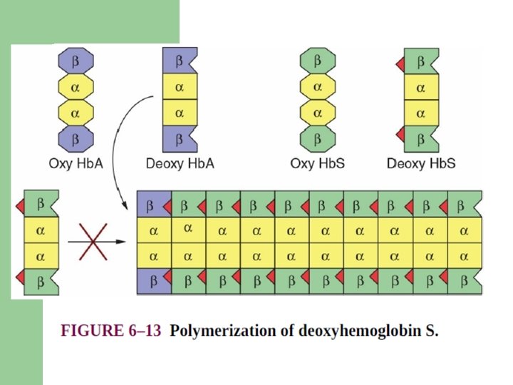
, nonpolar amino acid valine")
Hemolytic Anemia ü Sickle cell anemia: abnormal hemoglobin (Hb S), nonpolar amino acid valine has replaced the polar surface Glu 6 of the β subunit, causing a hydrophobic “sticky patch” on the surface of the β subunit. in low oxygen it precipitates into crystals inside the RBC. Cell is fragile.

Hemolytic Anemia ü Thalassemia: genetic defect that result from the partial or total absence of one or more α or β chains of hemoglobin.

Hemolytic Anemia by infectious and toxic agents ü ü ü Insect and reptile venoms: have phospholipases or proteases that catalyze the hydrolytic breakdown of membrane. infectious bacteria: have lytic factors called hemolysins. Parasitic infections (eg, the plasmodia causing malaria): cause of hemolytic anemias in certain geographic areas

Glucose-6 -Phosphate Dehydrogenase Deficiency l l G 6 PD reduce NADP to NADPH which is needed for the production of glutathione (GSH). GSH protect RBC membranes from oxidant stress. Point mutations or deletions of the enzyme G 6 PD have show less activity than normal. Worldwide over 400 million people are G 6 PD deficient.


Glucose-6 -Phosphate Dehydrogenase Deficiency l Haemolytic anaemia can occur when cells are subjected to oxidant stress from: Ø Infections and other acute illnesses : diabetic ketoacidosis). Ø Drugs: Antimalarials, Sulphonamides, antibacterial, aspirin. Ø Possibly some vegetables: Fava beans

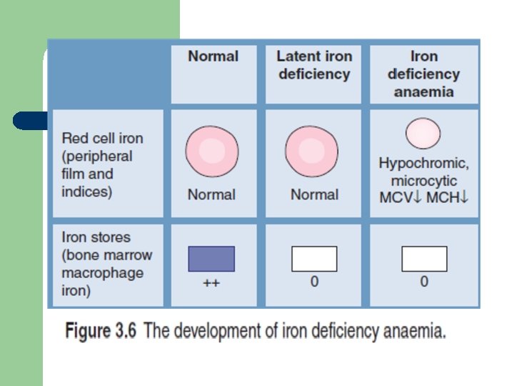
iron deficiency anemia l l l Iron deficiency will decrease hemoglobin synthesis, causing iron-deficient erythropoiesis. Resulting in accumulation of RBC protoporphyrin. Causes can be: Ø Dietary deficiency Ø Malabsorption Ø Gastrointestinal bleeding Ø Episodic blood (menstruation, pregnancy, lactation)

polycythemia l l Production of large quantities of extra RBCs. Occur when tissues become hypoxic because of: ü Little oxygen in the breathed air. ü Cardiac failure (low O 2 delivery). ü Genetic problem in the hemocytoblastic cells (excess RBC, WBC and platelets).

polycythemia l As a result: ü Entire vascular system becomes engorged. ü Blood capillaries become plugged by the viscous blood. ü Viscosity of the blood increases. ü Increase arterial pressure, hypertension develops.

References l l Victor A Hoffbrand, Paul Moss, J Pettit; Essential Haematology. Essentials Series Blackwell Science, New York; 2008. Victor W. Rodwell, David A. Bender, Kathleen M. Botham, Peter J. Kennelly, P. Anthony Weil. Harper’s Illustrated Biochemistry. Mc. Graw-Hill Ed, 31 ed, 2018.
- Slides: 24