Fundamentals of Blood Biochemistry BCH 220 DR MANSOUR
 DR. MANSOUR GATASHEH Biochemistry Department, Science College King")
Fundamentals of Blood Biochemistry (BCH 220) DR. MANSOUR GATASHEH Biochemistry Department, Science College King Saud University
 T")
Class 8: White blood cells (granulocytes, monocytes) T

Objectives for this lecture l Discuss the structure and functions of White blood cells. l understand the abnormalities in white blood cells.
 l l Protecting the body against infection. Two broad groups:")
White blood cells (leukocytes) l l Protecting the body against infection. Two broad groups: - Phagocytes cells of innate immune system. granulocytes (neutrophils, eosinophils, basophils, monocytes). - Lymphocytes adaptive immune response. develop immunological memory.
 Ø Blood count = 1. 8– 7. 5 × 10⁹/L")
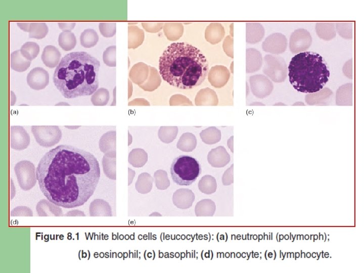
Granulocytes l Neutrophil (polymorph) Ø Blood count = 1. 8– 7. 5 × 10⁹/L (in normal pregnancy= 11 × 109/L ) Ø Ø Ø nucleus of between two and five lobes. Have granule of lysozyme. lifespan is only 6– 10 hours.

Granulocytes l Eosinophils: Ø Blood count = 0. 04– 0. 4 × 10⁹/L Ø more than three nuclear lobes. Ø in allergic responses, defence against parasites and removal of fibrin formed during inflammation.

Granulocytes l Basophils Ø Blood count = 0. 01– 0. 1 × 10⁹/L Ø contain heparin and histamine. Ø Have Ig. E attachment sites. Ø their degranulation is associated with histamine release.

Granulocytes l Monocytes Ø Blood count = 0. 2– 0. 8 × 10⁹/L Ø Large cell, large nucleus, contains many fine vacuoles.

Granulopoiesis l l Granulocytes and monocytes are formed in the bone marrow. Large numbers of neutrophils (10– 15 times than in the blood) are held in the marrow. After being released, they pend only 6– 10 hours in the circulation, then enter tissues to perform phagocytic function. After 4– 5 days in the tissues they are destroyed during defensive action.

Granulopoiesis l l l Monocytes spend a short time in the marrow. Circulate in blood for 20– 40 hours, then enter the tissues. In tissues they mature and carry out their functions. Lifespan may be several months or even years. Involved in antigen presentation to T cells.

Control of granulopoiesis l myeloid growth factors Ø e. g. interleukin‐ 1, granulocyte– macrophage colony‐stimulating factor (GM‐CSF), granulocyte CSF (G‐CSF). Ø Stimulate proliferation and differentiation, function. And also inhibit apoptosis. Ø Growth factors are produced from stromal cells and T lymphocytes.
 Ø chemotactic")
Normal function of neutrophil and monocyte 1. Chemotaxis (cell mobilization and migration) Ø chemotactic substances released from damaged tissues. Ø interaction of leucocyte adhesion molecules with ligands on the damaged tissues.

Normal function of neutrophil and monocyte 2. Phagocytosis Ø The foreign material (e. g. bacteria, fungi) or dead or damaged cells of the host are phagocytosed. Ø Recognition of a foreign particle is aided by binding with immunoglobulin. Ø secrete growth factors and chemokines which regulate inflammation and immune responses.

Normal function of neutrophil and monocyte 3. Killing and digestion Ø By oxygen‐dependent reactions, superoxide (O 2−), hydrogen peroxide (H 2 O 2) and other activated (O 2) species. Ø By oxygen‐independent reactions, Nitric oxide (NO). Ø non‐oxidative mechanisms, cathepsin G, lysozyme, elastase.

Defects of phagocytic cell function Chemotaxis Ø ‘lazy leucocyte’ syndrome due to corticosteroid therapy or myeloid leukaemia, myelodysplasia. l Phagocytosis Ø lack of complement components. l Killing Ø abnormality affecting oxidase, so patients have recurring infections. l

Causes of neutrophil leucocytosis l l levels greater than 7. 5 × 10⁹/L. Causes: - Bacterial infections - Inflammation and tissue necrosis - Metabolic disorders (uraemia, gout) - haemorrhage or haemolysis - Pregnancy - Neoplasms (carcinoma, leukaemia) - Drugs (corticosteroid therapy)

Neutropenia neutrophil level falls below 0. 5 × 10⁹/L l patient will have recurrent infections. l Causes: - Congenital - Drug‐induced (Anti‐inflammatory, Antibacterial drugs) - Autoimmune - Hypersensitivity - Infections (Viral (HIV), bacterial) l

Leukemia l l l Uncontrolled production of abnormal WBCs in the circulating blood. Caused by cancerous mutation of a myelogenous or lymphogenous cell. Types : - Lymphocytic: by cancerous production of lymphoid cells, in a lymph node. - Myelogenous: cancerous

Leukemia l l Acute leukemia often lead to death within a few months if untreated. Leukemic cells, especially the undifferentiated cells, are nonfunctional for providing normal protection against infection. spread to the spleen, lymph nodes, liver, and other vascular regions. Develop infection, anemia, and a bleeding tendency caused by lack of platelets.
 Chronic Myeloid Leukaemia")
Myeloid malignancies Acute Myeloid Leukaemia (AML M-3) Chronic Myeloid Leukaemia

References l l Victor A Hoffbrand, Paul Moss, J Pettit; Essential Haematology. Essentials Series Blackwell Science, New York; 2008. Victor W. Rodwell, David A. Bender, Kathleen M. Botham, Peter J. Kennelly, P. Anthony Weil. Harper’s Illustrated Biochemistry. Mc. Graw-Hill Ed, 31 ed, 2018.
- Slides: 22