Fundamentals of Blood Biochemistry BCH 220 DR MANSOUR
 DR. MANSOUR GATASHEH Biochemistry Department, Science College King")
Fundamentals of Blood Biochemistry (BCH 220) DR. MANSOUR GATASHEH Biochemistry Department, Science College King Saud University

Class 13: Plasma proteins: types, functions and variation in different diseases. T

Objectives for this lecture l Discuss the different type of plasma proteins. l understand the effect of liver and kidney disease on the level of plasma proteins.

Plasma contains a complex mixture of proteins § Concentration 65 – 80 g L Ø groups on the basis solubility (ethanol): Ø Fibrinogen 4% Ø Albumin 60% Ø Globulins 35% Ø Other plasma proteins : 1% Ø rich in disulfide bonds Ø contain carbohydrate (glycoprotein) or lipid (Lipoprotein).

Plasma proteins l electrophoresis within a cellulose acetate for serum protein give: ü Albumin ü α 1 -, α 2 -, β-, and γ- globulins.
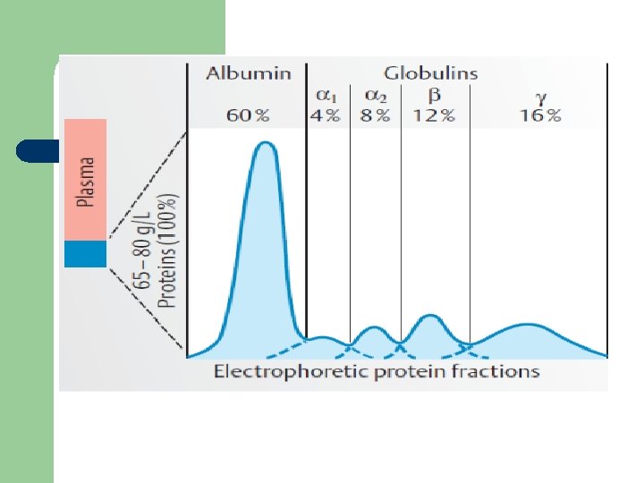

Major Components of globlin bands

Plasma proteins participate in: 1. Blood coagulation 2. Maintenance of homeostasis (p. H, osmotic pressure) 3. Defence against infection 4. Transport of: - nutrients - hormones - Metabolites - metabolic waste - drugs

properties of plasma proteins l Synthesized in the liver. (Eexception: -globulins – synthesized in plasma cells). l have characteristic half-life in the circulation (albumin – 20 days)
 l Class of proteins whose plasma levels change (increase or")
Acute phase reactants (APRs) l Class of proteins whose plasma levels change (increase or decrease) during acute inflammatory response 1. APRs concentration changes in: 1. 2. 3. 4. infection surgery injury cancer
: ~1000 -fold increase!")
Types of APRs Positive Negative 1 -antitrypsin albumin C-reactive protein (CRP): ~1000 -fold increase! transferrin fibrinogen haptoglobin (HP) C 3, C 4

Acute inflammatory response l Immediate response occurs with stress or inflammation caused by infection, injury or surgical trauma l l Normal or ↓ albumin ↑ α 1 and α 2 globulins α 1 α 2 -globulins

Chronic inflammatory response l Late response is correlated with chronic infection (autoimmune diseases, chronic liver disease, chronic infection, cancer) l l l Normal or ↓ albumin ↑α 1 or α 2 globulins ↑↑ γ globulins α 1 α 2 γ-globulins

Nephrotic syndrome l The kidney damage illustrates the long term loss of lower molecular weight proteins l l ↓ albumin and Ig. G – they are filtered in kidney Retention of higher molecular weight proteins l ↑↑ α 2 -macroglobulin and ↑β-globulin) α 2 -globulin β-globulin fractions

Liver damage - Cirrhosis l Cirrhosis can be caused by chronic alcohol abuse or viral hepatitis l l l ↓ albumin ↓ α 1, α 2 and β globulins ↑ Ig A in γ-fraction γ-globulins

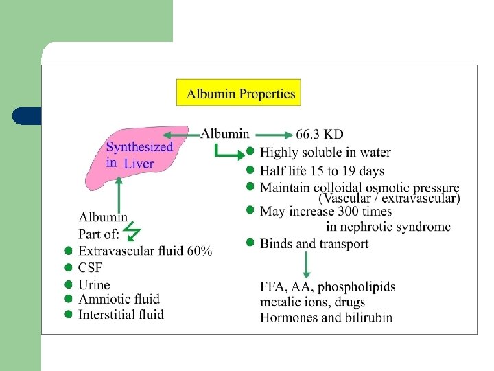
Albumin Not a glycoprotein l has lowest molecular weight of plasma proteins l Liver produces 12 g /day (25% of total hepatic protein synthesis and 50% of secreted protein) l Half-life: 20 days l – For this reason, measurement of serum albumin concentration is used to assays liver function test

Albumin Functions Maintenance of the osmotic pressure of plasma 1. gives greater osmotic effect at the p. H 7. 4 of blood o responsible for about 75 - 80 % of the osmotic effect of plasma because: o • • It constitutes slightly> half the plasma proteins by weight It has the lower molecular weight of the major plasma proteins.

Albumin Functions 2. Transport: It can bind and transport many diverse molecules and serve as low-specificity transport protein, which include: o free fatty acids o steroid hormones o bilirubin o drugs (sulfonamides, aspirin) o Ca 2+, Cu 2+
 – decrease in the ratio of")
Causes of Albumin Deficiency l Liver diseases (cirrhosis) – decrease in the ratio of albumin to globulins l Protein malnutrition l Excessive excretion by kidneys (renal disease) (proteinuria) l Mutation causing analbuminemia (little or no circulating albumin) • There will be a reduction in osmotic pressure, leading to

Transferrin l Beta globulin l Concentration in plasma: 3 g l l Functions: 1. Transport of iron: from catabolism of heme and from food (gut) to the sites where iron is required, i. e. to the bone marrow and other organs 2. 2 moles of Fe 3+ per 1 mole of transferrin

Causes of transferrin deficiency: l Burns l Infections l Malignancies l Liver and kidney diseases l Pregnancy

Ferritin l Intracellular protein; only small portion in plasma l Function: – l Stores iron that can be called upon for use when needed Primary hemochromatosis: – genetic disorder – characterized by increased absorption of iron from the intestine accumulated iron damages organs such as the liver, skin, heart, and pancreas. – concentration of ferritin is elevated.

Ceruloplasmin l 2 -globulins l Conc. in plasma: 300 mg l l Functions: o Carries 90% of copper in plasma (copper – cofactor for a variety of enzymes); o o 1 molecule binds 6 atoms of copper; binds copper more tightly than albumin that carries other 10% of copper Albumin may be more important in copper transport (donates copper to tissues more readily)

Causes of ceruloplasmin decrease l Liver diseases, in particular Wilson´s disease: o o o Genetic disease in which copper fails to be excreted into the bile and accumulates in liver, brain, kidney, and red blood cells Cause: mutations in the gene encoding for copperbinding ATPase Consequences: accumulation of copper in liver, brain, kidneys… liver disease, neurologic symptoms

Causes of ceruloplasmin increase l Inflammatory states l Carcinomas, leukaemia l Rheumatoid arthritis

Haptoglobin l 2 - globulin, tetrameric l Functions: o o binds free hemoglobin and delivers it to the reticuloendothelial cells complex Hb-Hp is too large to pass through glomerulus prevention of loss of free Hb in the urine kidney damage

Causes of Hp increase l Inflammation, infection l Injury l Malignancies

Causes of Hp decrease l Haemolytic anaemia half-life of Hp = 5 days x of complex Hp-Hb = 90 min (the complex is being rapidly removed from plasma) Hp levels fall when Hb is constantly being released from red blood cells (as in haemolytic anaemia)

Hemopexin l -globulins l Binds free heme liver and transfers it to the prevent its urinary excretion
 and thus")
transferrin ferritin ceruloplasmin haptoglobin hemopexin act as antioxidants: remove Fe 2+ (iron) and thus prevent the Fenton reaction: H 2 O 2 + Fe 2+ Fe 3+ + OH· + OHFree radicals Oxidative stress cellular damage eventual cellular death
")
1 - Antitrypsin l A glycoprotein with 394 a. a (52 k. Da) l Synthesized by hepatocytes and macrophages l Major component ( 90 %) of the 1 -fraction l Highly polymorphic, the most common is M type l Function: principal plasma inhibitor of serine protease (inhibits trypsin, elastase)

1 - Antitrypsin l Genetic deficiency of α 1 -Antitrypsin – Synthesis of the defective α 1 -Antitrypsin occurs in the liver but it cannot secrete the protein – α 1 -Antitrypsin accumulates in hepatocytes and is deficient in plasma

1 - Antitrypsin l Deficiency has a role in emphysema –proteolytic damage of the lung l Methionine involved in antitrypsin (AT) binding to proteases is oxidized by smoking AT no longer inhibits proteases increased proteolytic damage of the lung, devastating in patients with AT-deficiency
 l l l Major protein in the human fetal plasma")
α 1 Fetoglobulin (AFP) l l l Major protein in the human fetal plasma and amniotic fluid (glycoprotein) AFP levels decrease gradually during intra-uterine life and reach adult levels at birth Very low amounts in adults Function is unknown but it may protect fetus from immunologic attack by the mother or has same function of albumin in adult Sequences of fetoglobulin and albumin are homologous
 l Elevated maternal AFP levels associated with: l Neural tube defect,")
α 1 Fetoglobulin(AFP) l Elevated maternal AFP levels associated with: l Neural tube defect, anencephaly l Decreased maternal AFP levels associated with: – Increased risk of Down’s syndrome l AFP is a tumor marker for: l Hepatoma and testicular cancer
,")
Fibrinogen l Structure – MW 340 000 – 6 polypeptide chains, 2α (67, 000), 2β (56, 000), 2 g a (47, 000) b g Disulfide bond
 Fibrinogen Fibrin Thrombin Plasmin Fibrin Degradation")
Fibrinogen l Function – Blood coagulation (clotting) Fibrinogen Fibrin Thrombin Plasmin Fibrin Degradation

Lipid transport in blood l The plasma lipoprotein are spherical macromolecular complex of lipids and specific proteins (apolipoproteins ) l Lipoproteins function both to keep their component lipid soluble as they transport them in the plasma (to and from the tissues)

Plasma Lipoproteins Structure l LP core – – Triglycerides Cholesterol esters l LP surface – – – Phospholipids Proteins cholesterol
 3. intermediate density lipoproteins")
Lipoprotein classes 1. Chylomicrons 2. very low density lipoproteins (VLDL) 3. intermediate density lipoproteins (IDL) 4. low density lipoproteins (LDL) 5. high density lipoproteins (HDL)

They are identified and classified on basis of: l Chemical composition l Physical properties including density and floatation characteristics l Mobility upon electrophoresis

Chemical composition Lipoprotein Chemical % Chylomicron VLDL HDL Triglyceride 90 65 10 2 Cholesterol 5 13 43 18 Phospholipid 4 12 22 30 Protein 1 10 25 50 Percent 10 0 80 Protein 60 40 Cholesterol 20 Phospholipid Triglyceride 0 Chylomicron VLDL HDL

Increasing density Properties and functions of lipoproteins

l l Note that o high LDL values are bad o high HDL values are good High LDL Cholesterol and Low HDL Cholesterol Atherosclerosis
 into")
Separation of lipoproteins l Plasma lipoproteins are separated by 2 methods (ultracentrifugation, electrophoresis) into different fractions Comparison between Ultracentrifugatio Electrophoresis _ n Chylomicrons VLDL HDL Albumin+FFA +

References l l Victor A Hoffbrand, Paul Moss, J Pettit; Essential Haematology. Essentials Series Blackwell Science, New York; 2008. Victor W. Rodwell, David A. Bender, Kathleen M. Botham, Peter J. Kennelly, P. Anthony Weil. Harper’s Illustrated Biochemistry. Mc. Graw-Hill Ed, 31 ed, 2018.
- Slides: 47