Functions Supports the body Protects soft body parts
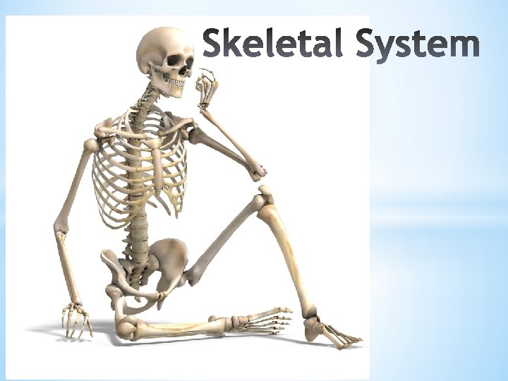

Functions: • • • Supports the body Protects soft body parts Produces blood cells Stores minerals and fat Skeleton + muscle – allow body to move

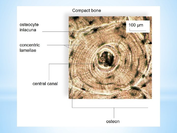
Tissues of the skeletal system: 1. Bone 2. Cartilage 3. Dense fibrous connective tissue

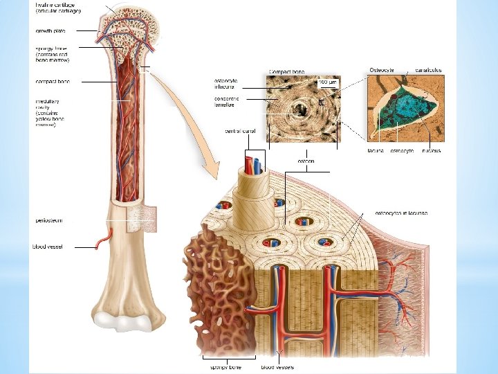
 chondrocytes inlacunae growth plate spongy bone (contains")
Hyaline cartilage matrix hyaline cartilage (articular cartilage) chondrocytes inlacunae growth plate spongy bone (contains red bone marrow) compact bone medullary cavity (contains yellow bone marrow) periosteum 50 µm
 compact bone Medullary cavity")
hyaline cartilage growth plate spongy bone (contains red bone marrow) compact bone Medullary cavity (contains yellow bone marrow) Endosteum Diaphysis Periosteum blood vessel Epiphysis

Bone classification 1. By shape – long, short, flat, irregular 2. By location – axial and appendicular

parietal bone maxilla palatine bone frontal bone zygomatic bone vomer bone sphenoid bone nasal bone ethmoid bone lacrimal bone temporal bone zygomatic bone maxilla occipital bone external auditory canal foramen magnum styloid process occipital bone mandible *

frontal bone temporal bone nasal bone zygomatic bone maxilla mandible

larynx hyoid bone

1 2 3 4 5 6 spinous process of vertebra Seven cervical vertebrae in neck region 7 1 2 3 4 5 Twelve thoracic vertebrae - Ribs attach here. 6 7 8 9 10 11 transverse process of vertebra intervertebral disks 12 1 2 3 Five lumbar vertebrae in small of back 4 5 Sacrum: Five fused vertebrae in adult Coccyx: Usually three to five fused vertebrae form the “tailbone. ”

thoracic vertebra 1 2 3 4 true ribs 5 sternum 6 7 8 false ribs 9 10 11 12 floating ribs costal cartilage

Skull: frontal bone zygomatic bone maxilla mandible Skull: parietal bone temporal bone occipital bone clavicle scapula Pectoral girdle: clavicle scapula humerus Rib cage: sternum ribs costal cartilages vertebral column ulna radius Pelvic girdle: coxal bones carpals metacarpals sacrum coccyx phalanges femur patella * fibula tibia metatarsals phalanges tarsals a. b.

Copyright © The Mc. Graw-Hill Companies, Inc. Permission required for reproduction or display. clavicle acromion process coracoid process greater tubercle glenoid cavity scapula deltoid tuberosity humerus capitulum head of radius trochlea radius ulna head of ulna carpals metacarpals phalanges

Copyright © The Mc. Graw-Hill Companies, Inc. Permission required for reproduction or display. ilium acetabulum head of femur coxal bone pubis neck ischium greater trochanter lesser trochanter femur medial condyle patella (kneecap) tibial tuberosity lateral epicondyle head of fibula tibia fibula medial malleolus tarsals metatarsals phalanges lateral malleolus talus

Copyright © The Mc. Graw-Hill Companies, Inc. Permission required for reproduction or display. Skull: frontal bone zygomatic bone maxilla mandible Skull: parietal bone temporal bone occipital bone clavicle scapula Pectoral girdle: clavicle scapula humerus Rib cage: sternum ribs costal cartilages vertebral column ulna radius Pelvic girdle: coxal bones carpals metacarpals sacrum coccyx phalanges femur patella * fibula tibia metatarsals phalanges tarsals a. b.

a. b. c. d. e. p. q. f. g. h. i. r. j. k. l. s. t. u. v. w. m. x. y. n. o. z.

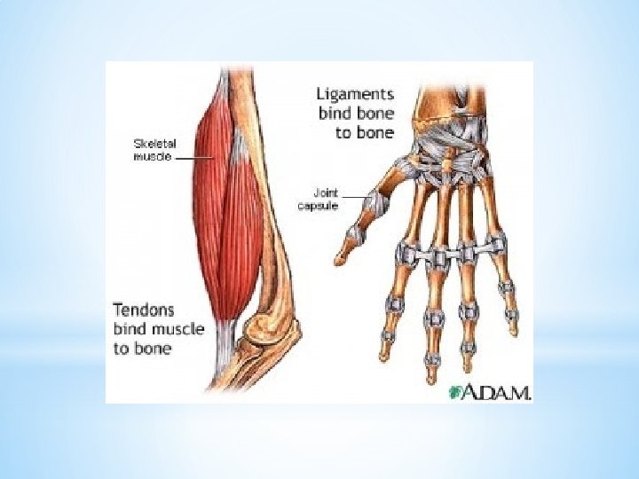
bursae joint cavity filled with synovial fluid articular cartilage meniscus ligament head of humerus b. Generalized synovial joint ulna humerus c. Ball-and-socket joint a. A gymnast depends on flexible joints. d. Hinge joint scapula

extension flexion abduction flexion extension abduction Inversion: Sole of foot turns inward. adduction flexion adduction extension abduction adduction Flexion: Joint angle decreases. Extension: Joint angle increases. Adduction: Body part moves toward midline. Rotation: Body part moves around its own axis. Abduction: Body part moves away from midline. . Circumduction: Body part moves so that a cone shape is outlined. Eversion: Sole of foot turns outward. .
- Slides: 22