Functions of the Nervous System Sensory input monitor


 · Brain ·")
 · CNS develops from the embryonic neural tube · The")


 · Paired (left and right) superior parts of the brain ·")


")


- Slides: 27
Functions of the Nervous System Sensory input – monitor changes occurring inside and outside the body · Changes = stimuli · Done by a sensory receptor (Ex. - Rods and cones of eye, olfactory neurons of nose, touch receptors in integument…) http: //www. colorado. edu/intphys/Class/IPHY 3430 -200/image/10 -4. jpg
Functions of the Nervous System · Integration · To process and interpret sensory input and decide if action is needed · Done in brain or spinal cord http: //www. faqs. org/health/images/uchr_02_img 0126. jpg
Basic Tasks of the Nervous System Sensory Input: Monitor both external and internal environments. Integration: Process the information and often integrate it with stored information. Motor output: If necessary, signal effector organs to make an appropriate response.
Functional Properties of Nervous Tissue · Irritability – ability to respond to stimuli · Conductivity – ability to transmit an impulse Copyright © 2003 Pearson Education, Inc. publishing as Benjamin Cummings http: //media. photobucket. com/image/synapse 79/neuronen-m. Slide 7. 17 synapse. jpg#!o. ZZ 2 QQcurrent. ZZhttp%3 A%2 F%2 Fmedia. photobucket. com%2 Fimag e%2 Fpink_deity%2 FSynapse. jpg%3 Fo%3 D 2
Structural Classification of the Nervous System · Central nervous system (CNS) · Brain · Spinal cord · Peripheral nervous system (PNS) · Nerves outside the brain and spinal cord Copyright © 2003 Pearson Education, Inc. publishing as Benjamin Cummings Slide 7. 2
Central Nervous System (CNS) · CNS develops from the embryonic neural tube · The neural tube becomes the brain and spinal cord · The opening of the neural tube becomes the ventricles · Four chambers within the brain · Filled with cerebrospinal fluid Copyright © 2003 Pearson Education, Inc. publishing as Benjamin Cummings Slide 7. 26
Protection of the Central Nervous System Layers 1. Scalp and skin 2. Skull and vertebral column 3. Meninges Figure 7. 16 a Copyright © 2003 Pearson Education, Inc. publishing as Benjamin Cummings Slide 7. 44 a
Protection of the Central Nervous System · Cerebrospinal fluid- CSF Produced by which cells? · Blood brain barrier Figure 7. 16 a Copyright © 2003 Pearson Education, Inc. publishing as Benjamin Cummings Slide 7. 44 b
Meninges · Dura mater · Double-layered external covering · Periosteum – attached to surface of the skull · Meningeal layer – outer covering of the brain · Folds inward in several areas Slide 7. 45 a
Meninges · Arachnoid layer · Middle layer · Web-like · Pia mater · Internal layer · Clings to the surface of the brain Copyright © 2003 Pearson Education, Inc. publishing as Benjamin Cummings Slide 7. 45 b
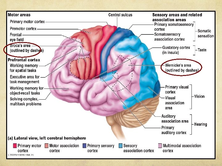
Higher Brain function Spatial Vision Speech, memory, hearing Motor control-Posture & equilibrium Autonomic Functions: breathing, heartbeat, respiration, etc,
Regions of the Brain · Cerebral hemispheres · Diencephalon · Brain stem · Cerebellum Copyright © 2003 Pearson Education, Inc. publishing as Benjamin Cummings Figure 7. 12 Slide 7. 27
Cerebral Hemispheres (Cerebrum) · Paired (left and right) superior parts of the brain · Include more than half of the brain mass Figure 7. 13 a Copyright © 2003 Pearson Education, Inc. publishing as Benjamin Cummings Slide
Language: aphasias: loss of language ability due to damage to specific areas of the brain Broca’s Area: speaking and forming words- damage = difficulty speaking, not understanding Wernicke’s area: understanding of words damage= “word salad”
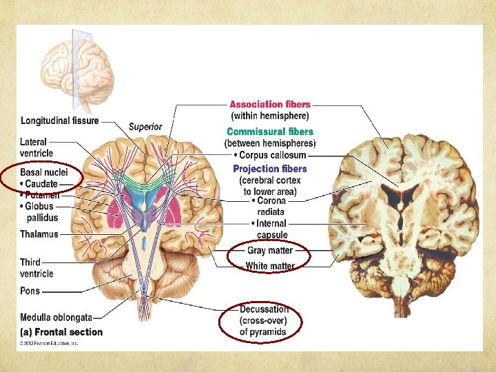
·Basal nuclei – receive information from cerebral cortex ·Regulates voluntary motor activities by modifying info sent to the motor cortex ·Problems = ie unable to control muscles, spastic, jerky ·Involved in Huntington’s and Parkinson’s Disease • Crossover of Pyramids in Medulla: Each cerebral hemisphere controls voluntary movement in opposite side of the body
Diencephalon
Brain Stem
Traumatic Brain Injury National TBI Estimates Every year, at least 1. 7 million TBIs occur either as an isolated injury or along with other injuries. TBI is a contributing factor to a third (30. 5%) of all injury-related deaths in the United States. About 75% of TBIs that occur each year are concussions or other forms of mild TBI.
Did you know? 1 - 19 yrs • Each year, U. S. emergency departments (EDs) treat an estimated 173, 285 sports- and recreation-related TBIs • During the last decade, ED visits for sports- and recreationrelated TBIs, including concussions, among children and adolescents increased by 60%. • Overall, the activities associated with the greatest number of TBI-related ED visits included bicycling, football, playground activities, basketball, and soccer. • National surveillance in 9 high school sports: • TBI represents almost 9% of all injuries reported in the 9 sports • Numbers and rates are highest in football (55, 007; 0. 47 per 1000 athlete exposures) and girl’s soccer (29, 167; 0. 36 per 1000 athlete exposures)
Spinal Cord · Extends from the foramen magnum to the region of L 2 · Below is the cauda equina (a collection of spinal nerves) · Enlargements occur in the cervical and lumbar regions Figure 7. 18 Copyright © 2003 Pearson Education, Inc. publishing as Benjamin Cummings Slide 7. 52
Spinal Cord Anatomy · Exterior white mater – conduction tracts Figure 7. 19 Copyright © 2003 Pearson Education, Inc. publishing as Benjamin Cummings Slide 7. 53 a
Spinal Cord Anatomy · Internal gray matter - mostly cell bodies · Dorsal (posterior) horns · Anterior (ventral) horns Figure 7. 19 Copyright © 2003 Pearson Education, Inc. publishing as Benjamin Cummings Slide 7. 53 b
Spinal Cord Anatomy · Central canal filled with cerebrospinal fluid- continuous with CFS in Brain Figure 7. 19 Copyright © 2003 Pearson Education, Inc. publishing as Benjamin Cummings Slide 7. 53 c
Spinal Cord Anatomy · Meninges · Nerves leave at the level of each vertebrae · Dorsal rootsensory neurons · Ventral rootcontains motor neurons Copyright © 2003 Pearson Education, Inc. publishing as Benjamin Cummings Slide 7. 54