Functions of the digestive system Ingest food using


 Endocrine gland - secretes hormones (insulin and glucagon) 2) Exocrine gland")
 •")


 a. Urea")


- Slides: 27
Functions of the digestive system: • Ingest food – using mouth, teeth, tongue • Digest food – using mouth, stomach, small intestine • Movement of GI tract contents – usually by peristalsis • Absorb nutrients – using small intestine • Eliminate remains – large intestine Types of digestion (2 types) • Mechanical –chew and mixing food –mouth and stomach • Chemical – enzyme breaks down macromolecules into subunit molecules
1. Mouth - mixes food with saliva, forms chewed food into bolus for swallowing 2. Pharynx- back of throat 3. Epiglottis – stops food going down trachea 4. Esophagus-uses peristalsis -No chemical digestion –has sphincter btwn esophagus and stomach 5. Stomach –store food, churns, mixes food with gastric juice - chyme • Gastric glands–gastric juice -Contains pepsin • HCL –activates pepsin 6. Small intestine – *main nutrient absorption slightly basic p. H (>7) -Duodenum 7. Large Intestine ***Main job is to reabsorb water! Also absorbs salts, vitamins, stores and gets rid of indigestible material- Does not produce digestive enzymes • Regions: Cecum, colon (ascending, transverse, descending, sigmoid colon), rectum, anal canal • Rectum – (last 20 cm) opens at anus – where defecation occurs
Small intestine Section of intestinal wall villus lumen lacteal blood capillaries goblet cell lymph nodule venule lymphatic vessel arteriole
transverse colon ascending colon small intestine descending colon cecum orifice of appendix rectum internal anal sphincter anal canal sigmoid colon external anal sphincter anus
A. Pancreas 1) Endocrine gland - secretes hormones (insulin and glucagon) 2) Exocrine gland –secretes pancreatic juice via duct B. Liver – largest organ, Liver functions: 1. Monitors blood. 2. Stores iron, vitamin ADEK 3. Makes plasma proteins from AA 4. Excess glucose in blood stored in liver *glucose=quickest, most readily available source of energy 5. Produces urea after converting aa to glucose 6. Produces bile 7. Makes lipids from fatty acids C. Gall bladder Stores excess bile produced by liver
Carbohydrates: • salivary amylase -salivary glands-mouth • pancreatic amylase -pancreas into Duodenum (SI) • maltase -SI wall Proteins: • pepsin -gastric glands in stomachactivated by HCL • trypsin -pancreas into Duodenum (SI) • Peptidase -SI wall Nucleic acids: • Nuclease –pancreas into duodenum (SI) • Nucleosidase -SI wall Lipids: • Bile – gall bladder from liver into SI– emulsifies fat • Lipase – pancreas into duodenum (SI)
BIOLOGY 067: Macromolecules
CH 2 OH O H OH H H O H O CH 2 OH H H O CH 2 OH O H OH H O OH branched nonbranched A) Carbohydrates The subunit of a carbohydrate = monosaccharide Monosaccharide’s are bonded together through a dehydration reaction to form polysaccharides starch granule cell wall potato cells
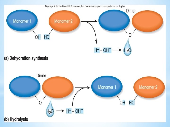
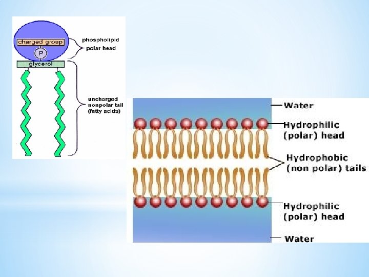
Lipids • The subunits of lipids = glycerol and 3 fatty acids • fat molecules are joined together through a dehydration reaction and are broken down back into their subunits through hydrolysis • a specialized lipid is a phospholipids =glycerol, phosphate, 2 fatty acids H H C O OH HO + H C OH C H H C C H H H H H C C H HO C C H H H H H C C C H H H O dehydration reaction O C C H H C OH HO H H H C C C H H O H H H C C C C H H H O H H H C C C hydrolysis reaction O H C H O H H + 3 H 2 O H H H glycerol 3 fatty acids fat molecule 3 water molecules
Proteins • The subunit of a protein=amino acid • An amino acid always has an amino group and an acidic group • Amino acids join to form a polypeptide through a dehydration reaction (= dehydration synthesis) • Amino acids are joined together at a peptide bond • Denaturation is the process of changing the 3 dimensional shape of proteins – often caused by extremes in heat or p. H H
Level Shape Bonding Primary Linear chain of aa Peptide bonds between aa Secondary Helix (coiling) Hydrogen bonds between aa Pleated sheet (folding) Tertiary globular Final 3 D shape and function: Ionic, covalent, hydrogen bonds btwn R groups Quaternary (rare) All shapes All bonding – 2 or more associated polpeptides
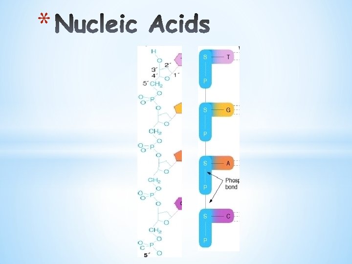
Parts of a nucleotide phosphate C P 5' Nucleotide O S 4' 1' 2' 3' sugar nitrogencontaining base
Nucleic Acids • The subunit of nucleic acids is nucleotides • The 3 components of a nucleotide =a phosphate group, a sugar, and a nitrogen base • The nitrogen bases found in DNA are: Adenine, Thymine, Guanine, and Cytosine. • The nitrogen bases found in RNA are: Adenine, Uracil, Guanine, and Cytosine • ATP is a nucleotide that carries energy in the form of a high-energy phosphate bond. • ATP is adenosine (adenosine=adenine + ribose) + 3 phosphate groups
Specialized Nucleotide: ATP – Adenine triphosphate ATP = adenine + ribose + 3 phosphate groups Adenosine ATP P ADP + energy
Organic molecules Subunits Examples Functions CH 2 OH Carbohydrates O H Monosaccharides, disaccharides, polysaccharides OH Immediate energy and stored energy; structural molecules H H HO OH H OH Glucose H Lipids Fats, oils, phospholipids, steroids H O C OH H C OH C HO H H H C C C H H H R Fatty acid H Glycerol Proteins Structural, enzymatic, carrier, hormonal, contractile amino group H 2 N acid group COOH H C R group Long-term energy storage; membrane components Support, metabolic, transport, regulation, motion Amino acid base phosphate C P Nucleic acids DNA, RNA O S Nucleotide Storage of genetic information
adrenal glands – produce hormones renal artery 1. Kidneys produce urine. renal vein aorta Inferior vena cava 2. Ureters transport urine. 3. Urinary bladder stores urine. 4. Urethra passes urine to outside.
Functions of the Urinary system Excretion of metabolic waste (esp nitrogen waste) a. Urea – end product of amino acid breakdown = Protein breakdown – made in the liver b. Uric acid formed from breakdown of nucleotides c. Creatinine – nitrogen waste formed from the end product of creatine phosphate breakdown d. Ammonium Maintains salt/water balance Maintains acid-base (p. H) balance Contributes to hormonal function • Secretes erythropoietin
Macroscopic Structure – 3 regions • Renal cortex renal cortex • Renal medulla • renal pelvis Renal pyramid in renal medulla renal pelvis ureter
Blood supply: Renal artery afferent arteriole glomerulus efferent arteriole peritubular capillary network venule renal vein Parts of the Nephron Glomerular capsule proximal convoluted tubule loop of nephron (descending limb ascending limb) distal convoluted tubule collecting duct (not part of nephron) Proximal has microvilli and mitochondria/distal has only mitochondria
Glomerular filtration – Blood to nephron How and why: • a passive process • works on blood pressure • Glomerular Filtrate: water, nitrogenous waste, nutrients and salts Controls – juxtaglomerular apparatus responds to conctrn of Na. Cl in filtrate – • Renin from juxtaglomerulus apparatus when blood pressure and volume decreases too low for filtration – this leads to secretion of • Aldosterone from adrenal glands promotes reabsorption of Na+ followed by reabsorption of water ∴ increase blood press and vol.
Tubular reabsorption – Nephron to Blood • 100% of glucose and amino acids and ~99% of water and salts– Na actively, Cl passively follows • descending limb of the loop, only water is passively transported into the blood through aquaporins • as you go back up the ascending limb – there are no aquaporins but Na. Cl is passively (in lower part), then actively (in upper part) reabsorbed into the tissues, • some reabsorption also happens in the distal convoluted tubule Control: • reabsorption of salt precedes reabsorption of water • urea leaks from collecting duct into medulla – add to salt concentration in inner medulla tissue a. Aldosterone –– promote excretion of K+ and reabsorb of Na+ from DCT into blood b. Renin– hormone released when blood pressure and blood volume drops c. Antidiuretic hormone (ADH) -secreted by pituitary (in brain) depending on osmolarity of the blood d. ANH hormone - inhibits secretion of renin and aldosterone
Tubular Secretion – Blood to Nephron – • mostly distal convoluted tubule • selectively and actively moves undesirable substances from blood to nephron. • Controls blood p. H by secreting H+ into nephron • i. e, Hydrogen ions (H+), creatinine, and excess drugs.
Maintaining acid base balance in blood 1. Regulated by acid-base buffering system • **hemoglobin important buffer = reduced hemoglobin HHb • These buffers are temporary solution to the change in H+ concentration 2. Also regulated by respiratory centre in brain • If concentration of H+ ions rises, increases ventilation, and as CO 2 exhaled, concentration of H+ decreases because rxn shifts to the right H+ + HCO 3 H 2 CO 3 H 2 O + CO 2 (released 3. • • Acid base balance also regulated in the kidneys Kidneys are slower acting but more powerful effect on p. H If blood is acidic, excretes H+ ions and reabsorbs bicarbonate If blood is basic, H+ not excreted and HCO 3 not reabsorbed Ammonia, acts as a buffer and removes H+ ions capillary HCO 3– H+ Kidney tubule HCO 3– H+ + NH 3+ NH 4 +