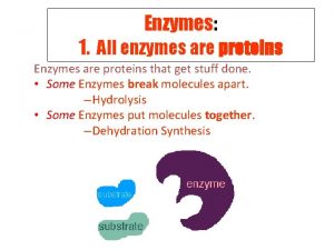
Functions of some Proteins Catalysis enzymes Structural collagen


![Acid Ionization Constant [CH 3 COO-][H+] Ka = ---------[CH 3 COOH] [CH 3 COO-] Acid Ionization Constant [CH 3 COO-][H+] Ka = ---------[CH 3 COOH] [CH 3 COO-]](https://slidetodoc.com/presentation_image_h/c344e5e07000ab702291c1c8eb3ca162/image-22.jpg)


- Slides: 83


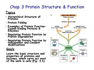
Functions of some Proteins Catalysis (enzymes) Structural (collagen) Contractile (muscle) Transport (hemoglobin) Storage (myoglobin) Electron transport (cytochromes) Hormones (insulin) Growth factor (EGF) DNA binding (histones) Ribosomal proteins Toxins and venoms (cholera & melittin) Vision (opsins) Immunoglobins

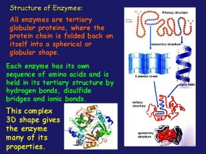
Levels of Protein Structure • Primary structure (1 o) – sequence of amino acids starting from the N-terminus of the peptide. • Secondary structure (2 o) – conformations of the peptide chain from rotation about the a-Cs, e. g. a-helices and b-sheets, etc. • Tertiary structure (3 o) – three dimensional shape of the fully folded polypeptide chain. • Quaternary structure (4 o) - arrangement of two or more protein chains into multisubunit molecule

Levels of Protein Structure Bonding: 1 o = covalent 2 o = H-bond 3 o = covalent & noncovalent 4 o = noncovalent

Amino Acids The 20 common are those used in making protein on a ribosome using m. RNA and t. RNA. These are called a-amino acids since each has a carboxyl group and an amino group attached to an a-carbon atom. They differ by the sidechain or “R” group. a +NH -CH-COOH 3 l R

Amino Acid Classification is made using the structure of the side chain, R. (* = essential) 1. None (hydrogen): 2. Non-polar: Aliphatic: Alicyclic: 3. Aromatic: 4. Polar uncharged: 5. Thiol: 6. Acidic: 7. Basic: Gly Ala, Val*, Leu*, Ile*, Met* Pro Phe*, Tyr, Trp* Ser, Thr*, Asn, Gln Cys Asp, Glu Lys*, His*, Arg*

ball & stick model wedge Fischer projection


Amino Acid Names & Codes

All are Chiral at the a-carbon atom except Gly

D, L Assignments The convention for making D, L assignments is to draw a Fischer projection with the carboxyl at the top and the R at the bottom. +NH 3 to the left = L to the right = D

L stereochemistry = S, for this case

Acids and Bases, p. H, p. Ka • p. H is defined as the negative logarithm of the concentration of H+ p. H = - log [H+] = log 1/ [H+] • p. Ka is defined as the negative logarithm of an acid ionization constant Ka. p. Ka = - log [Ka] = log 1/ [Ka]

Acetic Acid is a Weak Acid • Weak acids and bases do not ionize (dissociate) completely in H 2 O. CH 3 COOH <==> CH 3 COO- + H+ acetic acid acetate anion conjugate acid form conjugate base form
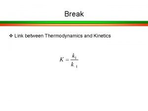
![Acid Ionization Constant CH 3 COOH Ka CH 3 COOH CH 3 COO Acid Ionization Constant [CH 3 COO-][H+] Ka = ---------[CH 3 COOH] [CH 3 COO-]](https://slidetodoc.com/presentation_image_h/c344e5e07000ab702291c1c8eb3ca162/image-22.jpg)
Acid Ionization Constant [CH 3 COO-][H+] Ka = ---------[CH 3 COOH] [CH 3 COO-] -log Ka = -log [H+] + -log --------[CH 3 COOH] [CH 3 COO-] p. H = p. Ka + log --------[CH 3 COOH]

Henderson-Hasselbalch Written in a more general form this expression is called the Henderson-Hassebalch equation. This relates the p. H of a solution to the p. Ka of the weak acid in solution and the ratio of its conjugate base/conjugate acid forms. [A-] p. H = p. Ka + log -----[HA]

Buffers Resist Change in p. H • Buffer capacity is the ability of a solution to resist changes in p. H. • Most effective buffering occurs where: solution p. H = buffer p. Ka • At this point: [conjugate acid] = [conjugate base] • Effective buffering range is usually at p. H values equal to the p. Ka ± 1 p. H unit

Amino Acid Ionization, p. Kas Each of the 20 common a-amino acids has two p. Ka values, for the carboxyl group and the amino group attached to the a-carbon. +NH -CH -COO- + H+ -CH -COOH < === > 3 2 +NH - < === > NH -COO- + H+ -CH -COO 3 2 2 2 Seven of the 20 have an ionizable sidechain and therefore have a third p. Ka value.

Amino Acid p. Kas Gly Ala Val Leu Ile Met Pro Phe Trp Ser Thr Asn Gln Cys Asp Glu Tyr Lys His Arg a-COOH 2. 34 2. 32 2. 36 2. 28 1. 99 1. 83 2. 21 2. 63 2. 02 2. 17 1. 71 2. 09 2. 19 2. 20 2. 18 1. 82 2. 17 a-+NH 3 9. 60 9. 69 9. 62 9. 60 9. 21 10. 60 9. 13 9. 39 9. 15 10. 43 8. 80 9. 13 10. 78 9. 82 9. 67 9. 11 8. 95 9. 17 9. 04 R (sidechain) 8. 33 3. 86 4. 25 10. 07 10. 79 6. 00 12. 48

Peptide 3. 86 D, 4. 25 E Peptide 10. 0

Effect of Change in p. H

The Structure of an Amino Acid An amino acid can never exist as an uncharged compound. © 2014 Pearson Education, Inc.

The p. I of Alanine The isoelectric point (p. I) of an amino acid is the p. H at which it has no net charge. © 2014 Pearson Education, Inc.

The p. I of Lysine © 2014 Pearson Education, Inc.

The p. I of Glutamic Acid © 2014 Pearson Education, Inc.

Formation of a Peptide Two amino acids are joined together to form a peptide (amide) bond with a loss of HOH. After becoming part of a peptide or protein these are called “residues” due to loss of HOH.

Primary Structure (1 o) The sequence of amino acids (N-term to C-term) in a peptide or protein is its primary structure. A pentapeptide

Disulfide Bond Formation

Disulfide Bridges Disulfide bridges contribute to the overall shape of the protein. © 2014 Pearson Education, Inc.

Straight and Curly Hair © 2014 Pearson Education, Inc.

Insulin has • two interchain disulfide bridges and • one intrachain disulfide bridge. © 2014 Pearson Education, Inc.

Peptide Bond Resonance Due to resonance participation of the unshared pair of electrons on N, amides are neutral. . .

Peptide Bond Planarity 6 atoms are coplanar

Peptide Bond Structures Less crowded in the favored trans arrangement

Rotation Sites, y and f The rotational arrangements about a-carbons of a peptide or protein gives its secondary (2 o) structure.

f View from N-term to a-carbon

y View from a -carbon to C-term

Ramachandran Plot

Secondary Structure (2 o) The two most common secondary structures are the a-helix and the b-sheet. Each of these 2 o structures have fairly specific y and f angles. All other rotational angles represent “random” secondary structure. Secondary structure is maintained by hydrogen bonding. a-helix by intramolecular H-bonds b-sheet by intermolecular H-bonds

a-helix in a Ramachandran Plot y = -47 f = -57

The a-helix, a 3. 613 helix

The a-helix

Hydrogen Bond Contacts

a-helix in a Protein

b-sheet in a Ramachandran Plot y f = +135 = -139

A b-sheet strand

Anti-parallel b-sheet

Parallel b-sheet

Anti-parallel b-sheet

b-sheet in a Protein

b-turn or hairpin turn A turn is 4 -5 aa residues. A b-turn (hairpin) is 4 aa residues.

Loops are usually larger than turns. >5 aa residues

a-Keratin, a fibrous protein, forms an a coiled coil Each strand is a modified a-helix, 3. 5 residues/turn. Right-handed helices form a left-handed supercoil.

Collagen, a triple helix Gly every third residue; Gly-Pro-HPro is frequent. Interstrand H-bond at Gly. No Cys, so, no -S-S-

Tertiary Structure (3 o) The three dimensional folding of a polypeptide is its tertiary structure. Both the a-helix and b-sheet may exist within the tertiary structure. Generally the distribution of amino acid sidechains in a globular protein finds mostly nonpolar residues in the interior of the protein and polar residues on the surface. Tertiary structure is maintained by noncovalent interactions and disulfide bonding.

Myoglobin, a globular protein

Myoglobin Surface Blue = charged Cross-section Yellow = hydrophobic

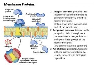
Porin, a membrane spanning protein

Domain A domain is a discrete globular area within protein. There are four general types: All a All b Mixed a/b a+b only a- helices and loops only b- sheet & loops or turns alternating abab cluster of a then cluster of b

Domain Multiple domains exist in the protein below.

Quaternary Structure (4 o) is an assembly of 3 o structures (two or more subunits). A dimer

Hemoglobin, a tetramer Quaternary structure is maintained by noncovalent interactions

Denaturing Proteins Denaturing agents destroy the protein 3 o structure (causes the protein to unravel). Methods: Heat; Extremes of p. H; Detergents; Mechanical agitation; Mercaptoethanol – breaks -S-S- bonds; 6 M guanidine HCl or 10 M urea: these are chaotropic agents that break up noncovalent interactions.

Denaturing agents

Disulfide oxidation-reduction Oxidized Reduced

Ribonuclease 4 disulfide bonds. -S-S-

Then removing urea and ME permits reoxidation.

Reoxidation reforms –S-S- but not necessarily in the correct place. A trace of ME allows reduction/oxidation to occur until the low free energy form is found and >98% of activity is restored.

Sharp Transition suggests an all or nothing effect in denaturing.

Lysozyme Ribbon diagrams 3 o structure Active site & -S-S-

Protein Cleavage Protein sequencing is most manageable with small polypeptides. Therefore, in order to sequence a large protein, it must be cleaved into smaller pieces. Cleavage is conducted using either chemical or enzymatic methods. The pieces must be separated and purified before sequencing.

Chemical and Enzymatic Cleavage

CNBr Cleavage at Met

Enzymatic cleavage by Trypsin


An Example, Peptide overlap
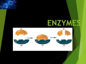
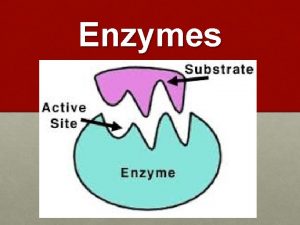
 All enzymes are globular proteins
 Not all enzymes are proteins
 Structural proteins function
 Thin
Thin Structural role of proteins
 What is the difference between hbv and lbv
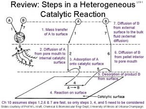
 7 steps of heterogeneous catalysis
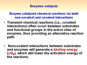
 Enzymatic catalysis
 Catalysis by approximation
 What is covalent catalysis
 What is covalent catalysis
 Specific acid base catalysis
 Specific acid base catalysis
 Specific acid base catalysis
 Mixed inhibitor km and vmax
 Langmuir-hinshelwood mechanism heterogeneous catalysis
 Erzeng
 Energy catalysis and biosynthesis
 Catalytic combustion
 Sodium hydroxide iupac id sodium oxidanide
 Structure of protein
 Glycoprotein vs glycolipid
 Transport
 Functions of membrane proteins
 Cell membrane selectively permeable
 Functions and importance of proteins
 Function of plasma protein
 Types of globulin
 Name the enzymes secreted by small intestine
 Digestive enzymes and their functions table
 Examples of enzymes
 Cocoa collagen sendayu tinggi
 Classification of connective tissue
 Collagen is a major component of
 Matrix vesicle theory of mineralization
 Connective tissue description
 Totally derma collagen
 Types of tissues
 Totally derma tub
 Fibrous proteins
 Collagen fibre structure
 Parkinson's disease
 Dr. alireza peyman
 Unicellular organisms
 They say sometimes you win some
 Sometimes you win some
 Ice cream is countable or uncountable
 Force and motion
 Fire and ice diamante poem
 Some say the world will end in fire some say in ice
 Some trust in chariots and some in horses song
 Intensity transformation functions
 Function of a chromosomes
 Carrier vs channel proteins
 Globular vs fibrous proteins
 Section 8-1 carbohydrates fats and proteins answer key
 Salting in salting out
 Complementary proteins
 Example of denatured protein
 Primary derived proteins
 Monomers protein
 Protein digestion
 Protein folding
 Proteins database
 Bradford method
 Dr nutrition
 Motifs and domains of proteins
 Peripheral vs integral proteins
 Precipitation of proteins by strong mineral acids
 Salting out proteins
 Salting out proteins
 Salt bridges in proteins
 Higher order structure of proteins
 Carrier vs channel proteins
 Salting in
 Biomedical importance of proteins
 Organic compounds such as proteins and starches are too
 Three peptide chains woven like a rope
 Three main types of rna
 Steroid hormones include
 Section 8-1 summary carbohydrates fats and proteins
 Protein elements
 Integral proteins hydrophobic
 Dna rna and proteins study guide answers
