Functions of Respiratory System Ventilation moves air to


")
 External nares > vestibule of nose > nasal cavity > pharynx")


 Alveolar Type I cells –")
 Alveolar Type II cells release surfactant, makes lungs more Compliant. (‘septal’ cells). 3)")


: Same muscles required as in Eupnea. Expiration requires: Internal Intercostals Rectus")


- Slides: 39
Functions of Respiratory System Ventilation - moves air to and from alveoli. Large surface area for gas exchange. Regulates p. H of body fluids. Permit vocal sounds (communication).
External nares
• Upper Respiratory Tract – Nose – Nasal cavity – Pharynx (3 parts) Functions: Warm, Filter and Humidify incoming air. • Lower Respiratory Tract - Larynx - Trachea Functions: - Bronchi Conduct air and exchange gases. - Bronchioles - Alveoli
Respiratory Tract (Passageway) External nares > vestibule of nose > nasal cavity > pharynx (naso-, oro- laryngo-) > larynx > trachea > 1 o bronchi > 2 o bronchi > 3 o bronchi > bronchioles > terminal bronchioles > respiratory bronchioles > alveolar duct > alveolar sac > alveoli (end).
There are 2 Zones of this tract The Respiratory Tract is divided into: – Conducting Zone – Respiratory Zone
“Respiratory Epithelium” • Lines conducting portions of tract. • Pseudostratified ciliated columnar epithelium (with goblet cells) – Produces mucus to trap foreign particles • Lamina propria is the connective tissue layer (Epithelium and lamina propria = mucus membrane)
Respiratory Epithelium
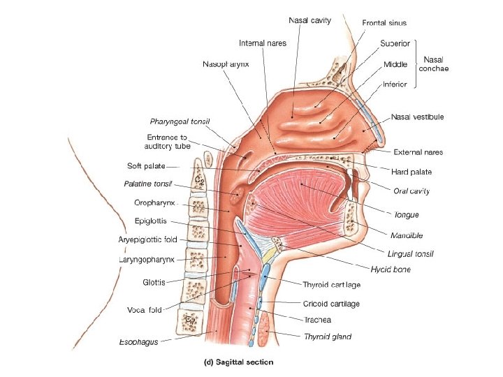
The Journey of Air • External nares – Entrance into vestibule guarded by hairs • Nasal cavity – Superior, middle and inferior meatuses • Narrow grooves and conchal surfaces • Hard palate – Divides nasal and oral cavities • Soft palate – Superior nasopharynx and pharynx • Internal nares – Between nasal cavity and nasopharynx
The Pharynx – 3 Parts 1. Nasopharynx • Superior to food entry - air only • Closed off during swallowing • Pharyngeal tonsil (adenoids) – Located on posterior wall • Opening to the auditory tube
2. Oropharynx • From soft palate to the epiglottis • Stratified squamous epithelium • Two types of tonsils in oropharynx Palatine tonsils – in the lateral walls of fauces. Lingual tonsils – covers posterior surface of tongue
3. Laryngopharynx • Shared respiratory and digestive passageway • Stratified squamous epithelium • Continuous with the esophagus and larynx
The Larynx • Surrounds glottis - air passes through glottis to reach lungs • Epiglottis - prevents solids from entering respiratory system
Elevation of larynx folds epiglottis over glottis when swallowing.
Trachea • About 4. 5 inches in length. • About 1 inch in diameter. Submucosa has “C” rings of cartilage Posterior wall created by the trachealis (smooth muscle), this distorts to allow food passage in esophagus.
Trachea • ~ 4. 5 inches long • ~ 1 inch in diameter “C” cartilage rings (submucosa) Trachealis is posterior wall – allows food passage in esophagus.
Bronchi and Bronchioles
Bronchioles • Do not contain cartilage • Have smooth muscle • Are Innervated by ANS Parasympathetic – constrict airways Sympathetic – dilate airways
The Bronchiole Tree
Bronchopulmonary Segments
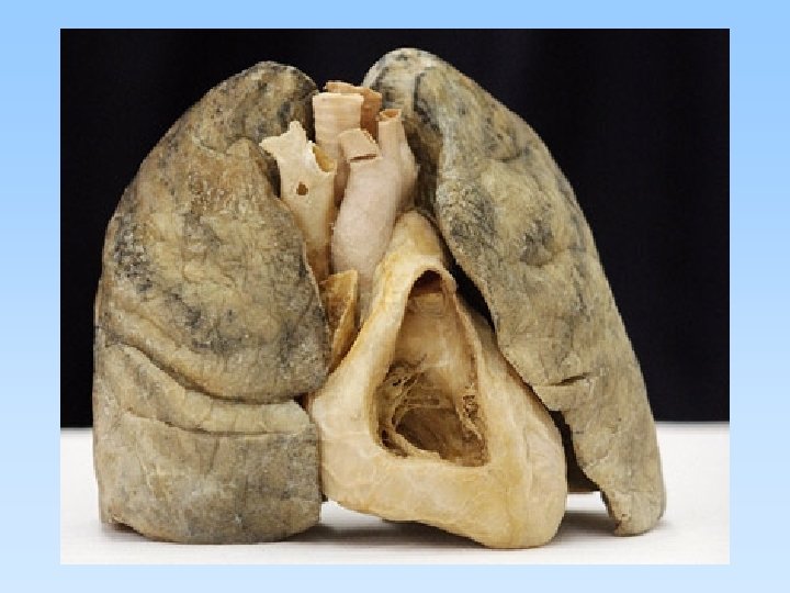
The Lungs • Separated by fissures – Right lung has three lobes. – Left lung has two lobes. • Costal surface – Anterior surface – Follows inner contours of rib cage • Mediastinal surface – Contains hilus • Costal notch - Left lung
Right Lung Left Lung
Medial aspect of each Lung
Each Lung is in a ‘Bag’ Visceral pleura Parietal pleura
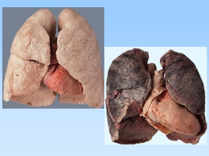
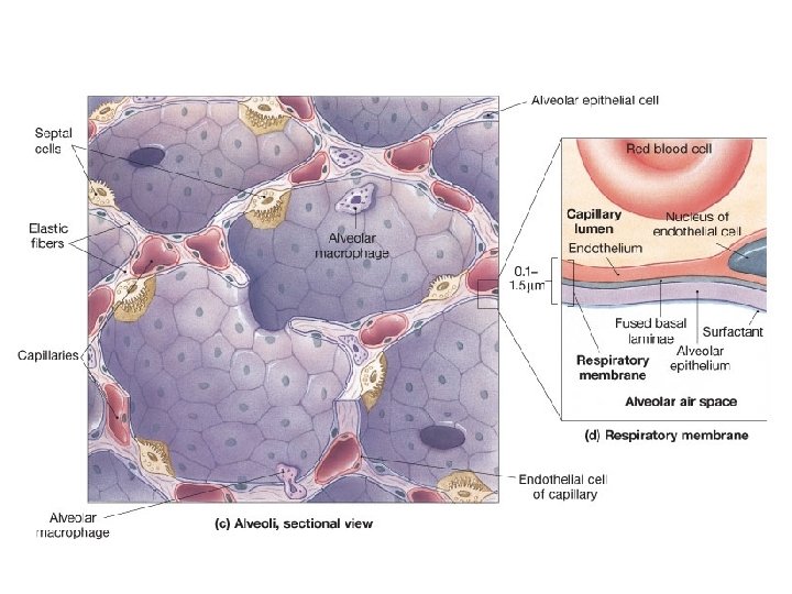
There about 150 million alveoli in each Lung The alveoli is where gas exchange occurs with the pulmonary capillaries
An alveolus consist of 3 Types of cells 1) Alveolar Type I cells – thin (simple squamous epithelium); makes ‘walls’ of alveoli, provides surface area for gas exchange.
2) Alveolar Type II cells release surfactant, makes lungs more Compliant. (‘septal’ cells). 3) Alveolar Macrophages – for protection of alveolar surface. Release trypsin – an enzyme that degrades proteins. Alveoli also contain Elastic fibers Elastic Recoil - Push air out (assists ventilation). Capillaries cover 90% of surface
Respiratory Muscles Ventilation - movement of air into and out of lungs.
Eupnea = normal quite breathing at rest. For Inspiration Muscle activity required: Diaphragm External Intercostals Accessory muscles Sternocleidomastoid Scalenes
Expiration in Eupnea: No Muscular activity required!!! Restful Breathing ~ 10% Basal Metabolic Rate (BMR) normally What is Emphysema? With emphysema this increases to ____ %
Forceful breathing (hypereupnea): Same muscles required as in Eupnea. Expiration requires: Internal Intercostals Rectus abdominis Transverse abdominis, Internal and External obliques.
Three pairs of nuclei in reticular formation of pons and medulla oblongata Respiratory rhythmicity center - Sets respiratory pace. Located in the medulla oblongata. Apneustic center - Strong, sustained inspiratory movements, used for ‘overdrive’ when breathing deep. Pons Pneumotaxic center - Inhibits apneustic and inspiratory centers, sets limits to over inflation of lung. Pons
Locations of these centers in the brain
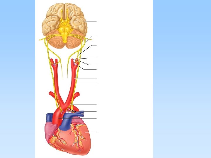
Sensory Receptors - regulate respiration. Mechanoreceptors detect changes in lung volume or arterial blood pressure Chemoreceptors Changes in PCO 2, p. H, PO 2 of blood and CSF Central chemoreceptors - in medulla Peripheral chemoreceptors 1) Aortic bodies (in aortic arch) 2) Carotid bodies (in carotid sinus)
The major respiratory centers and other factors important to respiratory control.