Functions of bones Support Protection Movement Blood cell

Functions of bones • • Support Protection Movement Blood cell formation • Storage

Support • Strong • Rigid

Protection • Surround organ • Protect against damage

Movement • Attach to bones • Provide levers

Blood Cell Formation • All blood cells formed in the bones • Red marrow (hematoposiesis)

Storage • Calcium • Phosphate
 • Dense connective tissue • Cartilage • Blood")
Bone Tissues • Bone (osseous tissue) • Dense connective tissue • Cartilage • Blood forming tissue • Nervous tissue

Types of Bones • Bones can be divided into four classes – – Flat bones Long bones Short bones Irregular bones

Flat Bones • Thin and flat – Cranium – Ribs – Sternum

Short Bones • About equal in length and width – Wrist bones – Ankle bones

Long Bones • Greater in length than width • Absorb stress from body weight – – Thighs Legs Arms Forearms

Irregular bones • Complex shapes – Vertebrae – Facial bones

Skeleton • Divided into two parts – Axial – Appendicular
")
Axial Skeleton • Skull • Vertebral column • Bony thorax (thoracic cage)

Skull • Cranium • Facial bones
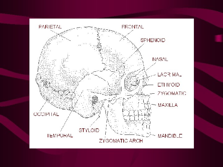

Fetal Skull • Newborn infant skull isn’t complete • Not all the hyaline cartilage has yet ossified • Cartilage meets at fontanels

Vertebral Column • Divided into three regions – Cervical – Thoracic – Lumbar Sacrum Coccyx

Fetal Vertebral Column • Originally a convex curve • As baby learns to lift it’s head, the cervical curve develops • As baby learns to walk, Lumbar curve develops

Bony Thorax • Consists of the ribs, sternum and thoracic vertebrae • Provides a bony, protective cage around the organs of the thoracic cavity • Heart, Lungs and major blood vessels

Appendicular Skeleton • • • Pelvic Girdle Lower Limbs Pectoral Girdles Clavicles Scapulae Upper limbs

Pelvic Girdle • Strong frame • Supports lower limbs

Pelvis • Two coxial bones • Sacrum – Three separate bones in the fetus – The three join anteriorly

Female and Male Pelvis are Different

Pelvic Girdle Differences Feature Male Female Orientation Tilted backward Tilted forward Pelvic inlet Narrow, heart shaped Wide, oval-shaped Pubic arch Less than 90 o Greater than 90 o Sacrum Narrow and long Wide and short, greater curvature Bone thickness Thick and heavy Thin and light Coccyx Immovable Movable

Bone Growth I • Bone begins as a model of hyaline cartilage in an embryo

Bone Growth II • A collar forms around the midsection

Bone Growth III • Primary ossification center as cartilage is replaced by spongy bone

Bone Growth IV • Spongy bone expands as more blood vessels penetrate

Bone Growth V • Central cavity is formed and secondary ossification centers develop

Bone Growth VI • Ossification continues until spongy bone fills epiphyses and compact bone surrounds the entire structure
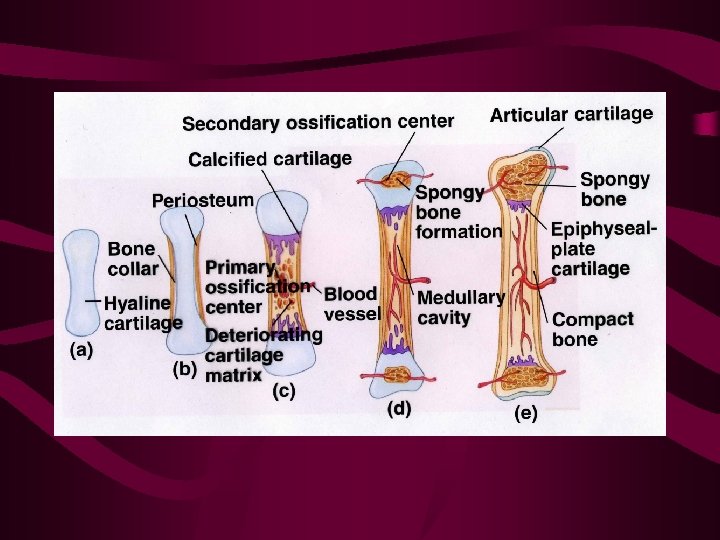

Bone Growth VII • Hyaline cartilage remains in the epiphyseal plates and articular cartilages

Diaphysis • Shaft of the bone – Most of the bones length – Dense bone – Covered by fibrous connective tissue (periosteum)

Epiphyses • Ends of long bones – Thin layer of compact bone surrounding spongy bone – Covered by Articular Cartilage – hyaline cartilage – Slippery and smooth

Periosteum • Dense, connective tissue firmly attached to bone • Covers everything except the articular cartilage • Large supply of blood vessels

Joints or Articulations • There are three groups of joints or articulations binding bones together in the body • They are classified by the amount of movement they allow and the materials forming the joining
 tissue between articulating bone • Little or")
Fibrous Joints • Consist of fibrous (dense) tissue between articulating bone • Little or no movement allowed – Sutures in the skull

Cartilaginous Joints • Binds bones together with cartilage • Allows little or no movement • Shock absorbers – Symphysis Pubis – Intervertebral disks

Synovial Joints • There are five types of freely moving or Synovial joints in the human body • • • Hinge Ball and Socket Pivot Saddle Gliding

Synovial Joint • Permit the greatest freedom of movement • Synovial fluid between bones • Enclosed in the Articular Capsule • The outside layer is the tough fibrous capsule

Hinge Joint • Allows movement in only one plane • Convex surface of one bone fits into the concave surface of another – Knee – phalanges

Ball and Socket • Maximum allowable movement • Ball-shaped process of one bone fits into the cup-shaped socket of another – Hip joint – Shoulder joint

Pivot Joint • Allows rotation around a central axis • Cylindrical surface of one bone rotates in the ring of another – Vertebral column – elbow

Saddle Joint • Allows back and forth, side to side and some pivoting • Convex surface of one bone fits concave surface of complimentary bone – Trapezium and metacarpal of the thumb

Gliding Joint • Permit a sliding movement • Surfaces are nearly flat – Clavicle and sternum – Some wrist and ankle bones

Types of Bone Fracture • There are seven types of fracture – – – – Greenstick Simple Compound Spiral Comminuted Impacted depressed

Greenstick Fractures • Also called an incomplete fracture • Break does not extend all the way through the bone • Common in children. Their bones are more flexible

Simple Fracture • No tear in the skin • Break extends all the way through the bone

Compound Fracture • Broken end of the bone tears skin from the inside • Bone may break into several fragments

Comminuted Fractures • Bone has broken into several pieces • Common in the elderly with brittle bones

Spiral Fracture • A ragged break that occurs when excessive twisting forces are applied to the bone • Common sports fracture • Common childabuse fracture

Impacted Fractures • Ends of the bones are forced into each other • Most common cause: Falling with out streched arms

Depressed Fracture • Bone is broken and forced inward • Most common in skull fractures

Bone Disorders • Congenital diseases – Spina bifida

Abnormal Curvatures of the Spine • Lordosis – Exaggerated curvature of the lumbar region swayback

Abnormal Curvatures of the Spine • Kyphosis – Exaggerated curing out of the thoracic region

Abnormal Curvatures of the Spine • Scoliosis – Lateral deviation of the spine from the midline

Gout • A type of acute arthritis – Accumulation of uric acid crystals in the synovial fluid

Osteitis Also called Paget’s Disease • Progressive bone disease – Gross deformation – Bone pain

Osteoarthritis • Degerative joint disease – Sclerosis – Loss of articular cartilage – Growth of bone/cartilage within the joint – Inflammation of the synovial membrane

Rickets & Osteomalacia • Vitamin deficiency • Lack of Vitamin D – Thinning and softening of the bones – In children • Bowed limbs • Soft skulls

Osteomyelitis • Bacterial infection usually caused by compound fracture Staphylococcus aureus – May spread through bloodstream – Usually treeted by surgery and antibiotics

Osteoporosis • Decreased bone mass • Imbalance betweenbone formation and bone reabsorbtion – Post menopausal women – Elderly men and women

Rheumatoid Arthritis • Degeneration of cartilage and dense connective tissue in a synovial joint

Bone Tumor • Grow in the marrow of the bone • Rare, but extremely painful – Malignant myeloma – Osteochondroma – Osteogenic sarcoma

Bone Tumor • Most types undergo malignant change
- Slides: 67