Functional Types of Movements in the Gastrointestinal Tract

 Propulsive Movements, which cause")


 are")


- Slides: 24
Functional Types of Movements in the Gastrointestinal Tract
Two types of movements occur in the gastrointestinal tract: (1) Propulsive Movements, which cause food to move forward along the tract at an appropriate rate to accommodate digestion and absorption, and (2) Mixing Movements, which keep the intestinal contents thoroughly mixed at all times.
Propulsive Peristalsis Movements- The basic propulsive movement of the gastrointestinal tract is peristalsis, which is illustrated in Figure. A contractile ring appears around the gut and then moves forward; this is analogous to putting one’s fingers around a thin distended tube, then constricting the fingers and sliding them forward along the tube. Any material in front of the contractile ring is moved forward.
Peristalsis is an inherent property of many syncytial smooth muscle tubes; stimulation at any point in the gut can cause a contractile ring to appear in the circular muscle, and this ring then spreads along the gut tube. The usual stimulus for intestinal peristalsis is distention of the gut. That is, if a large amount of food collects at any point in the gut, the stretching of the gut wall stimulates the enteric nervous system to contract the gut wall 2 to 3 centimeters behind this point, and a contractile ring appears that initiates a peristaltic movement. Other stimuli that can initiate peristalsis include chemical or physical irritation of the epithelial lining in the gut. Also, strong parasympathetic nervous signals to the gut will elicit strong peristalsis.
Function of the Myenteric Plexus in Peristalsis occurs only weakly or not at all in any portion of the gastrointestinal tract that has congenital absence of the myenteric plexus. Also, it is greatly depressed or completely blocked in the entire gut when a person is treated with atropine to paralyze the cholinergic nerve endings of the myenteric plexus. Therefore , successful peristalsis requires an active myenteric plexus.
Directional Movement of Peristaltic Waves Toward the Anus. Peristalsis, theoretically, can occur in either direction from a stimulated point, but it normally dies out rapidly in the oral direction while continuing for a considerable distance toward the anus. The exact cause of this directional transmission of peristalsis has never been ascertained, although it probably results mainly from the fact that the myenteric plexus itself is “polarized” in the anal direction, which can be explained as follows.
Peristaltic Reflex and the “Law of the Gut. ” When a segment of the intestinal tract is excited by distention and thereby initiates peristalsis, the contractile ring causing the peristalsis normally begins on the oral side of the distended segment and moves toward the distended segment, pushing the intestinal contents in the anal direction for 5 to 10 centimeters before dying out. At the same time, the gut sometimes relaxes several centimeters downstream toward the anus, which is called “receptive relaxation, ” thus allowing the food to be propelled more easily anally than oral. This complex pattern does not occur in the absence of the myenteric plexus. Therefore, the complex is called the myenteric reflex or the peristaltic reflex. The peristaltic reflex plus the anal direction of movement of the peristalsis is called the “law of the gut. ”
Mixing Movements Mixing movements differ in different parts of the alimentary tract. In some areas, the peristaltic contractions themselves cause most of the mixing. This is especially true when forward progression of the intestinal contents is blocked by a sphincter, so that a peristaltic wave can then only churn the intestinal contents, rather than propelling them forward. At other times, local intermittent constrictive contractions occur every few centimeters in the gut wall. These constrictions usually last only 5 to 30 seconds; then new constrictions occur at other points in the gut, thus “chopping” and “shearing” the contents first here and then there. These peristaltic and constrictive movements are modified in different parts of the gastrointestinal tract for proper propulsion and mixing.
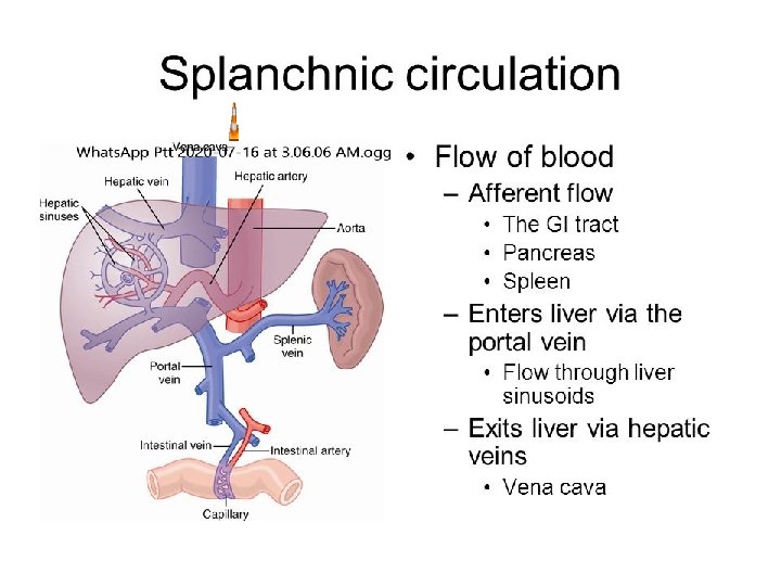
Gastrointestinal Blood Flow- “Splanchnic Circulation”
The blood vessels of the gastrointestinal system are part of a more extensive system called the splanchnic circulation. It includes the blood flow through the gut itself plus blood flows through the spleen, pancreas, and liver. The design of this system is such that all the blood that courses through the gut, spleen, and pancreas then flows immediately into the liver by way of the portal vein. In the liver, the blood passes through millions of minute liver sinusoids and finally leaves the liver by way of circulation. hepatic veins that empty into the vena cava of the general This flow of blood through the liver, before it empties into the vena cava, allows the reticuloendothelial cells that line the liver sinusoids to remove bacteria and other particulate matter that might enter the blood from the gastrointestinal tract, thus preventing direct transport of potentially harmful agents into the remainder of the body
The non-fat, water-soluble nutrients absorbed from the gut (such as carbohydrates and proteins) are transported in the portal venous blood to the same liver sinusoids. Here, both the reticuloendothelial cells and the hepatic cells, absorb and store temporarily from one half to three quarters of the nutrients. Also, much chemical intermediate processing of these nutrients occurs in the liver cells. Almost all of the fats absorbed from the intestinal tract are not carried in the portal blood but instead are absorbed into the intestinal lymphatics and then conducted to the systemic circulating blood by way of the thoracic duct, bypassing the liver.
Anatomy of Gastrointestinal Supply the Blood Figure shows the general arterial blood supply to the gut, including the superior mesenteric and inferior mesenteric arteries supplying the walls of the small and large intestines by way of an arching arterial system. Not shown in the figure is the celiac artery, which provides a similar blood supply to the stomach.
Effect of Gut Activity and Metabolic Factors on Gastrointestinal Blood Flow
Under normal conditions, the blood flow in each area of the gastrointestinal tract, as well as in each layer of the gut wall, is directly related to the level of local activity. For instance, during active absorption of nutrients, blood flow in the villi and adjacent regions of the submucosa is increased as much as eightfold. Likewise, blood flow in the muscle layers of the intestinal wall increases with increased motor activity in the gut. For instance, after a meal, the motor activity, secretory activity, and absorptive activity all increase; likewise, the blood flow increases greatly but then decreases back to the resting level over another 2 to 4 hours.
Possible Causes of the Increased Blood Flow During Gastrointestinal Activity.
First, several vasodilator substances are released from the mucosa of the intestinal tract during the digestive process. Most of these are peptide hormones, including cholecystokinin , vasoactive intestinal peptide, gastrin, and secretin. Second, some of the gastrointestinal glands also release into the gut wall two kinins, kallidin and bradykinin, at the same time that they secrete their secretions into the lumen. These kinins are powerful vasodilators that are believed to cause much of the increased mucosal vasodilation that occurs along with secretion. Third, decreased oxygen concentration in the gut wall can increase intestinal blood flow at least 50 to 100 per cent; therefore, the increased mucosal and gut wall metabolic rate during gut activity probably lowers the oxygen concentration enough to cause much of the vasodilation. The decrease in oxygen can also lead to as much as a fourfold increase of adenosine, a well-known vasodilator that could be responsible for much of the increased flow.
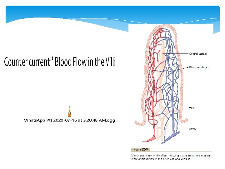
the arterial flow into the villus and the venous flow out of the villus are in directions opposite to each other, and that the vessels lie in close apposition to each other. Because of this vascular arrangement, much of the blood oxygen diffuses out of the arterioles directly into the adjacent venules without ever being carried in the blood to the tips of the villi. As much as 80 per cent of the oxygen may take this short-circuit route and therefore not be available for local metabolic functions of the villi. The reader will recognize that this type of counter current mechanism in the villi is analogous to the counter current mechanism in the vasa recta of the kidney medulla. Under normal conditions, this shunting of oxygen from the arterioles to the venules is not harmful to the villi, but in disease conditions in which blood flow to the gut becomes greatly reduced, such as in circulatory shock, the oxygen deficit in the tips of the villi can become so great the villus tip or even the whole villus suffers ischemic death and can disintegrate. Therefore, for this reason and others, in many gastrointestinal diseases the villi become seriously blunted, leading to greatly diminished intestinal absorptive capacity.
Nervous Control of Gastrointestinal Blood Flow
Stimulation of the parasympathetic nerves going to the stomach and lower colon increases local blood flow at the same time that it increases glandular secretion. This increased flow probably results secondarily from the increased glandular activity and not as a direct effect of the nervous stimulation. Sympathetic stimulation, by contrast, has a direct effect on essentially all the gastrointestinal tract to cause intense vasoconstriction of the arterioles with greatly decreased blood flow. After a few minutes of this vasoconstriction, the flow often returns almost to normal by means of a mechanism called “autoregulatory escape. ” That is, the local metabolic vasodilator mechanisms that are elicited by ischemia become dominant over the sympathetic vasoconstriction and, therefore, redilate the arterioles, thus causing return of necessary nutrient blood flow to the gastrointestinal glands and muscle.
Importance of Nervous Depression of Gastrointestinal Blood Flow When Other Parts of the Body Need Extra Blood Flow.
A major value of sympathetic vasoconstriction in the gut is that it allows shut-off of gastrointestinal and other splanchnic blood flow for short periods of time during heavy exercise, when increased flow is needed by the skeletal muscle and heart. Also, in circulatory shock, when all the body’s vital tissues are in danger of cellular death for lack of blood flow—especially the brain and the heart— sympathetic stimulation can decrease splanchnic blood flow to very little for many hours. Sympathetic stimulation also causes strong vasoconstriction of the largevolume intestinal and mesenteric veins. This decreases the volume of these veins, thereby displacing large amounts of blood into other parts of the circulation. In haemorrhagic shock or other states of low blood volume, this mechanism can provide as much as 200 to 400 milliliters of extra blood to sustain the general circulation.