Functional Neuroanatomy of ObsessiveCompulsive Disorder Sanjaya Saxena M

Functional Neuroanatomy of Obsessive-Compulsive Disorder Sanjaya Saxena, M. D. Director, UCSD Obsessive-Compulsive Disorders Program Associate Professor, UCSD Department of Psychiatry UCSD School of Medicine

Functional Neuroanatomy of Obsessive-Compulsive Disorder I. Structural Neuroimaging in OCD II. Functional Neuroimaging in OCD A. Resting State B. Pre- to Post-Treatment C. Symptom Provocation III. Heterogeneity in OCD A. OCD Symptom Factors B. Neural Correlates of OCD Symptom Factors IV. Questions Raised and Directions for Future Research

Functional Neuroimaging in Neuropsychiatry Goals • To understand how the brain mediates complex behavior – Normal vs. Abnormal – Across Diagnostic Boundaries • To determine the pathophysiology of neuropsychiatric disorders • To determine the cerebral mechanisms of action of treatments (Saxena et al, 2003)
 • Symptom Induction")
Functional Neuroimaging Study Methods • Baseline Studies (compared to Normal Controls) • Symptom Induction or Provocation • Pre- to Post-Treatment Studies • Cognitive Activation • Pharmacological Challenge

Structural Neuroimaging in OCD Replicated CT and MRI Findings • Smaller Orbitofrontal Cortex Volumes • Greater Orbitofrontal Gray Matter Density • Greater Anterior Cingulate Volumes in Pediatric OCD • Abnormal Striatal Volumes and Asymmetry – Reduced caudate volume in TS and some studies of OCD – Enlarged basal ganglia volumes in some studies may be associated with anti-streptococcal antibodies – Less left-right asymmetry

Chemical Neuroimaging in OCD MR Spectroscopy Findings • Low NAA in Caudate, Anterior Cingulate, and Left DLPFC • High Glutamate in Caudate - decreased after SRI Rx • Low Glutamate in AC • High Choline in Medial Thalamus

Neuroanatomy of OCD
 - Brain Activity: Glucose Metabolism")
Functional Neuroimaging Imaging Modalities • Positron Emission Tomography (PET) - Brain Activity: Glucose Metabolism or Blood Flow (r. CBF) - Neurochemical: Receptor or Transporter Binding : Transmitter Synthesis • Single Photon Emission Computed Tomography (SPECT) • Functional MRI (f. MRI) - BOLD: r. CBF, Volume, Perfusion, CMRO
")
Positron Emission Tomography (PET)

FDG-PET: Cerebral Glucose Metabolism

Types of PET Scanning Studies in OCD 1. OCD subjects versus normal controls at baseline 2. OCD subjects scanned pre- and post- treatment 3. Symptom provocation studies 4. Cognitive activation studies 5. Neurotransmitter / receptor binding studies
")
(Baxter et al, 1987)
")
Pre- to Post-Treatment PET Studies of OCD (Baxter et al, 1992)

Pre- and Post-Treatment Neuroimaging of OCD Effects of Cognitive-Behavioral Therapy • UCLA Studies (Baxter, Schwartz, et al): – Both CBT and fluoxetine decreased right caudate activity in responders – Both treatments broke pathological pre-treatment correlations between OFC, caudate, and thalamus • Wayne State Studies (Rosenberg, Gilbert, Benazon, et al): – Caudate glutamate decreased after paroxetine, not after CBT – Decreased caudate choline concentration in responders to CBT – Thalamic volume decreased after paroxetine, not after CBT
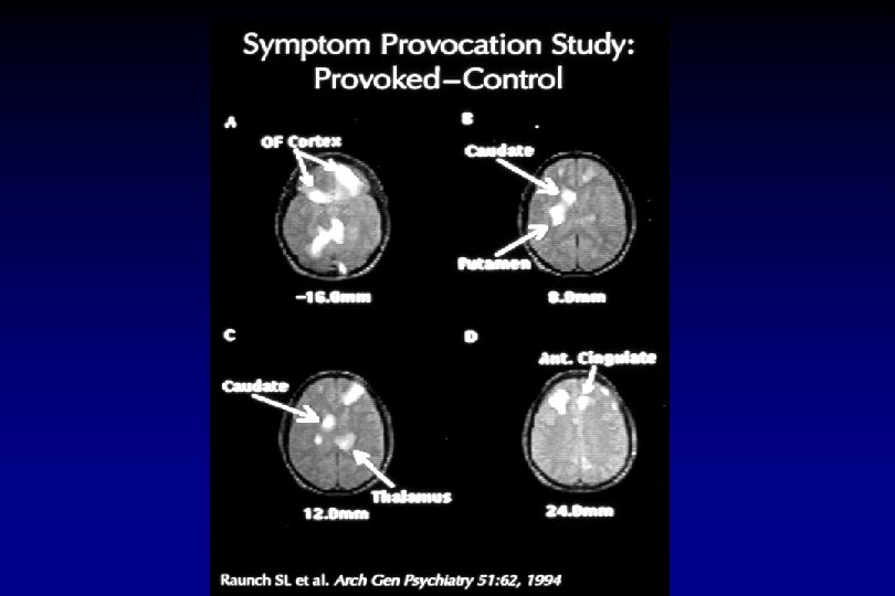

Functional Neuroimaging in OCD § Hyperactivity in orbitofrontal cortex, caudate, thalamus, and anterior cingulate gyrus. at rest, compared to controls further with symptom provocation with treatment (SRI’s, CBT, or surgery)

Orbitofrontal-Subcortical Circuit

“Classic” Conceptualization of Direct and Indirect Cortico-Basal Ganglia-Thalamo-Cortical Pathways Cerebral Cortex Striatum D 1 (+) Direct Pathway (+) D 2 (-) S. N. c G. P. int. and S. N. r Thalamus (-) (+) Indirect Pathway G. P. ext. (-) Subthalamic Nucleus (Saxena et al, 1998)
")
Frontal-Striatal Projections (Saxena et al, 1998)
 (+) (+) Striatum D 1")
Current Conceptualization of Frontal-Subcortical Circuitry Dorsal Cortical Structures (–) (+) (+) Striatum D 1 D 2 (+) Ventral/Limbic Structures (+) Direct Pathway (–) S. N. c (–) (+) G. P. i. and S. N. r Thalamus (–) Indirect Pathway (–) Indirect Basal Ganglia Control System (Saxena et al, 1998)

Model of OCD Pathophysiology Imbalance of Direct >> Indirect Pathway “Tone” in Orbitofrontal-Subcortical Circuit Orbitofrontal Cortex Ventromedial Caudate D 1 (+) Direct pathway (+) Medial Dorsal Thalamus (-) D 2 (-) GPi and SNr (-) Indirect pathway Indirect Basal Ganglia Control System (Saxena et al, 1998)
- Slides: 22