Function of Muscles Characteristics of Muscles n 3


(2 slides) heads n Myosin and actin overlap n")


- Slides: 22
Function of Muscles
Characteristics of Muscles n 3 types: n n muscle cell = muscle fiber n All muscles share some terminology ¨ Prefix myo refers to muscle ¨ Prefix mys refers to muscle ¨ Prefix sarco refers to flesh pg 179
1. Skeletal Muscle Characteristics
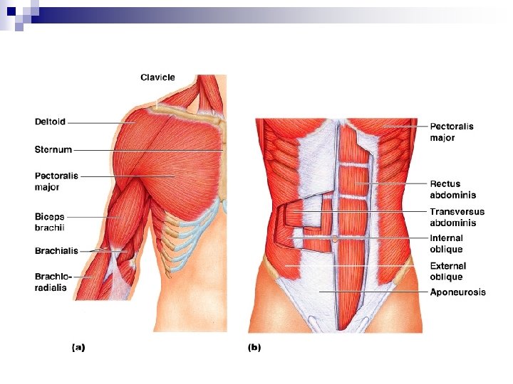
Connective Tissue Wrappings of Skeletal Muscle n Endomysium – n Perimysium – n Epimysium – Figure 6. 1
Skeletal Muscle Attachments n Muscle attachment sites: n n n connective tissue attachments ¨ Tendon – ¨ Aponeuroses –
2. Smooth Muscle Characteristics n n Figure 6. 2 a
3. Cardiac Muscle Characteristics n n Figure 6. 2 b
Quick Write n Fill in table distinguishing between the 3 muscle types: Smooth Skeletal Cardiac
Microscopic Anatomy of Skeletal Muscle 2 slides Cells are multinucleate n Nuclei are beneath the sarcolemma n Figure 6. 3 a
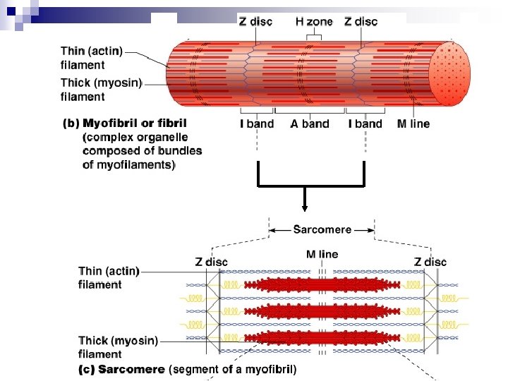
Microscopic Anatomy of Skeletal Muscle n Myofibril ¨Bundles of myofilaments n. I band = light band ¨Z n. A disc at midline band = dark band ¨H zone at midpoint ¨ M Line Figure 6. 3 b
Microscopic Anatomy of Skeletal Muscle n Sarcomere: ¨ Contractile unit (Z disc to Z disc) Figure 6. 3 b
Microscopic Anatomy of Skeletal Muscle n Organization of the sarcomere: ¨ Thick filaments = myosin filaments (protein) ¨ Thin filaments = actin filaments (protein) Figure 6. 3 c
Microscopic Anatomy of Skeletal Muscle (good)(2 slides) heads n Myosin and actin overlap n Figure 6. 3 d
Microscopic Anatomy of Skeletal Muscle n At rest: bare zone ¨ lacks n actin filaments (H zone) Sarcoplasmic reticulum (SR) – for storage of calcium Figure 6. 3 d
Mc. Graw Review Video
Draw a Sarcomere n Label: Z disc, H zone, A band, I band, M line, actin and myosin
So now we know the microscopic anatomy of muscle cell……. so what you may ask? ? Who cares? ? ? It will help us understand the ______
Muscle Cell Contraction Review website n Prezi: n