
From Gene to Protein Chapter 17 Metabolism and

From Gene to Protein Chapter 17
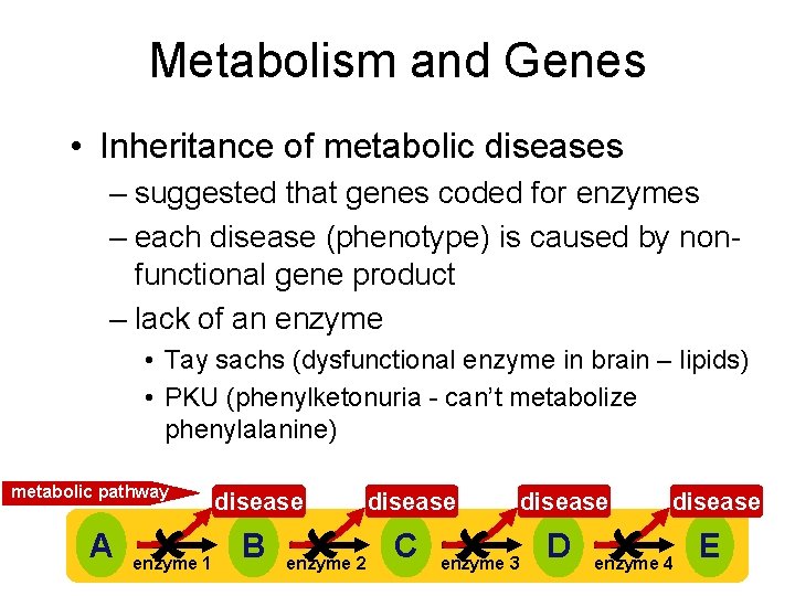
Metabolism and Genes • Inheritance of metabolic diseases – suggested that genes coded for enzymes – each disease (phenotype) is caused by nonfunctional gene product – lack of an enzyme • Tay sachs (dysfunctional enzyme in brain – lipids) • PKU (phenylketonuria - can’t metabolize phenylalanine) metabolic pathway A enzyme 1 disease B C D E enzyme 2 enzyme 3 enzyme 4
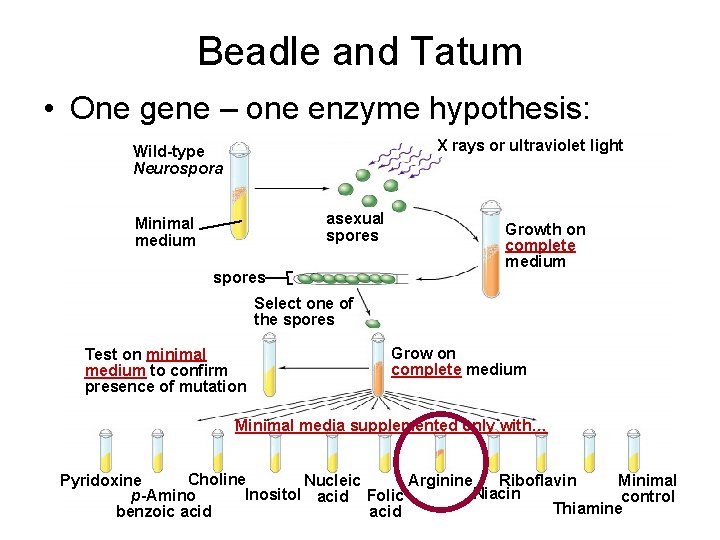
Beadle and Tatum • One gene – one enzyme hypothesis: X rays or ultraviolet light Wild-type Neurospora asexual spores Minimal medium spores Growth on complete medium Select one of the spores Test on minimal medium to confirm presence of mutation Grow on complete medium Minimal media supplemented only with… Choline Pyridoxine Riboflavin Minimal Nucleic Arginine Niacin Inositol acid Folic p-Amino control Thiamine acid benzoic acid
One gene-one polypeptide hypothesis – Why is the one gene-one enzyme hypothesis now called one gene-one polypeptide hypothesis? – Now: one gene – one polypeptide – Why?
Evolution of the Genetic Code • The genetic code is nearly universal, shared by the simplest bacteria to the most complex animals • Genes can be transcribed and translated after being transplanted from one species to another Copyright © 2008 Pearson Education Inc. , publishing as Pearson Benjamin Cummings
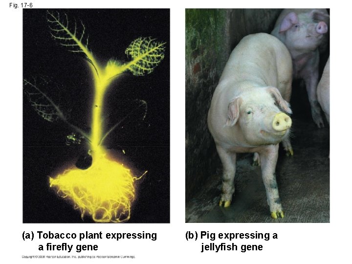
Fig. 17 -6 (a) Tobacco plant expressing a firefly gene (b) Pig expressing a jellyfish gene
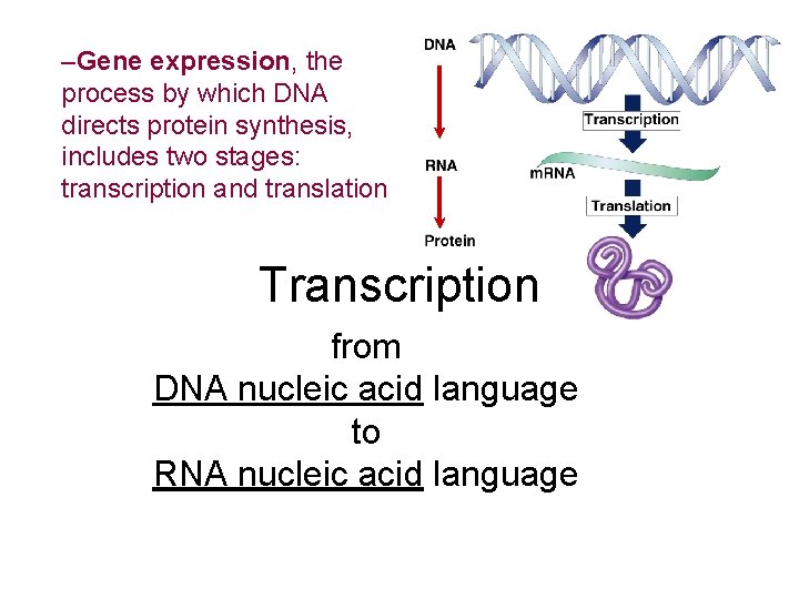
–Gene expression, the process by which DNA directs protein synthesis, includes two stages: transcription and translation Transcription from DNA nucleic acid language to RNA nucleic acid language
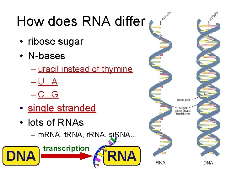
How does RNA differ from DNA • ribose sugar • N-bases – uracil instead of thymine – U : A – C : G • single stranded • lots of RNAs – m. RNA, t. RNA, r. RNA, si. RNA… DNA transcription RNA
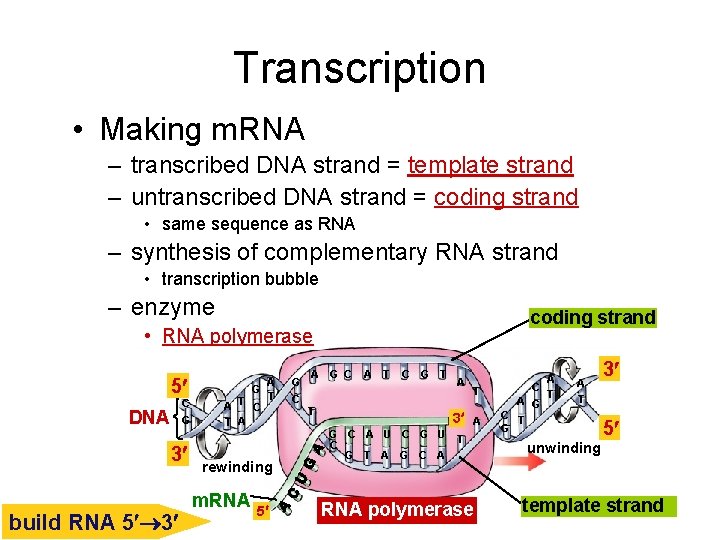
Transcription • Making m. RNA – transcribed DNA strand = template strand – untranscribed DNA strand = coding strand • same sequence as RNA – synthesis of complementary RNA strand • transcription bubble – enzyme coding strand • RNA polymerase 5 C DNA G 3 build RNA 5 3 A G T A T C T A rewinding m. RNA 5 G C A T C G T T A 3 G C A U C G T A G C A T RNA polymerase C A G C T G A T unwinding 3 5 template strand

RNA polymerases • 3 RNA polymerase enzymes – RNA polymerase I • only transcribes r. RNA genes • makes ribosomes – RNA polymerase II • transcribes genes into m. RNA – RNA polymerase III • only transcribes t. RNA genes – each has a specific promoter sequence it recognizes
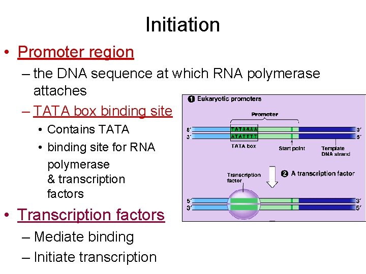
Initiation • Promoter region – the DNA sequence at which RNA polymerase attaches – TATA box binding site • Contains TATA • binding site for RNA polymerase & transcription factors • Transcription factors – Mediate binding – Initiate transcription
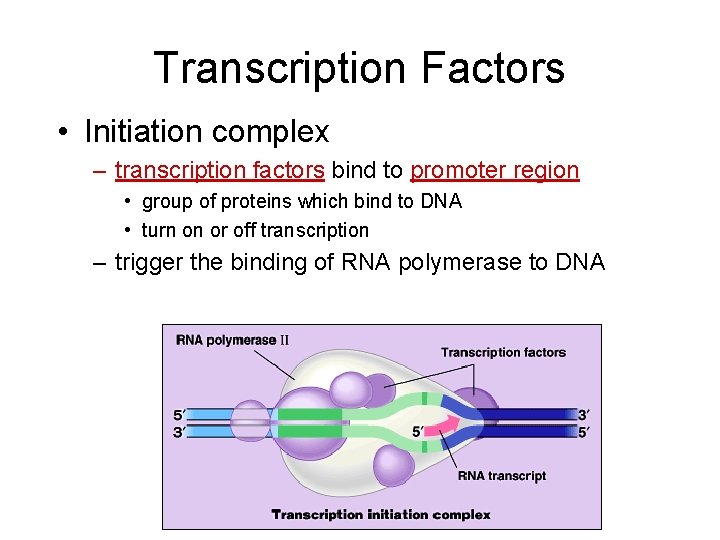
Transcription Factors • Initiation complex – transcription factors bind to promoter region • group of proteins which bind to DNA • turn on or off transcription – trigger the binding of RNA polymerase to DNA
Elongation and Termination • http: //bcs. whfreeman. com/thelifewire/conte nt/chp 12/1202001. html
Alteration of m. RNA Ends • Each end of a pre-m. RNA molecule is modified in a particular way: – The 5 end receives a modified nucleotide 5 cap – The 3 end gets a poly-A tail (AAA…AAA) • These modifications share several functions: – They seem to facilitate the export of m. RNA – They protect m. RNA from hydrolytic enzymes – They help ribosomes attach to the 5 end Copyright © 2008 Pearson Education Inc. , publishing as Pearson Benjamin Cummings
RNA Modification http: //www. youtube. com/watch? v=Yj. Wu. Vrzv. ZYA

Fig. 17 -3 DNA TRANSCRIPTION What differences are seen between bacteria and eukaryotic cells? m. RNA Ribosome TRANSLATION Polypeptide (a) Bacterial cell Nuclear envelope DNA TRANSCRIPTION Pre-m. RNA PROCESSING m. RNA TRANSLATION Ribosome Polypeptide (b) Eukaryotic cell
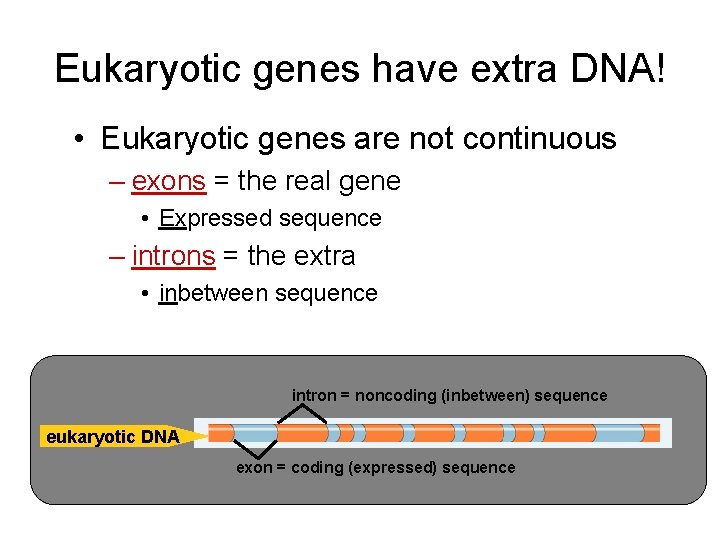
Eukaryotic genes have extra DNA! • Eukaryotic genes are not continuous – exons = the real gene • Expressed sequence – introns = the extra • inbetween sequence intron = noncoding (inbetween) sequence eukaryotic DNA exon = coding (expressed) sequence
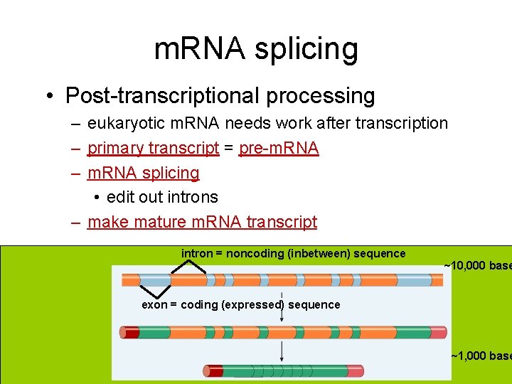
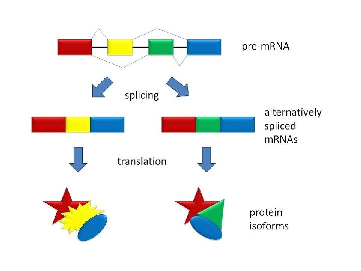
m. RNA splicing • Post-transcriptional processing – eukaryotic m. RNA needs work after transcription – primary transcript = pre-m. RNA – m. RNA splicing • edit out introns – make mature m. RNA transcript intron = noncoding (inbetween) sequence ~10, 000 base exon = coding (expressed) sequence ~1, 000 base
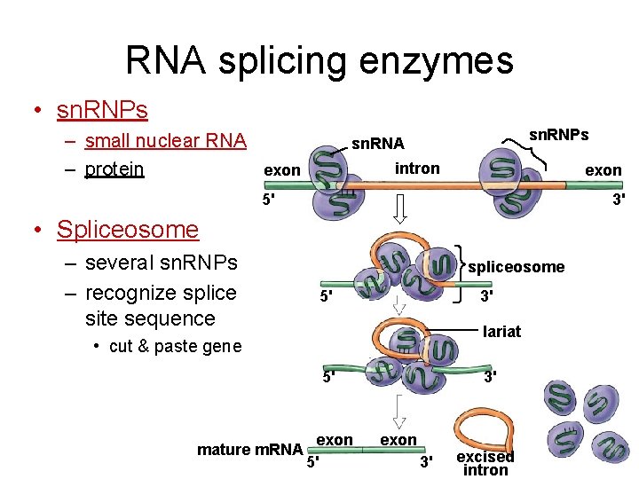
RNA splicing enzymes • sn. RNPs – small nuclear RNA – protein sn. RNPs sn. RNA intron exon 5' 3' • Spliceosome – several sn. RNPs – recognize splice site sequence spliceosome 5' 3' lariat • cut & paste gene 5' mature m. RNA exon 5' 3' exon 3' excised intron
RNA Splicing http: //www. youtube. com/watch? v=HSD 1 Al. A 1 r 4 Y http: //www. youtube. com/watch? v=FVu. Aw. BGw_p. Q http: //bcs. whfreeman. com/thelifewire/content/chp 14/1401 s. swf
Ribozymes • Ribozymes are catalytic RNA molecules that function as enzymes and can splice RNA • The discovery of ribozymes rendered obsolete the belief that all biological catalysts were proteins Copyright © 2008 Pearson Education Inc. , publishing as Pearson Benjamin Cummings
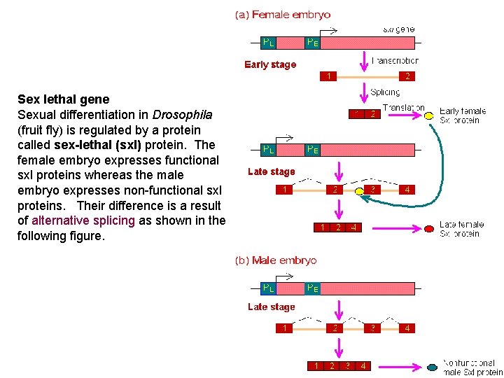
Sex lethal gene Sexual differentiation in Drosophila (fruit fly) is regulated by a protein called sex-lethal (sxl) protein. The female embryo expresses functional sxl proteins whereas the male embryo expresses non-functional sxl proteins. Their difference is a result of alternative splicing as shown in the following figure.

The Genetic Code • How are the instructions for assembling amino acids into proteins encoded into DNA? • There are 20 amino acids, but there are only four nucleotide bases in DNA • How many bases correspond to an amino acid? Copyright © 2008 Pearson Education Inc. , publishing as Pearson Benjamin Cummings
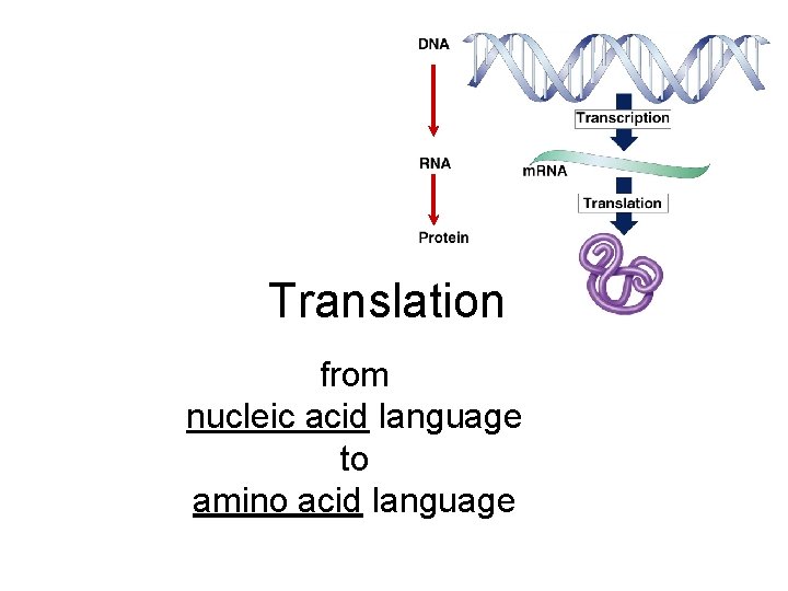
Translation from nucleic acid language to amino acid language
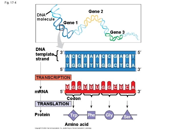
Fig. 17 -4 DNA molecule Gene 2 Gene 1 Gene 3 DNA template strand TRANSCRIPTION m. RNA Codon TRANSLATION Protein Amino acid
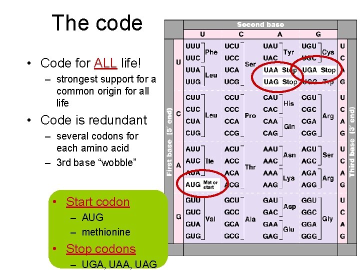
The code • Code for ALL life! – strongest support for a common origin for all life • Code is redundant – several codons for each amino acid – 3 rd base “wobble” • Start codon – AUG – methionine • Stop codons – UGA, UAG
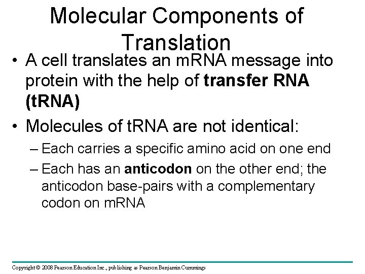
Molecular Components of Translation • A cell translates an m. RNA message into protein with the help of transfer RNA (t. RNA) • Molecules of t. RNA are not identical: – Each carries a specific amino acid on one end – Each has an anticodon on the other end; the anticodon base-pairs with a complementary codon on m. RNA Copyright © 2008 Pearson Education Inc. , publishing as Pearson Benjamin Cummings

Transfer RNA structure • “Clover leaf” structure – anticodon on “clover leaf” end – amino acid attached on 3 end
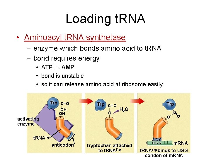
Loading t. RNA • Aminoacyl t. RNA synthetase – enzyme which bonds amino acid to t. RNA – bond requires energy • ATP AMP • bond is unstable • so it can release amino acid at ribosome easily Trp C=O H 2 O O activating enzyme t. RNATrp anticodon tryptophan attached to t. RNATrp AC C UGG =O O Trp C OH OH Trp C=O m. RNA t. RNATrp binds to UGG condon of m. RNA
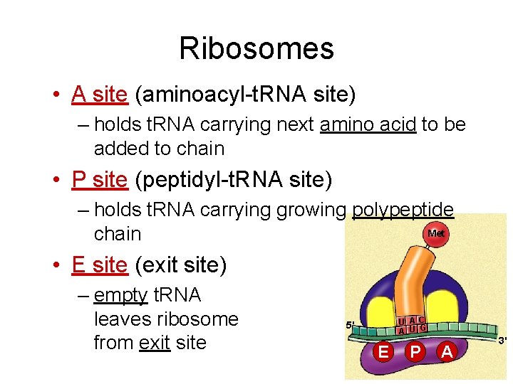
Ribosomes • A site (aminoacyl-t. RNA site) – holds t. RNA carrying next amino acid to be added to chain • P site (peptidyl-t. RNA site) – holds t. RNA carrying growing polypeptide Met chain • E site (exit site) – empty t. RNA leaves ribosome from exit site U A C A U G 5' E P A 3'

Building a polypeptide • Initiation – brings together m. RNA, ribosome subunits, initiator t. RNA • Elongation – adding amino acids based on codon sequence • Termination 3 2 1 – end codon Val Leu Met Met Leu Ala Leu release factor Ser Trp A C t. RNA G U AC 5' CUGAA U m. RNA A U G 3' E P A 5' U A C G A C AA U G CU G 5' 3' U A C GA C A U G C UG AAU 3' 5' U A C G A C AA U AU G C U G 3' A CC U GG U A A 3'
Initiation • The initiation stage of translation brings together m. RNA, a t. RNA with the first amino acid, and the two ribosomal subunits • First, a small ribosomal subunit binds with m. RNA and a special initiator t. RNA • Then the small subunit moves along the m. RNA until it reaches the start codon (AUG) • Proteins called initiation factors bring in the large subunit that completes the translation initiation complex

Fig. 17 -17 3 U A C 5 Met 5 A U G 3 Initiator t. RNA P site Met Large ribosomal subunit GTP GDP E m. RNA 5 Start codon m. RNA binding site 3 Small ribosomal subunit 5 A 3 Translation initiation complex
Elongation • During the elongation stage, amino acids are added one by one to the preceding amino acid • Each addition involves proteins called elongation factors and occurs in three steps: codon recognition, peptide bond formation, and translocation Copyright © 2008 Pearson Education Inc. , publishing as Pearson Benjamin Cummings
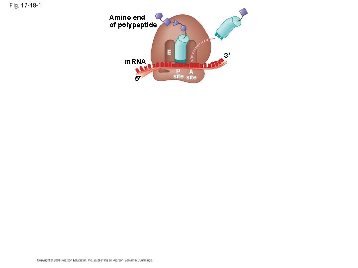
Fig. 17 -18 -1 Amino end of polypeptide E 3 m. RNA 5 P A site
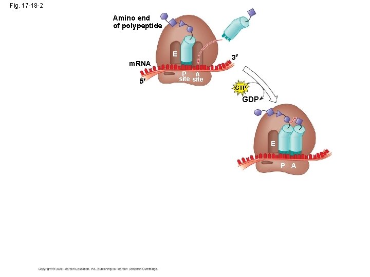
Fig. 17 -18 -2 Amino end of polypeptide E 3 m. RNA 5 P A site GTP GDP E P A
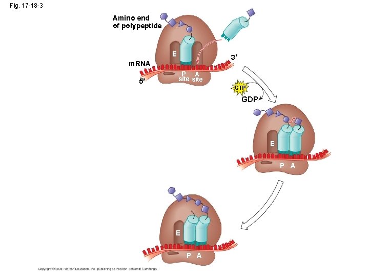
Fig. 17 -18 -3 Amino end of polypeptide E 3 m. RNA 5 P A site GTP GDP E P A
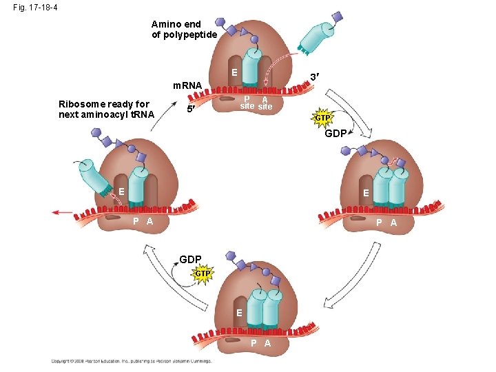
Fig. 17 -18 -4 Amino end of polypeptide E 3 m. RNA Ribosome ready for next aminoacyl t. RNA P A site 5 GTP GDP E E P A GDP GTP E P A
Termination of Translation • Termination occurs when a stop codon in the m. RNA reaches the A site of the ribosome • The A site accepts a protein called a release factor • The release factor causes the addition of a water molecule instead of an amino acid • This reaction releases the polypeptide, and the translation assembly then comes apart
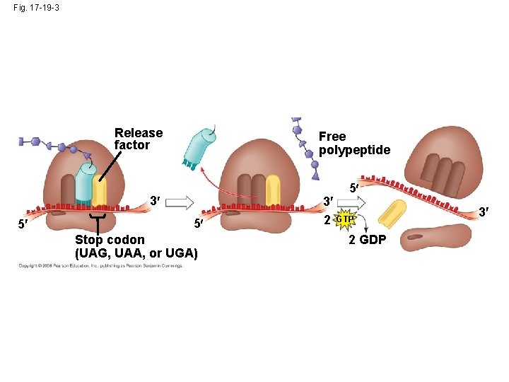
Fig. 17 -19 -3 Release factor Free polypeptide 5 3 5 5 Stop codon (UAG, UAA, or UGA) 3 2 GTP 2 GDP 3
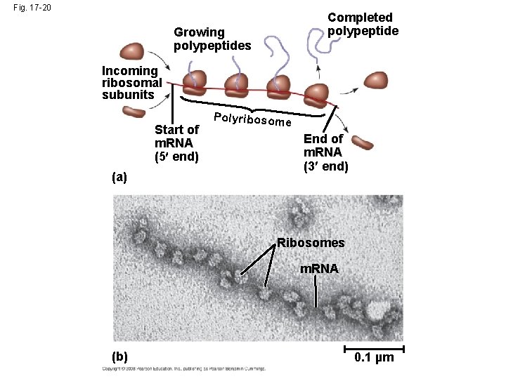
Fig. 17 -20 Completed polypeptide Growing polypeptides Incoming ribosomal subunits Start of m. RNA (5 end) (a) Polyribosome End of m. RNA (3 end) Ribosomes m. RNA (b) 0. 1 µm
Targeting Polypeptides to Specific Locations • Two populations of ribosomes are evident in cells: free ribsomes (in the cytosol) and bound ribosomes (attached to the ER) • Free ribosomes mostly synthesize proteins that function in the cytosol • Bound ribosomes make proteins of the endomembrane system and proteins that are secreted from the cell • Ribosomes are identical and can switch from free to bound
• Polypeptide synthesis always begins in the cytosol • Synthesis finishes in the cytosol unless the polypeptide signals the ribosome to attach to the ER • Polypeptides destined for the ER or for secretion are marked by a signal peptide Copyright © 2008 Pearson Education Inc. , publishing as Pearson Benjamin Cummings
• A signal-recognition particle (SRP) binds to the signal peptide • The SRP brings the signal peptide and its ribosome to the ER Copyright © 2008 Pearson Education Inc. , publishing as Pearson Benjamin Cummings

Fig. 17 -21 Ribosome m. RNA Signal peptide removed Signalrecognition particle (SRP) CYTOSOL ER LUMEN SRP receptor protein Translocation complex ER membrane Protein
Translation • http: //highered. mcgrawhill. com/sites/0072943696/student_view 0/ chapter 3/animation__how_translation_wor ks. html • http: //highered. mcgrawhill. com/sites/0072507470/student_view 0/ chapter 3/animation__protein_synthesis__ quiz_3_. html
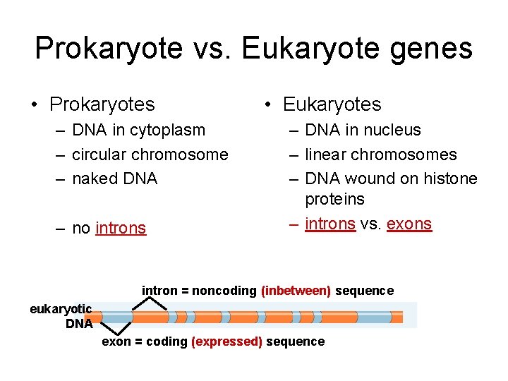
Prokaryote vs. Eukaryote genes • Prokaryotes – DNA in cytoplasm – circular chromosome – naked DNA – no introns • Eukaryotes – DNA in nucleus – linear chromosomes – DNA wound on histone proteins – introns vs. exons intron = noncoding (inbetween) sequence eukaryotic DNA exon = coding (expressed) sequence
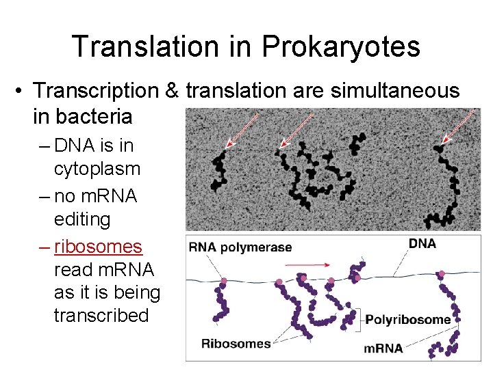
Translation in Prokaryotes • Transcription & translation are simultaneous in bacteria – DNA is in cytoplasm – no m. RNA editing – ribosomes read m. RNA as it is being transcribed
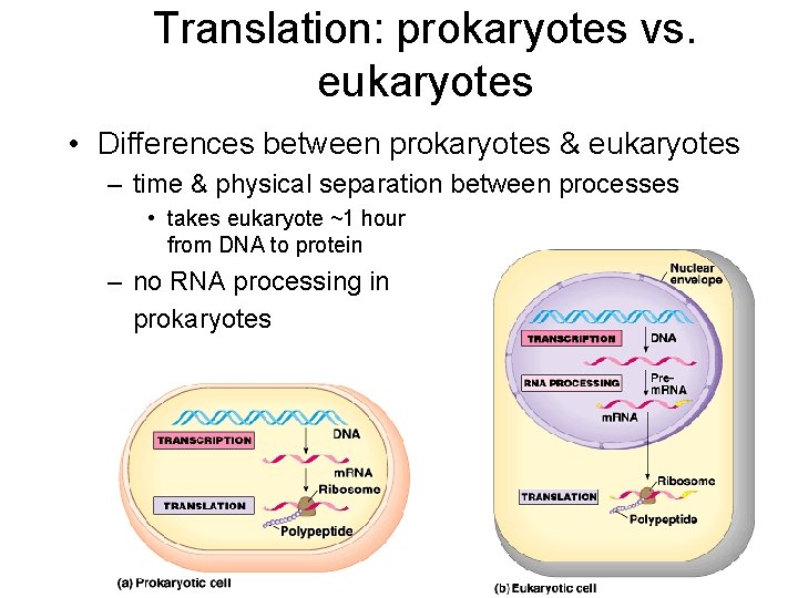
Translation: prokaryotes vs. eukaryotes • Differences between prokaryotes & eukaryotes – time & physical separation between processes • takes eukaryote ~1 hour from DNA to protein – no RNA processing in prokaryotes
Mutations • Mutations are changes in the genetic material of a cell or virus • Point mutations are chemical changes in just one base pair of a gene • The change of a single nucleotide in a DNA template strand can lead to the production of an abnormal protein • Point mutations within a gene can be divided into two general categories – Base-pair substitutions – Base-pair insertions or deletions
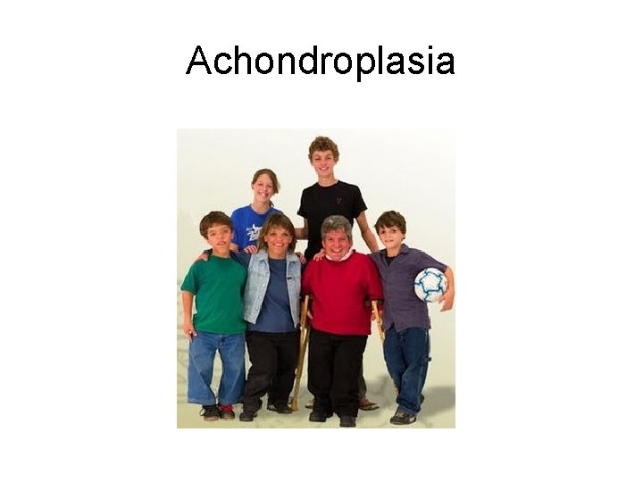
Achondroplasia
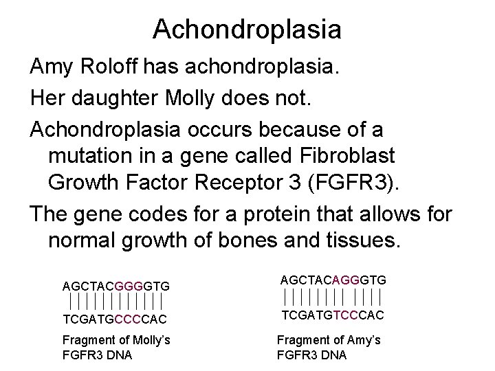
Achondroplasia Amy Roloff has achondroplasia. Her daughter Molly does not. Achondroplasia occurs because of a mutation in a gene called Fibroblast Growth Factor Receptor 3 (FGFR 3). The gene codes for a protein that allows for normal growth of bones and tissues. AGCTACGGGGTG AGCTACAGGGTG TCGATGCCCCAC TCGATGTCCCAC Fragment of Molly’s FGFR 3 DNA Fragment of Amy’s FGFR 3 DNA
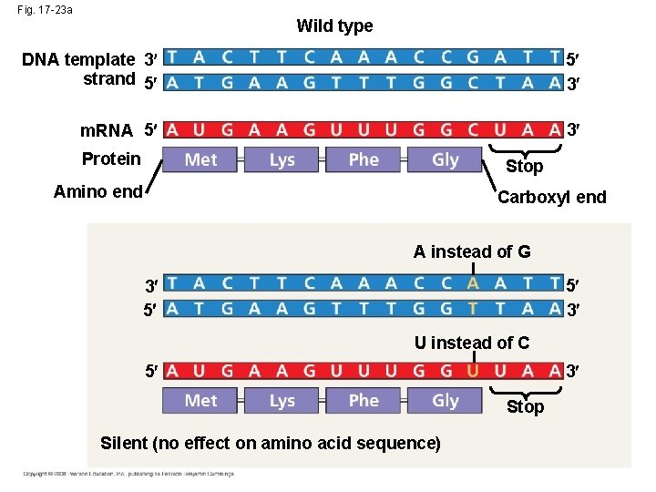
Fig. 17 -23 a Wild type DNA template 3 strand 5 5 3 m. RNA 5 3 Protein Stop Amino end Carboxyl end A instead of G 5 3 3 5 U instead of C 5 3 Stop Silent (no effect on amino acid sequence)
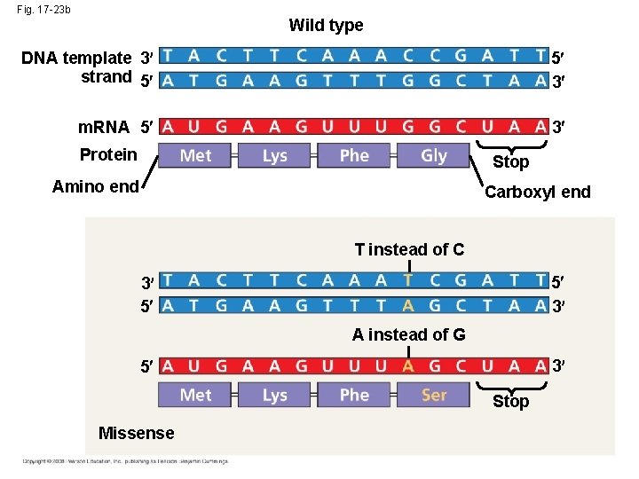
Fig. 17 -23 b Wild type DNA template 3 strand 5 5 3 m. RNA 5 3 Protein Stop Amino end Carboxyl end T instead of C 5 3 3 5 A instead of G 3 5 Stop Missense
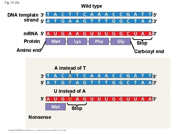
Fig. 17 -23 c Wild type DNA template 3 strand 5 5 3 m. RNA 5 3 Protein Stop Amino end Carboxyl end A instead of T 3 5 5 3 U instead of A 5 3 Stop Nonsense
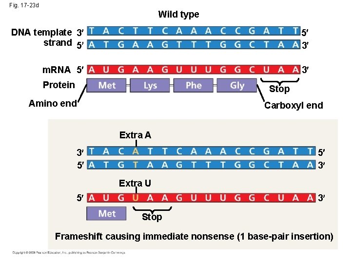
Fig. 17 -23 d Wild type DNA template 3 strand 5 5 3 m. RNA 5 3 Protein Stop Amino end Carboxyl end Extra A 5 3 3 5 Extra U 5 3 Stop Frameshift causing immediate nonsense (1 base-pair insertion)
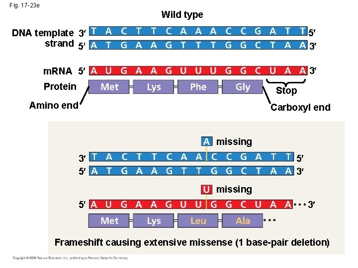
Fig. 17 -23 e Wild type DNA template 3 strand 5 5 3 m. RNA 5 3 Protein Stop Amino end Carboxyl end missing 5 3 3 5 missing 5 3 Frameshift causing extensive missense (1 base-pair deletion)
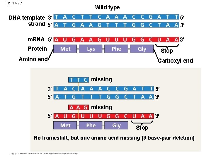
Fig. 17 -23 f Wild type DNA template 3 strand 5 5 3 m. RNA 5 3 Protein Stop Amino end Carboxyl end missing 5 3 3 5 missing 5 3 Stop No frameshift, but one amino acid missing (3 base-pair deletion)
- Slides: 59