Franz Joseph Gall 1758 1828 P Broca 1824
 (1758— 1828)")
Франц Галль (Franz Joseph Gall) (1758— 1828)
 (1824 -1880) К. Вернике (K. Wernicke) (1848 -1904)")
П. Брока (P. Broca) (1824 -1880) К. Вернике (K. Wernicke) (1848 -1904)

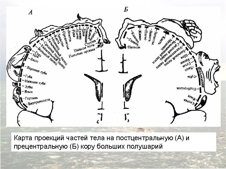
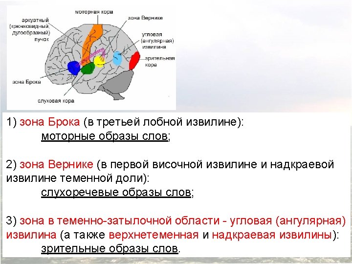
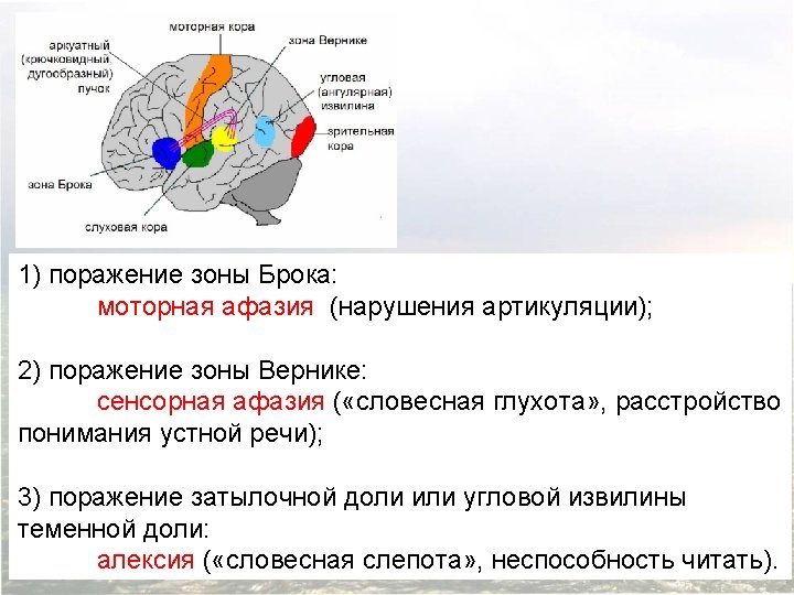
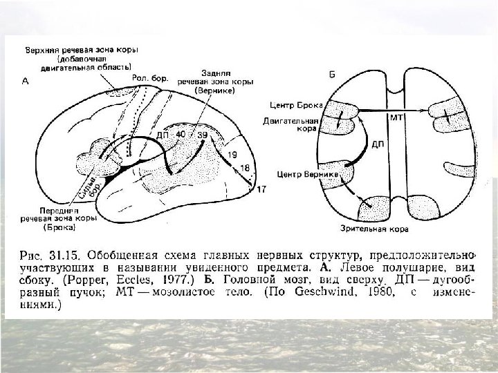
The relationship of the major language areas to the classical cytoarchitectonic map of the cerebral cortex. Whereas primary sensory and motor functions are sometimes coextensive with these areas, more general cognitive functions like attention, identification, and planning typically encompass a number of different cytoarchitectonic areas in one or more cortical lobes. The language functions described by Broca and Wernicke are associated with at least three of the cytoarchitectonic areas defined by Brodmann (area 22, at the junction of the parietal and temporal lobes [Wernicke’s area]; and areas 44 and 45, in the ventral and posterior region of the frontal lobe [Broca’s area]), and are not coextensive with any of them.
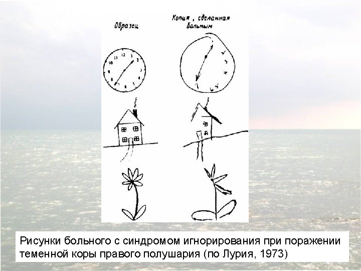

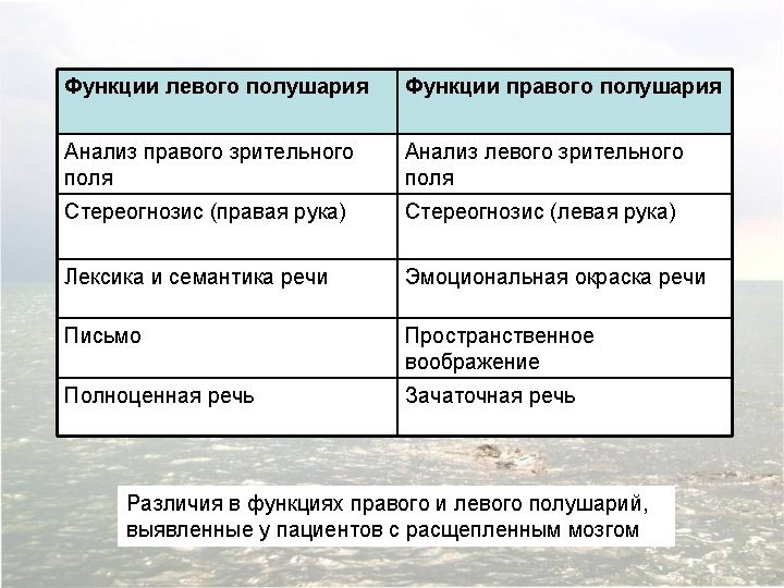
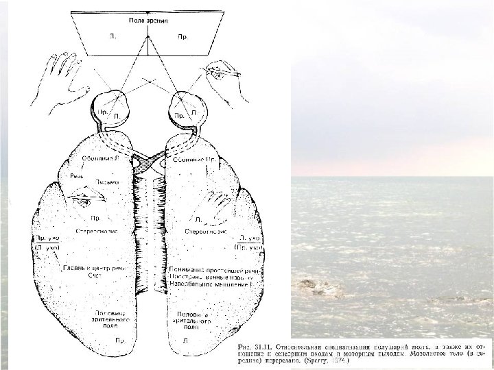
Confirmation of hemispheric specialization for language obtained by studying individuals in whom the connections between the right and left hemispheres have been surgically divided. Single-handed, visionindependent stereognosis can be used to evaluate the language capabilities of each hemisphere in split-brain patients. Objects held in the right hand, which provides somatic sensory information to the left hemisphere, are easily named; objects held in the left hand, however, are not readily named by these patients.
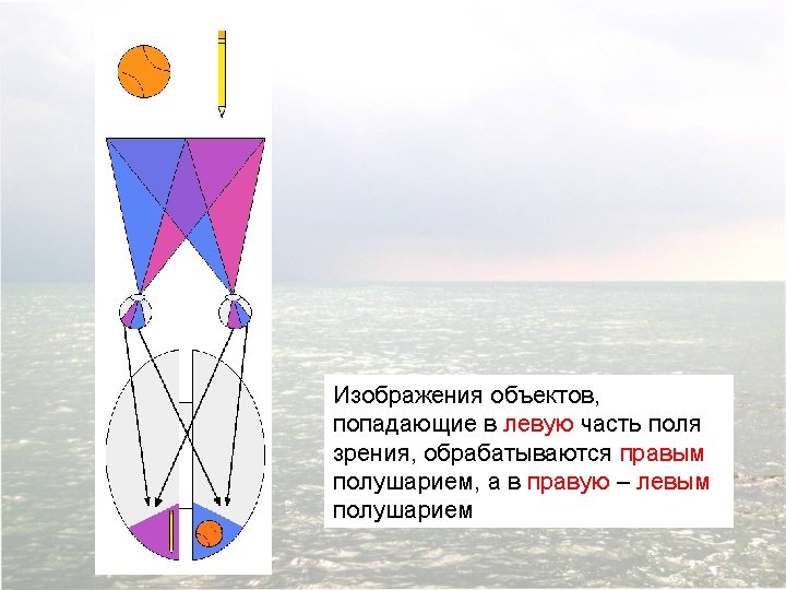
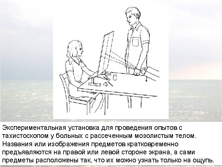
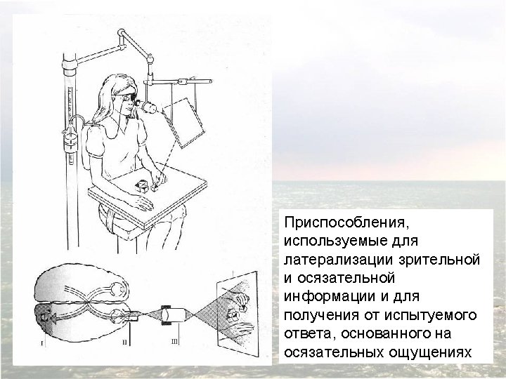

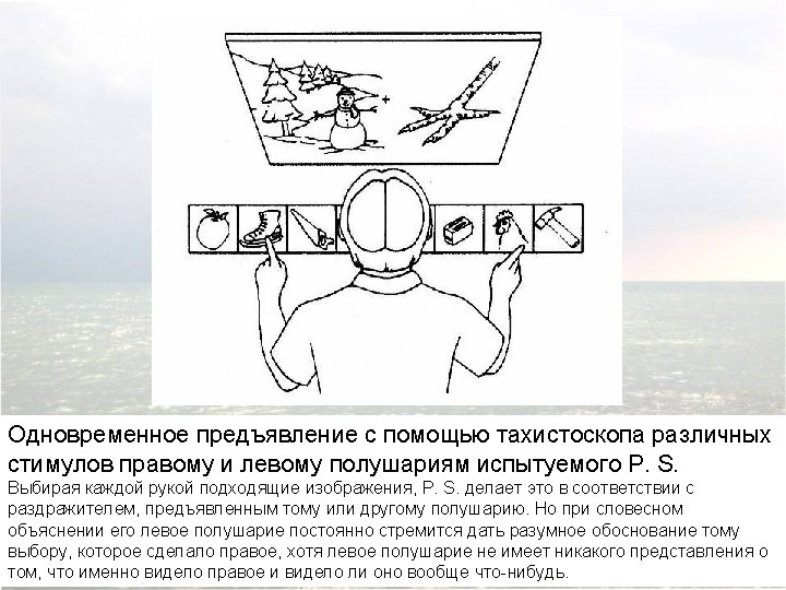
Visual stimuli or simple instructions can be given independently to the right or left hemisphere in normal and splitbrain individuals. Since the left visual field is perceived by the right hemisphere (and vice versa), a briefly presented (tachistoscopic) instruction in the left visual field is appreciated only by the right brain (assuming that the individual maintains fixation on a mark in the center of the viewing screen). In normal subjects, activation of the right visual cortex leads to hemispheric transfer of visual information via the corpus callosum to the left hemisphere. In split-brain patients, information presented to the left visual field cannot reach the left hemisphere, and patients are unable to produce a verbal report regarding the stimuli. However, such patients are able to provide a verbal report of stimuli presented to the right visual field. A wide range of hemispheric functions can be evaluated using this tachistoscopic method, even in normal subjects.
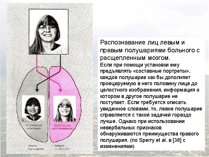
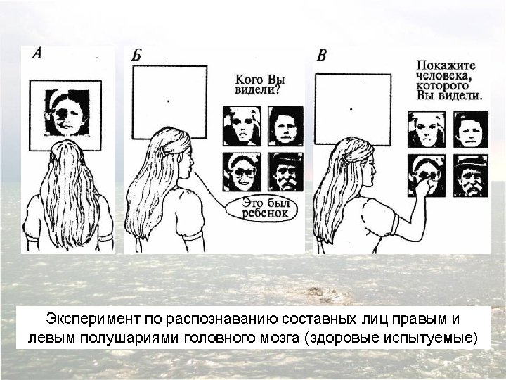
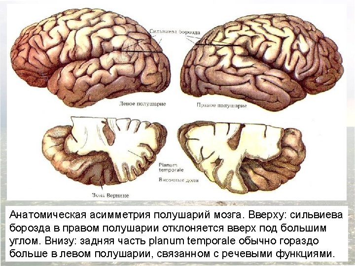

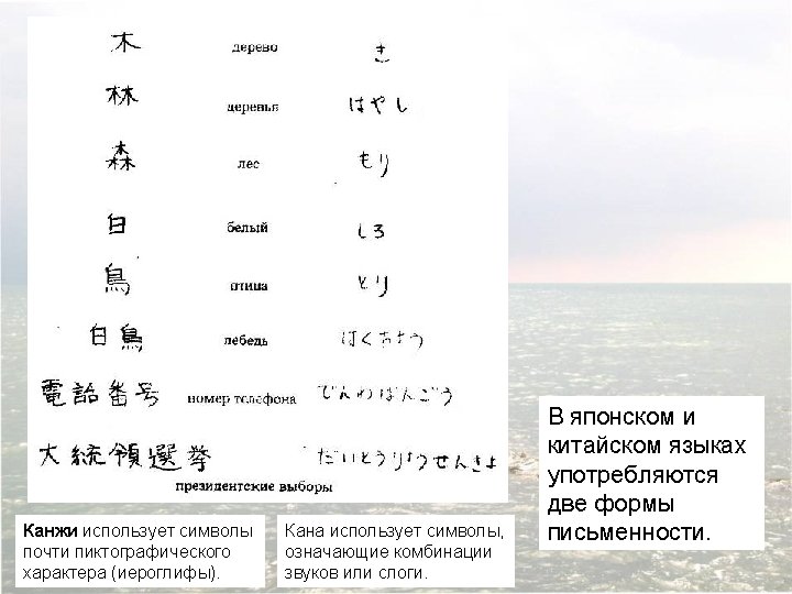
Asymmetry of the right and left human temporal lobes. A region of the surface of the temporal lobe called the planum temporale is significantly larger in the left hemisphere of most (but far from all) individuals. A magnetic resonance image in the frontal plane, showing this asymmetry (arrows) in a normal adult subject. (Такая асимметрия наблюдается лишь у 67% людей, в то время как центр речи расположен слева у 95 -97% людей)
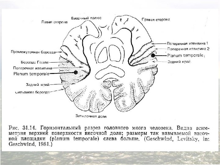
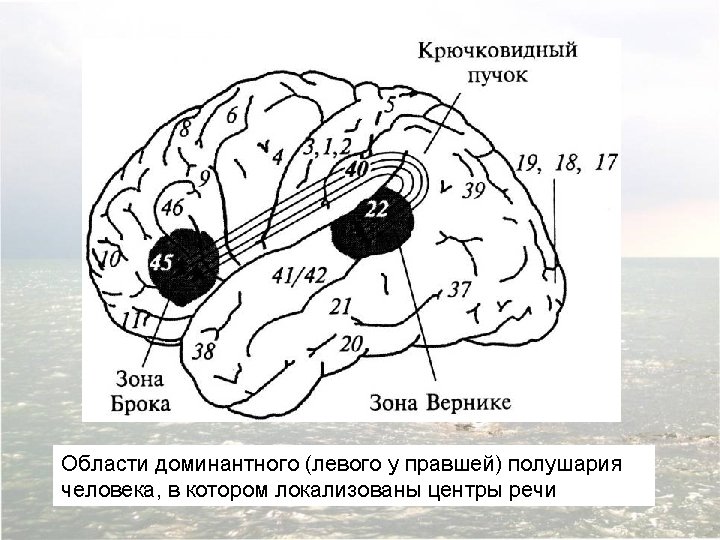
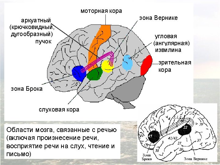

Diagram of the major brain areas involved in the comprehension and production of language. The primary sensory, auditory, visual, and motor cortices are indicated to show the relation of Broca’s and Wernicke’s language areas to these other areas that are necessarily involved in the comprehension and production of speech, albeit in a less specialized way.

Evidence for the variability of language representation among individuals, determined by electrical stimulation during neurosurgery. Diagram from Penfield’s original study illustrating sites in the left hemisphere at which electrical stimulation interfered with speech. (after Penfield and Roberts, 1959)

Evidence for the variability of language representation among individuals, determined by electrical stimulation during neurosurgery. Diagrams summarizing data from 117 patients whose language areas were mapped by electrical recording at the time of surgery. The number in each red circle indicates the (quite variable) percentage of patients who showed interference with language in response to stimulation at that site. Note also that many of the sites that elicited interference fall outside the classic language areas (after Ojemann et al. , 1989. )
 in a")
Language-related regions of the left hemisphere mapped by positron emission tomography (PET) in a normal human subject. Subjects reclined within the PET scanner and followed instructions on a special display (these details are not illustrated). The left panels indicate the task being practiced prior to scanning. The PET scan images are shown on the right. Language tasks such as listening to words and generating word associations elicit activity in Broca’s and Wernicke’s areas, as expected. However, there is also activity in primary and association sensory and motor areas for both active and passive language tasks. These observations indicate that language processing involves cortical regions in addition to the classic language areas. (From Posner and Raichle, 1994. )

Афазия Брока: ‘I am a sig…no…man…uh, well, …again. ’ These words were emitted slowly, and with great effort. The sounds were not clearly articulated; each syllable as uttered harshly, explosively, in a throaty voice. With practice, it was possible to understand him, but at first I encountered considerable difficulty in this. ‘Let me help you, ’ I interjected. ‘You were a signal…’ ‘A sig-nal man…right, ’ Ford completed my phrase triumphantly. ‘Were you in the Coast Guard? ’ ‘No, er, yes, …ship…Massachu…chusetts…Coastguard …years. ’ He raised his hands twice, indicating the number nineteen. ‘Oh, you were in the Coast Guard for nineteen years. ’ ‘Oh…boy…right, ’ he replied. ‘Why are you in the hospital, Mr. Ford? ’ Ford looked at me strangely, as if to say, Isn’t it patently obvious? He pointed to his paralyzed arm and said, ‘Arm no good, ’ then to his mouth and said, ‘Speech…can’t say…talk, you see. ’ Howard Gardner, 1974. (The Shattered Mind: The Person after Brain Damage, pp. 60– 61. )

Афазия Вернике: Boy, I’m sweating, I’m awful nervous, you know, once in a while I get caught up, I can’t mention the tarripoi, a month ago, quite a little, I’ve done a lot well, I impose a lot, while, on the other hand, you know what I mean, I have to run around, look it over, trebbin and all that sort of stuff. Oh sure, go ahead, any old think you want. If I could I would. Oh, I’m taking the word the wrong way to say, all of the barbers here whenever they stop you it’s going around, if you know what I mean, that is tying and tying for repucer, repuceration, well, we were trying the best that we could while another time it was with the beds over there the same thing… Howard Gardner, 1974. (The Shattered Mind: The Person after Brain Damage, p. 68. )

 Demonet et al. Renewal")
Основные области мозга, участвующие в речи (латеральная сторона левого полушария) Demonet et al. Renewal of the Neurophysiology of Language: Functional Neuroimaging. Physiol Rev 85: 49 -95, 2005.
 Demonet et al. Renewal")
Основные области мозга, участвующие в речи (медиальная сторона левого полушария) Demonet et al. Renewal of the Neurophysiology of Language: Functional Neuroimaging. Physiol Rev 85: 49 -95, 2005.

Synopsis of PET studies of auditory language processing. This figure depicts the impact of several factors on the distribution of language-related neural activities. Schematic activations are redrawn from the original studies using the significance threshold reported in each study. A: Belin et al. (22) showed that rapid acoustic transitions elicit more activity in the left STG than contralateral right-sided regions. B and C: Demonet et al. (106) showed that increasing difficulty of phoneme monitoring tasks induces a left-sided asymmetry in the activity of the STG. D: Demonet et al. (105) found increased activity in the dorsal pathway by contrasting phoneme monitoring (the difficult variant) with semantic categorization (the reverse contrast is shown in J). E: Scott et al. (355) showed that intelligible speech samples activate a specific pathway along the anterior part of the left superior temporal sulcus. Pale red depicts activations elicited by speechlike stimuli including unintelligible samples, and red depicts intelligible speech activations. The white cluster in the right hemisphere is interpreted as a correlate of dynamic pitch perception. Demonet et al. Renewal of the Neurophysiology of Language: Functional Neuroimaging. Physiol Rev 85: 49 -95, 2005.
 F and G: Thierry et al. (394) found two regions specifically activated for")
(Продолжение) F and G: Thierry et al. (394) found two regions specifically activated for accessing semantic contents from spoken words vs. environmental sounds (anterior part of the left STG) (F) and environmental sounds vs. spoken words (posterior part of the right STG) (G). Note the congruence of E, left, and F. H: Thierry et al. (394) found common activations for verbal and nonverbal meaningful inputs relative to matched noises in perisylvian regions. I and J: De´monet et al. (103 and 105, respectively) showed a set of regions activated during semantic processing compared with pure tone monitoring (I) or phoneme detection (J). In addition to the activity in the anterior part of the left superior temporal sulcus (E), the “ventral” pathway involved in lexical semantic processing included an area at the junction of the left inferior and middle temporal gyri. The right angular gyrus appears activated in J and not in I because it was activated to the same level in semantic categorization and pure tone monitoring in I. Demonet et al. Renewal of the Neurophysiology of Language: Functional Neuroimaging. Physiol Rev 85: 49 -95, 2005.

Signing deficits in congenitally deaf individuals who had learned sign language from birth and later suffered lesions of the language areas in the left hemisphere. Left hemisphere damage produced signing problems in these patients analogous to the aphasias seen after comparable lesions in hearing, speaking patients. In this example, the patient (upper panels) is expressing the sentence “We arrived in Jerusalem and stayed there. ” Compared to a normal control (lower panels), he cannot properly control the spatial orientation of the signs. The direction of the correct signs and the aberrant direction of the “aphasic” signs are indicated in the upper left-hand corner of each panel.

Different regions in the temporal lobe are activated by different word categories using PET imaging. Dotted lines show location of the relevant temporal regions in these horizontal views. Note the different patterns of activity in the temporal lobe in response to each stimulus category. (After Damasio et al. , 1996. )
- Slides: 70